Dual Functionalized Lactococcus lactis Shows Tumor Antigen Targeting and Cytokine Binding in Vitro
←
→
Page content transcription
If your browser does not render page correctly, please read the page content below
ORIGINAL RESEARCH
                                                                                                                                                    published: 26 January 2022
                                                                                                                                               doi: 10.3389/fbioe.2022.822823

                                             Dual Functionalized Lactococcus
                                             lactis Shows Tumor Antigen Targeting
                                             and Cytokine Binding in Vitro
                                             Abida Zahirović 1, Tina Vida Plavec 1 and Aleš Berlec 1,2*
                                             1
                                               Department of Biotechnology, Jožef Stefan Institute, Ljubljana, Slovenia, 2Faculty of Pharmacy, University of Ljubljana, Ljubljana,
                                             Slovenia

                                             Pro-inflammatory cytokines play an important role in the development and progression of
                                             colorectal cancer (CRC). Tumor-targeting bacteria that can capture pro-inflammatory
                                             cytokines in the tumor microenvironment and thus block their tumor-promoting effects
                                             might provide clinical benefits in inflammation-associated CRC. The aim of this study was
                           Edited by:
                     Michele Galluccio,
                                             to develop bacteria with dual functionality for selective delivery of cytokine-binding proteins
            University of Calabria, Italy    to the tumor by targeting specific receptors on cancer cells. We engineered a model lactic
                         Reviewed by:        acid bacterium, Lactococcus lactis, to co-display on its surface a protein ligand for tumor
                            Mario Tello,
                                             antigens (EpCAM-binding affitin; HER2-binding affibody) and a ligand for pro-inflammatory
           University of Santiago, Chile
Pamela Del Carmen Mancha-Agresti,            cytokines (IL-8-binding evasin; IL-6-binding affibody). Genes that encoded protein binders
   Federal University of Minas Gerais,       were cloned into a lactococcal dual promoter plasmid, and protein co-expression was
                                    Brazil
                             Priti Desai,
                                             confirmed by Western blotting. To assess the removal of IL-8 and IL-6 by the engineered
Institute of Advanced Research (IAR),        bacteria, we established inflammatory cell models by stimulating cytokine secretion in
                                    India
                                             human colon adenocarcinoma cells (Caco-2; HT-29) and monocyte-like cells (THP-1; U-
                   *Correspondence:
                                             937). The engineered L. lactis removed considerable amounts of IL-8 from the supernatant
                            Aleš Berlec
                      ales.berlec@ijs.si     of Caco-2 and HT-29 cells, and depleted IL-6 from the supernatant of THP-1 and U-937
                                             cells as determined by ELISA. The tumor targeting properties of the engineered bacteria
                    Specialty section:       were evaluated in human embryonic kidney epithelial cells HEK293 transfected to
         This article was submitted to
                     Synthetic Biology,      overexpress EpCAM or HER2 receptors. Fluorescence microscopy revealed that the
                a section of the journal     engineered L. lactis specifically adhered to transfected HEK293 cells, where the EpCAM-
       Frontiers in Bioengineering and
                                             targeting bacteria exhibited greater adhesion efficiency than the HER2-targeting bacteria.
                         Biotechnology
                                             These results confirm the concept that L. lactis can be efficiently modified to display two
       Received: 26 November 2021
        Accepted: 10 January 2022            proteins simultaneously on their surface: a tumor antigen binder and a cytokine binder.
        Published: 26 January 2022           Both proteins remain biologically active and provide the bacteria with tumor antigen
                             Citation:       targeting and cytokine binding ability.
 Zahirović A, Plavec TV and Berlec A
            (2022) Dual Functionalized       Keywords: cancer, probiotics, Lactococcus lactis, targeting, cytokines, IL-6, IL-8
    Lactococcus lactis Shows Tumor
       Antigen Targeting and Cytokine
                       Binding in Vitro.     Abbreviations: CFU, colony-forming-unit; CRC, colorectal cancer; EpCAM, epithelial cell adhesion molecule; HER2, human
Front. Bioeng. Biotechnol. 10:822823.        epidermal growth factor receptor 2; HRP, horseradish peroxidase; IL, interleukin; IRFP, infrared fluorescent protein; MCS,
     doi: 10.3389/fbioe.2022.822823          multiple cloning site; PBS, phosphate-buffered saline; PMA, phorbol 12-myristate 13-acetate; TBS, Tris-buffered saline.

Frontiers in Bioengineering and Biotechnology | www.frontiersin.org                    1                                            January 2022 | Volume 10 | Article 822823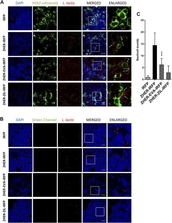
Zahirović et al.                                                                                           Engineered Bacteria With Dual Functionality

INTRODUCTION                                                               advantageous for intestinal drug delivery as it can survive the
                                                                           passage through the gastrointestinal tract (Plavec and Berlec,
Chronic, unresolved inflammation has been increasingly                      2019; Plavec and Berlec, 2020). L. lactis can serve as a protein
recognized as a key factor in the pathogenesis of many types               producer and a delivery vehicle at the same time, and it has
of cancers, including liver, pancreatic, gastric, and colorectal           previously been engineered for gastrointestinal delivery of various
cancers (CRC) (Terzic et al., 2010). CRC is the third most                 proteins, including viral antigens (Berlec et al., 2013), allergens
common cancer worldwide, and the second leading cause of                   (Zahirović and Lunder, 2018), immunomodulatory cytokines
cancer-related deaths (Sung et al., 2021). Studies have provided           (Steidler et al., 2003) and proteins with affinity for pro-
strong evidence that the pro-inflammatory cytokines interleukin             inflammatory cytokines (Ravnikar et al., 2010; Berlec et al.,
(IL)-6 and IL-8 (CXCL-8) have critical roles in the inflammatory            2017; Kosler et al., 2017; Škrlec et al., 2018; Plavec et al., 2019)
processes associated with CRC (Baier et al., 2005; West et al.,            and chemokines (Škrlec et al., 2017). Moreover, alternative
2015). IL-6 has been shown to promote tumor cell growth,                   methods to genetic modification of bacteria have been
invasion, and migration (Becker et al., 2004; Zeng et al., 2017),          introduced, including heterologous protein expression
while IL-8 increases proliferation, angiogenesis, and migration of         (Zadravec et al., 2015) and containment strategies (Steidler
malignant cells toward blood vessels, thus leading to tumor                et al., 2003). Some applications were also aimed at the
dissemination (Ning et al., 2011). Clinical data show that                 treatment of cancer (primarily CRC); these include L. lactis
patients with stage IV CRC have more than 10 times higher                  engineered to deliver antioxidant enzyme catalase (Del
serum level of IL-8 (1,089 pg/ml) than healthy individuals (79 pg/         Carmen et al., 2017) or pro-apoptotic peptide kisspeptin
ml) (Lee et al., 2012). Of the pro-inflammatory cytokines involved          (Zhang et al., 2016).
in CRC carcinogenesis, IL-6 shows the greatest increase in                     The wild-type lactic acid bacteria have shown protective
patients with CRC compared to healthy controls, particularly               effects against CRC through modulation of the gut microbiota,
in metastatic disease (Chung and Chang, 2003). Elevated IL-6               neutralization of carcinogens, and/or production of short-chain
correlates with a bad patient prognosis and poor clinical outcome          fatty acids (Uccello et al., 2012). These inherent anticancer effects
of CRC. Furthermore, pro-inflammatory cytokines have been                   can be enhanced by displaying cytokine-binding proteins on the
shown to reduce the responses to cancer immunotherapy by                   bacterial surface, which will result in the capture of pro-
inducing immunosuppression in the tumor microenvironment                   inflammatory cytokines by the bacteria, and neutralization of
(Albini and Sporn, 2007).                                                  their detrimental pro-tumorigenic effects in the tumor milieu.
    A growing body of data on the critical role of pro-                    Furthermore, expression of specific proteins directed towards
inflammatory cytokines at various stages of CRC development                 tumor antigens on the bacterial surface can enhance their
suggests that they represent new molecular targets in the                  selectivity for tumors (Duong et al., 2019). This was
treatment of CRC. Indeed, recent studies have demonstrated                 successfully achieved for attenuated Salmonella typhimurium
inhibitory effects of cytokine signaling blockers on cancer                by coating the bacterial surface with the RGD peptide that is
development and metastasis (Ning and Lenz, 2012; Johnson                   directed against αvβ3-integrin (Park et al., 2016), or with a single-
et al., 2018). These therapeutics will probably require                    domain antibody directed against the B-lymphocyte antigen
combinatorial approaches, and might therefore serve as                     CD20 (Massa et al., 2013).
adjuvant therapies following standard treatment with                           We have recently developed L. lactis strains that display small
radiotherapy, chemotherapy, or immune checkpoint inhibitors                protein binders of IL-8 (Škrlec et al., 2017) and IL-6 (Zahirović
(Wattenberg and Beatty, 2020).                                             et al., submitted) on their surface, and have demonstrated their
    When considered for cancer treatment, local delivery of                binding to cytokines in vitro. Both proteins that were displayed on
cytokine-binding proteins to the tumor is necessary to avoid               L. lactis, IL-8-binding evasin (Deruaz et al., 2008) and IL-6-
systemic cytokine blockade, and to thus circumvent the side                binding affibody (Yu et al., 2014), are non-immunoglobulin
effects associated with down-regulation of the normal immune               binders and have high specificity and affinity for their target
response. Site-specific drug delivery ensures maximal                       cytokines. We reasoned that the co-display of cytokine-binding
concentrations of therapeutic agents at the local site of action           proteins with ligands for tumor antigens can facilitate the specific
without negative side effects in healthy tissues. Sustained high           interactions of the bacteria with the cancer cells, which will
levels of cytokine-binding proteins in tumor tissue can be                 increase the accumulation and retention of the bacteria in the
achieved by targeting their delivery to the tumor using                    tumor tissue, and thus enable the targeted delivery of the
bacteria as the delivery vehicle. One of the main advantages of            cytokine-binding proteins to the tumors.
using bacteria for cancer treatments is their natural propensity for           For tumor antigen-targeting we used epithelial cell adhesion
the central hypoxic regions of a tumor. This is a prominent                molecule (EpCAM, CD326) (Das et al., 2015) and human
feature of anerobic or facultative anaerobic bacterial pathogens           epidermal growth factor receptor 2 (HER2, CD340) (Siena
such as Salmonella, Listeria, and Clostridium. However, even               et al., 2018); both are up-regulated in CRC, but not in healthy
when these bacteria are used in an attenuated form, there is a risk        cells, and therefore represent CRC markers. Small non-
of reversion to a virulent state (Toso et al., 2002).                      immunoglobulin scaffold proteins, EpCAM-binding affitin
    Lactococcus lactis (L. lactis), a model lactic acid bacterium,         (Kalichuk et al., 2018), and HER2-binding affibody (Feldwisch
raises no safety concerns and is considered a suitable host strain         et al., 2010), that possess high specificity and picomolar affinity
for development of biotherapeutics. L. lactis is particularly              for their receptors served as the targeting ligands. L. lactis bacteria

Frontiers in Bioengineering and Biotechnology | www.frontiersin.org    2                                    January 2022 | Volume 10 | Article 822823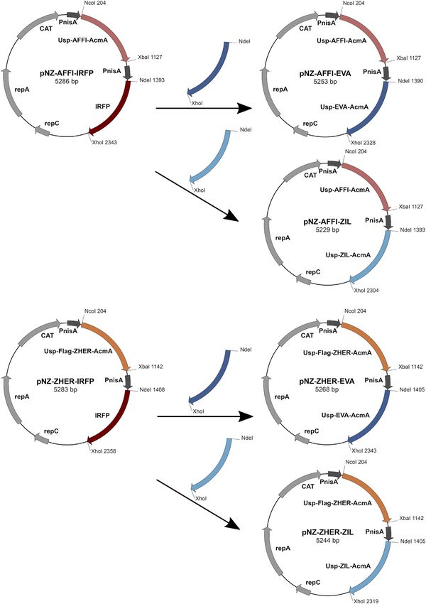
Zahirović et al.                                                                                                         Engineered Bacteria With Dual Functionality

TABLE 1 | The strains, plasmids and primers used in this study.

Strain, plasmid or primer                                 Relevant features or sequence                                                  Reference

Strain
  L. lactis NZ9000            MG1363 nisRK ΔpepN                                                                        NIZO

Plasmids
  pNZ8148                     pSH71 derivative, PnisA, Cmr, nisin-controlled expression                                 de Ruyter et al. (1996), Mierau and
                                                                                                                        Kleerebezem. (2005)
  pNZ-AFFI-IRFP               pNZ8148-containing gene fusion of spUsp45, affi and acmA3b on MCS1 and irfp gene on MCS2   Plavec et al. (2021b)
  pNZ-ZHER-IRFP               pNZ8148-containing gene fusion of spUsp45, flag-zher and acmA3b on MCS1 and irfp gene on   Plavec et al. (2021b)
                              MCS2
  pNZ-IRFP                    pNZ8148-containing irfp gene                                                              Berlec et al. (2015)
  pSD-EVA                     pNZ8148-containing gene fusion of spUsp45, evasin-3 and acmA3b                            Škrlec et al. (2017)
  pSD-ZIL                     pNZ8148-containing gene fusion of spUsp45, zil and acmA3b                                 Zahirović et al., submitted
  pNZ-AFFI-EVA-IRFP           pNBBX-containing AFFI, EVA and IRFP cassettes                                             Plavec et al. (2021a)
  pNZ-AFFI-ZIL-IRFP           pNBBX-containing AFFI, ZIL and IRFP cassettes                                             Plavec et al. (2021a)
  pNZ-ZHER-EVA-IRFP           pNBBX-containing ZHER, EVA and IRFP cassettes                                             Plavec et al. (2021a)
  pNZ-ZHER-ZIL-IRFP           pNBBX-containing ZHER, ZIL and IRFP cassettes                                             Plavec et al. (2021a)
  pNZ-AFFI-EVA                pNZ8148-containing gene fusion of spUsp45, affi and acmA3b on MCS1 and gene fusion of      This study
                              spUsp45, eva and acmA3b on MCS2
  pNZ-AFFI-ZIL                pNZ8148-containing gene fusion of spUsp45, affi and acmA3b on MCS1 and gene fusion of      This study
                              spUsp45, zil and acmA3b on MCS2
  pNZ-ZHER-EVA                pNZ8148-containing gene fusion of spUsp45, flag-zher and acmA3b on MCS1 and gene fusion    This study
                              of spUsp45, eva and acmA3b on MCS2
  pNZ-ZHER-ZIL                pNZ8148-containing gene fusion of spUsp45, flag-zher and acmA3b on MCS1 and gene fusion    This study
                              of spUsp45, zil and acmA3b on MCS2

Primers
  USP-F-Nde2                  TTATTTCATATGGCTAAAAAAAAGATTATCTCAG                                                        Škrlec et al. (2017)
  A3b-R-Xho2                  TTATTTCTCGAGTTATTTTATTCGTAGATACTGACC                                                      Škrlec et al. (2017)

NIZO, www.nizo.com; MCS, multiple cloning site.

that displayed these tumor antigen ligands on their surface were                          was added to the growth medium. Biliverdin HCl (15.5 μg/ml;
recently shown to specifically bind to tumor surface antigens                              Sigma-Aldrich) was added for expression of infrared fluorescent
(Plavec et al., 2021b). Lectin-displaying L. lactis that target                           protein (IRFP).
carbohydrate receptors on cancer cells were also developed
(Plavec et al., 2021c).
    In the present study, we sought to develop bacteria for targeted                      Construction of Dual Protein Expression
delivery of cytokine-binding proteins to tumors. We engineered                            Plasmids
L. lactis to display a combination of a tumor antigen-recognizing                         Restriction enzymes and T4 DNA ligase were from New
protein (EpCAM-binding affitin AFFI or HER2-binding affibody                                England Biolabs (Ipswich, MA, United States). PCR
ZHER) and a cytokine binding protein (IL-8–binding evasin EVA                             amplifications were performed with Taq polymerase (New
or IL-6–binding affibody ZIL) on their surface. The engineered                             England Biolabs), according to the manufacturer protocols.
bacteria were tested for their ability to remove IL-8 and IL-6 from                       Plasmid DNA was isolated (NucleoSpin plasmid; Macherey-
the supernatant of cancer cells and to bind tumor antigens                                Nagel, Düren, Germany), with addition of lysozyme (Sigma-
EpCAM or HER2 on HEK293 cells.                                                            Aldrich). Electroporation of L. lactis was performed according
                                                                                          to (Holo and Nes, 1995) (Gene Pulser II apparatus; BioRad,
                                                                                          Hercules, CA, United States). Nucleotide sequencing was
MATERIALS AND METHODS                                                                     performed by Eurofins Genomics (Ebersberg, Germany).
                                                                                          Primers (Integrated DNA Technologies, Leuven, Belgium)
Bacterial Strains, Media, and Growth                                                      and plasmids are listed in Table 1. Protein binders were
Conditions                                                                                introduced into the pNZDual plasmid using restriction
The bacterial strains used in this study are shown in Table 1.                            enzyme-based cloning. Plasmid pNZDual contains two
Lactococcus lactis NZ9000 was grown at 30°C in M17 medium                                 multiple cloning sites (MCS), each preceded by a nisin
(MilliporeSigma, Burlington, MA, United States) supplemented                              promoter, which enables expression of two proteins in L.
with 0.5% glucose (Fluka AG, Buchs, Switzerland) (GM-17                                   lactis (Berlec et al., 2018). To construct dual expression
medium) without agitation, or in the same medium solidified                                plasmids that encode tumor antigen binder along with
with 1.5% agar (Formedium, Hunstanton, United Kingdom). To                                cytokine binder, we used pNZDual plasmids which already
maintain selection pressure on L. lactis transformants, 10 μg/ml                          contained expression cassette with the tumor antigen binder
chloramphenicol (Sigma-Aldrich, St. Louis, MO, United States)                             gene (affi or zher) in MCS 1 and IRFP in MCS 2 [prepared

Frontiers in Bioengineering and Biotechnology | www.frontiersin.org                 3                                    January 2022 | Volume 10 | Article 822823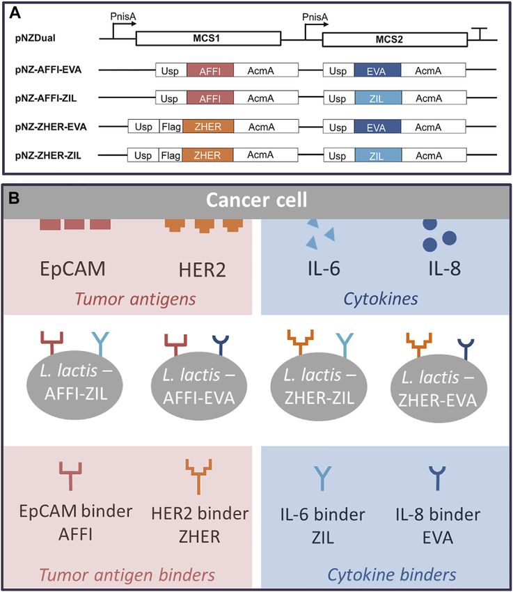
Zahirović et al.                                                                                                                       Engineered Bacteria With Dual Functionality

  FIGURE 1 | The scheme of the construction of dual expression plasmids. For construction of dual plasmids for surface display of tumor antigen binders and
  cytokine binders, we used previously prepared pNZDual plasmids which already contained expression cassette with the tumor antigen binder gene (ZHER or AFFI) in multiple
  cloning site 1 and IRFP in multiple cloning site 2. IRFP was substituted by the expression cassette containing the cytokine binder gene (EVA or ZIL). Thus, tumor antigen binder
  was paired with a cytokine binder, resulting in four dual plasmids pNZ-AFFI-EVA, pNZ-AFFI-ZIL, pNZ-ZHER-EVA, and pNZ-ZHER-ZIL. In each expression cassette, the
  binder gene was fused with the gene for Usp45 secretion signal and the gene for peptidoglycan binding domain of the AcmA protein, to enable surface display of the binder
  following its expression. AFFI, gene encoding EpCAM-binding affitin (183 bp); ZHER, gene encoding HER2-binding affibody (174 bp); EVA, gene encoding IL-8-binding
  evasin-3 (243 bp); ZIL, gene encoding IL-6-binding affibody (174 bp); Usp, gene encoding Usp45 secretion signal peptide (84 bp); AcmA, gene encoding the C-terminal
  portion of the AcmA surface anchoring protein containing three LysM repeats (642 bp); FLAG, epitope tag sequence; PnisA, nisin promoter.

Frontiers in Bioengineering and Biotechnology | www.frontiersin.org                       4                                            January 2022 | Volume 10 | Article 822823
Zahirović et al.                                                                                          Engineered Bacteria With Dual Functionality

previously in (Plavec et al., 2021b)], referred to as pNZ-AFFI-           Systems); for EVA, human IL-8 conjugated with Fc region
IRFP and pNZ-ZHER-IRFP, respectively. We replaced IRFP in                 (Sinobiological, China); and for ZIL, biotinylated human IL-6
the MCS 2 with the expression cassette containing the cytokine            (ImmunoTools, Friesoythe, Germany). Proteins conjugated to Fc
binder gene (eva or zil) via NdeI/XhoI restriction sites. Thus,           region were subsequently incubated with goat anti-human Fc
tumor antigen binder was paired with a cytokine binder,                   antibody (1:1,000, Jackson ImmunoResearch, West Grove, PA,
resulting in four dual plasmids pNZ-AFFI-EVA, pNZ-AFFI-                   United States), whereas IL-6 conjugated to biotin was incubated
ZIL, pNZ-ZHER-EVA, and pNZ-ZHER-ZIL (Table 1). A                          with mouse anti-biotin antibody (1:1,000, Abcam, Waltham, MA,
construction of dual protein expression plasmids is depicted              United States). After three washes with TBST, the membranes
in Figure 1. In each expression cassette, the binder gene was             were incubated with horseradish peroxidase (HRP)-conjugated
fused with the gene for Usp45 secretion signal and the gene for           secondary antibodies diluted 1:5,000 in blocking buffer, for 1.5 h.
peptidoglycan binding domain of the AcmA protein, to enable               The secondary antibodies used were HRP-conjugated anti-mouse
its release into the growth medium and subsequent surface                 (1:5,000; Jackson ImmunoResearch) and HRP-conjugated anti-
anchoring. The expression cassette containing cytokine binder             goat (1:5,000; Jackson ImmunoResearch). Following three washes
gene eva or zil was amplified from pSD-EVA (Škrlec et al.,                 with TBST, the membranes were incubated with a
2017) or pSD-ZIL plasmid (Zahirović et al., submitted),                   chemiluminescent reagent (Clarity Western ECL Substrate;
respectively. For generation of pSD-ZIL plasmid, zil gene                 Bio-Rad). Images were acquired using an imaging system
was back-translated and codon-optimized for use in L. lactis              (ChemiDoc MP; BioRad).
based on the amino acid sequence of IL-6-binding affibody
ZIL-6_13 (Yu et al., 2014). Zil gene was cloned into a
lactococcal plasmid pSDBA3b in the same way as evasin 3                   ELISA for Determination of Cytokine
following the protocol described in Škrlec et al. (2017). For             Binding by L. lactis
visualization of engineered bacteria, IRFP-encoding gene was              The cytokine concentrations were determined using commercially
subsequently inserted into the plasmid along with the protein             available ELISA kits (Mabtech, Nacka Strand, Sweden), according to
binders, using BglBrick cloning, which allowed easier assembly            the manufacturer recommendations. Here, 96-well plates (Maxisorp
of multiple gene cassettes (Plavec et al., 2021a).                        Nunc; Thermo Fisher Scientific) were coated with cytokine-binding
                                                                          antibodies at 4°C overnight. The wells were washed five times with
Expression of Fusion Proteins in L. lactis                                200 μl PBS containing 0.05% Tween-20 (wash buffer) and blocked
Overnight cultures of L. lactis harboring constructed plasmids            for 1 h at room temperature with PBS with 0.05% Tween and 0.1%
were diluted (1:50) in 10 ml fresh GM-17 medium, and grown to             bovine serum albumin (incubation buffer). L. lactis cells at 6 × 10⁹
an optical density (A600) of 0.50–0.80. Expression of the fusion          colony forming units (CFU)/ml were centrifuged (5,000 × g, 5 min,
proteins was induced by addition of 25 ng/ml nisin (Fluka AG)             4°C) and resuspended in 250 µl incubation buffer spiked with
(de Ruyter et al., 1996; Mierau and Kleerebezem, 2005). After a           300 pg/ml cytokine standards (Mabtech). The optical density of
3 h incubation, 1 ml culture was stored at 4°C for flow cytometric         bacterial suspension at 600 nm (OD600) was used to calculate the
analysis, and the remaining cell culture was centrifuged at 5,000×        number of L. lactis per ml using a factor determined earlier by serial
g for 10 min. The cell pellet was resuspended in 400 µl phosphate-        dilutions (1 OD600 = 1 ×10⁹ L. lactis/ml). Following 2 h at room
buffered saline (PBS; 10 mM Na2HPO4, 1.8 mM KH2PO4,                       temperature with gentle shaking, the bacterial cells were removed by
137 mM NaCl, and 2.7 mM KCl; pH 7.4) and stored at −20°C                  centrifugation (7,500× g, 7 min, room temperature), and 200 μl cell-
for SDS-PAGE analysis, or resuspended in different volumes of             free bacterial supernatant was added to the wells and incubated for
PBS for analysis of cytokine binding by ELISA.                            2 h at room temperature. The remaining cytokine levels in the
                                                                          supernatant were determined using a calibration curves, that were
SDS-PAGE and Western Blotting                                             generated by addition of different concentrations (0–1,200 pg/ml) of
SDS-PAGE was performed with a Mini-Protean II apparatus                   recombinant cytokine standards from the ELISA kit (Mabtech). The
(BioRad). Samples were thawed in an ice bath, briefly sonicated            standards were incubated in the same buffer and under the same
(UPS200S; Hielscher, Teltow, Germany), mixed with 2× Laemmli              conditions as the samples, to ensure equal matrix effect. Following
sample buffer and dithiothreitol, and denatured by heating at             incubation and washing of the wells, 100 μl biotinylated monoclonal
100°C before loading. Pre-stained standards (Page Ruler Plus;             antibodies against the cytokines were added in incubation buffer at
Thermo Fisher Scientific, Waltham, MA, United States) were                 their recommended concentrations, and incubated at room
used for molecular weight comparisons. Proteins were transferred          temperature for 1 h. Following washing, 100 μ streptavidin-HRP
onto nitrocellulose membrane (GE Healthcare Life Sciences,                (diluted 1:1,000 in incubation buffer) was added into the wells and
Marlborough, MA, United States) using semi-dry transfer at                incubated for 1 h at room temperature. The wells were washed again,
100 V for 90 min. Membranes were blocked with 5% non-fat                  and 50 μl 3,3′,5,5′-tetramethylbenzidine substrate (Sigma-Aldrich)
dried milk in TBS (50 mM Tris-HCl, 150 mM NaCl; pH 7.5) with              was added. The reaction was terminated after 20 min by addition of
0.05% Tween-20 (TBST) and incubated overnight at 4°C in                   50 μl 2 M sulfuric acid. Absorbances were read at 450 nm using a
blocking buffer with target proteins. For detection of protein            microplate reader (Infinite M1000; Tecan, Grödig, Austria), with
binders, the following target proteins were used: for AFFI,               wavelength correction at 650 nm. The binding is given as the
EpCAM/TROP1 Fc chimera (R&D Systems, Minneapolis, MN,                     proportions (%) of the cytokine removed from the solution by
United States); for ZHER, ErbB2/Her2 Fc chimera (R&D                      the bacteria.

Frontiers in Bioengineering and Biotechnology | www.frontiersin.org   5                                   January 2022 | Volume 10 | Article 822823
Zahirović et al. Engineered Bacteria With Dual Functionality
Cell Lines and Culturing with 1.5 µl PolyJet (SignaGen Laboratories, Rockville, MD,
Human embryonic kidney epithelial HEK293 cells (CRL-1573; United States) and 0.5 µg plasmid [for expression of the
American Type Culture Collection [ATCC], Manassas, Virginia, EpCAM-sfGFP fusion protein, pcDNA3-EpFL-sfGFP (Gaber
United States) and human colon adenocarcinoma Caco-2 cells et al., 2018); for expression of the HER2-mEmerald fusion
(HTB-37; ATCC) were cultured in Dulbecco’s modified Eagle’s protein, mEmerald-ERBB2-N-18 (Addgene plasmid #62755;
medium with high glucose and GlutaMAX (Gibco, Thermo http://n2t.net/addgene:62755; RRID:Addgene_62755)],
Fisher Scientific). Human colon adenocarcinoma HT29 according to the manufacturer protocols. The medium was
epithelial intestinal cells (HTB-38; ATCC) were maintained in replaced 24 h after transfection.
McCoy’s medium with 1.5 mM L-glutamine and 2,200 mg/L
sodium bicarbonate (ATCC). Human monocytic cell lines Bacterial Cell Adhesion Assay
THP-1 (TIB-202; ATCC) and U-937 (CRL-1593.2; ATCC) Bacterial cell adhesion assays were carried out as reported
were cultured in RPMI1640 medium (Gibco). Each medium previously (Plavec et al., 2021b). Here, the medium from
was supplemented with 10% (v/v) fetal bovine serum (Gibco), transfected HEK293 cells was aspirated and 500 μl 8 ×
100 U/mL penicillin, and 100 μg/ml streptomycin (Gibco). For 108 CFU/ml engineered L. lactis bacteria (diluted in pre-warmed
Caco-2 cell culturing, 25 mM HEPES (Sigma-Aldrich) and 1% RPMI; A600, 0.8) were added to each well, and incubated for 2 h at
Eagle’s minimum essential medium nonessential amino acids 37°C. Following incubation, the wells were gently washed twice
solution (Sigma-Aldrich) were added. Unless otherwise stated, with PBS to remove unattached L. lactis, fixed with 4%
the cells were seeded in 24-well tissue culture plates (Corning, paraformaldehyde in PBS for 20 min at room temperature,
Thermo Fisher Scientific) at 1 ×10⁵ or 3 ×10⁵ cells/well and washed twice with PBS, and mounted on a microscope slide
incubated at 37°C in a humidified atmosphere containing with DAPI-containing mounting agent (Thermo Fisher
5% CO2. Scientific), for confocal microscopy. Images were acquired with
a confocal microscope (LSM-710; Carl Zeiss, Germany) using a
×63 oil-immersion objective, with settings for bright-field, DAPI
(blue channel, 405 nm), GFP/mEmerald (green channel, 488 nm),
Cell Differentiation, Stimulation of Cytokine and IRFP (red channel, 633 nm). Images were processed and
Production, and Incubation With Bacteria analyzed using the ImageJ software. The adhesion of L. lactis
THP-1 and U-937 cells were differentiated into macrophage- cells to the HEK293 cells was quantified by counting the
like cells by incubation with 50 nM phorbol 12-myristate 13- numbers of L. lactis in 10 representative microscopy images
acetate (PMA; Sigma-Aldrich) for 48 h. Before stimulation of using ImageJ (Schneider et al., 2012). The results are expressed
cytokine secretion, PMA-containing medium was replaced as mean numbers of L. lactis per human cell ±standard deviation.
with fresh RPMI medium without PMA, and the cells were Human cells were counted using a cell counter plugin for ImageJ
incubated for another 48 h, to allow recovery. Cell (https://imagej.nih.gov/ij/plugins/cell-counter.html), where the
differentiation was verified by evaluation of their cells on the edges were excluded. L. lactis cells were counted
morphological changes under microscope. IL-6 production with the particle analysis function using the Otsu threshold
was induced by treating the cells with 1 μg/ml algorithm. To avoid background fluorescence being included as
lipopolysaccharide (LPS) from Escherichia coli serotype particles, the minimum size was set to 1 μm2. The area of a single L.
O55:B5 (L6529; Sigma-Aldrich) for 24 h. IL-8 secretion lactis bacterium was determined by averaging areas of putative
was induced by exposing the cells to 25 ng/ml human single bacteria for each set of 10 images. To determine the number
recombinant IL-1β (Cell Genix, Freiburg, Germany) for of individual L. lactis cells, the areas of aggregates were divided by
6 h. Cytokine levels were measured in the conditioned the calculated area of a single L. lactis bacterium.
media from the stimulated cells using ELISA development
kits (Mabtech), as described above. For cytokine removal Statistical Analysis
experiments, the engineered bacteria (6 × 10 9 CFU/ml) were Statistical analyses were performed using the Graph-Pad Prism
incubated with the conditioned media from the stimulated 7.00 software (San Diego, CA, United States). All of the data
cells or were co-cultured with the stimulated cells for 2 h. are presented as means ± standard deviation (SD). The
Before or after incubation with bacteria, conditioned media concentrations of cytokines obtained by ELISA were
were collected from stimulated cells, centrifuged (5 min compared using student’s two-tailed t-tests. Differences in
2,000 g at 4 °C and 15 min 1,000 g at 4 °C), and stored at cytokine-binding between relevant pairs of bacteria are
−80 °C until analysis. The contents of the remaining considered significant for p < 0.05.
cytokines in the cell culture supernatants were measured
with ELISA, and the proportions (%) of cytokines removed
by the engineered bacteria were calculated. Conditioned RESULTS
media from untreated cells were used as controls.
Design of Plasmids for Dual Protein
Cell Transfection Expression
HEK293 cells were seeded in 24-well plates at 2 ×10⁵ cells/well Four dual plasmids containing expression cassettes for
and transfected 24 h later, at approximately 70% confluency, simultaneous surface display of tumor antigen binders
Frontiers in Bioengineering and Biotechnology | www.frontiersin.org 6 January 2022 | Volume 10 | Article 822823Zahirović et al. Engineered Bacteria With Dual Functionality FIGURE 2 | Schematic representation of the gene constructs that were cloned into the lactococcal pNZDual plasmid for co-expression and simultaneous display of tumor antigen binding proteins and cytokine binding proteins on the surface of the Lactococcus lactis bacteria (A), with the resulting recombinant L. lactis strains (B). AFFI, gene encoding EpCAM-binding affitin (183 bp); ZHER, gene encoding HER2-binding affibody (174 bp); EVA, gene encoding IL-8-binding evasin-3 (243 bp); ZIL, gene encoding IL-6-binding affibody (174 bp); Usp, gene encoding Usp45 signal peptide for secretion into the growth medium (84 bp); AcmA, gene encoding the C-terminal portion of the AcmA protein containing three LysM repeats for surface anchoring (642 bp); FLAG, epitope tag sequence; MCS, multiple cloning site; PnisA, nisin promoter. and cytokine binders were constructed. The expression fusion protein due to negative effect on its display and cassette of each individual binders contained the protein functionality (Plavec et al., 2021b). binder gene fused to gene for signal peptide Usp45 and the gene for the surface anchor AcmA to enable its surface display. Based on protocol described in the Tumor Antigen Targeting and Cytokine Materials and methods (depicted in Figure 1), tumor Binding Proteins Are Co-expressed in L. lactis antigen binder gene (zher; affi) was paired with cytokine To examine the co-expression of the protein binders in the binder gene (eva; zil), resulting in dual plasmids pNZ- engineered L. lactis, whole bacterial cell lysates were analyzed by AFFI-EVA, pNZ-AFFI-ZIL, pNZ-ZHER-EVA, and pNZ- Western blotting. L. lactis transformed with the pSD plasmids that ZHER-ZIL (Table 1). The L. lactis strains transformed with encoded the individual binders were used as positive controls. the constructed dual plasmids were designated as L. lactis- Immunoblotting revealed the bands at 35 kDa, which AFFI-EVA, L. lactis-AFFI-ZIL, L. lactis-ZHER-EVA, and L. corresponds to the molecular weight of the full-length fusion lactis-ZHER-ZIL. The schematic of the dual gene constructs proteins (protein binder ~7 kDa, AcmA ~25 kDa). A and the resulting L. lactis strains are shown in Figure 2. A combination of protein binders (AFFI and EVA; AFFI and ZIL; FLAG-tag consensus sequence was added previously between ZHER and EVA; ZHER and ZIL) was present in the cell lysates from the secretion signal and the ZHER coding sequence to L. lactis strains carrying dual plasmids (Figure 3). Contrary to facilitate detection, whereas it was excluded from the AFFI distinct bands representing binders ZHER, AFFI and ZIL, the Frontiers in Bioengineering and Biotechnology | www.frontiersin.org 7 January 2022 | Volume 10 | Article 822823
Zahirović et al. Engineered Bacteria With Dual Functionality
FIGURE 3 | Tumor antigen targeting and cytokine binding proteins are co-expressed in L. lactis. Detection of tumor antigen binders AFFI (A) or ZHER (B) in combination
with cytokine binders EVA (C) or ZIL (D) in the cell lysates of the dual-plasmid-containing L. lactis by Western blotting. Cont, L. lactis carrying empty plasmid pNZ8148. L. lactis
expressing single binders (harboring pSD plasmids) served as the positive control. All binders were expressed in fusion with the Usp45 secretion signal and the AcmA surface
anchor. The bands at 35 kDa correspond to the calculated molecular weights of the full-length fusion proteins (protein binder ~7 kDa, AcmA ~25 kDa). Recombinant
human receptor EpCAM/TROP1 Fc chimera protein was used for detection of AFFI fusion protein, recombinant human receptor ErbB2/Her2 Fc chimera protein was used for
detection of ZHER, recombinant human IL-8 conjugated to Fc region was used for detection of EVA, and recombinant biotinylated human IL-6 was used for detection of ZIL.
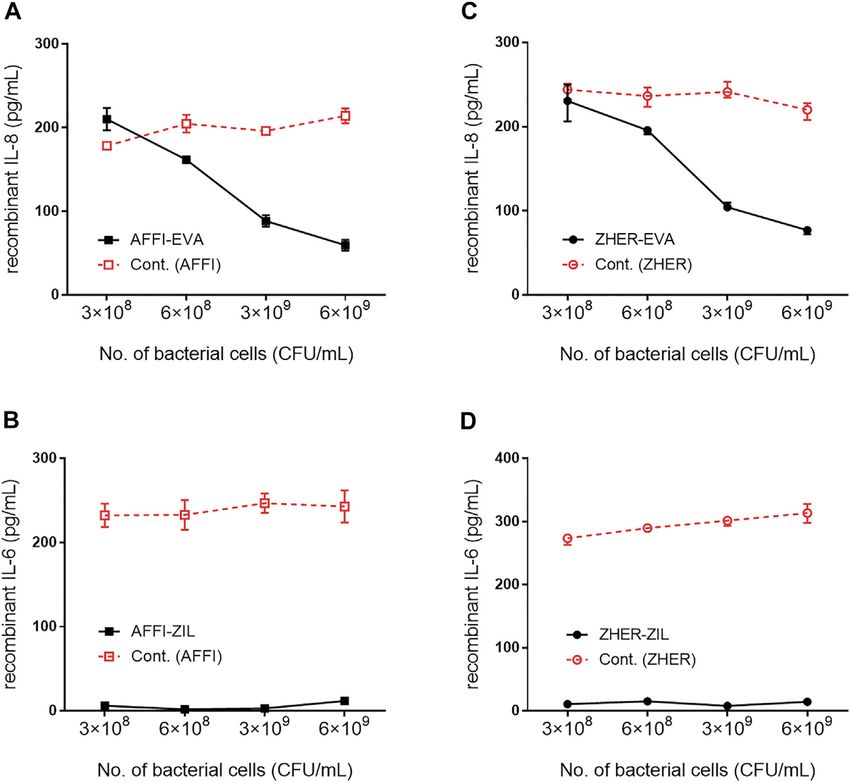
bands representing EVA were faint or invisible for all variants, binders) was assessed by incubation of increasing numbers
suggesting low functionality of EVA following SDS PAGE and of bacterial cells with recombinant cytokines added to the
western blot under denaturing conditions. Successful expression solutions at 300 pg/ml. The levels of the cytokines that
of EVA from pSD-EVA has been demonstrated previously remained in the solution were determined by ELISA. The
(Škrlec et al., 2017) and therefore this plasmid was used in the calculated recoveries were 102% for IL-8 and 98% for IL-6.
present study as a positive control. To facilitate detection, we tagged The results showed that 6 ×10⁹, 3 ×10 9, 6 ×10⁸, and 3 ×
EVA with either cmyc or flag and confirmed EVA expression in both 10 8 CFU/ml L. lactis-AFFI-EVA removed 81, 71, 47, and
single and dual plasmid carrying L. lactis strains by Western blotting 32%, respectively, of IL-8 from the solution (Figure 4A).
using anti-cmyc and anti-flag antibodies (data not shown). The Similarly, 6 ×10 9, 3 ×10⁹, 6 ×10⁸ and 3 × 10⁸ CFU/ml of
fusion proteins were consistently detected as double bands, with L. lactis-ZHER-EVA removed 75, 66, 36, and 25%,
additional slightly higher bands that probably represent their respectively, of IL-8 from the solution (Figure 4C). Thus,
unprocessed forms (with signal sequence). In the bacterial growth the extent of IL-8 removal correlated directly with the
medium, only one band was identified, corresponding to the mature number of bacterial cells. In contrast, the extent of IL-6
form of the fusion proteins (data not shown). No bands were seen for removal was uniformly high (>95%) regardless of the
lysates of control L. lactis cells carrying the empty plasmid pNZ8148. number of bacterial cells tested (Figures 4B,D). The
estimated minimal number of bacterial cells able to reduce
a 50% of IL-8 was 108 CFU/ml, whereas the minimal number
Removal of Recombinant IL-8 and IL-6 by of cells able to reduce a 50% of IL-6 was at least one order of
Engineered L. lactis as a Function of magnitude lower. L. lactis displaying only tumor antigen
Bacterial Cell Number binder (AFFI or ZHER) were used as the negative control
The cytokine-binding ability of the engineered bacteria (i.e., L. and showed negligible or low binding of recombinant
lactis co-expressing tumor antigen binders and cytokine cytokines.
Frontiers in Bioengineering and Biotechnology | www.frontiersin.org 8 January 2022 | Volume 10 | Article 822823Zahirović et al. Engineered Bacteria With Dual Functionality
FIGURE 4 | Removal of recombinant IL-8 and IL-6 by engineered L. lactis as a function of bacterial cell number. The concentration of recombinant IL-8 and recombinant
IL-6 that remained in the solution after incubation with increasing numbers of engineered L. lactis cells, as determined by ELISA. Bacterial strains tested: L. lactis co-expressing
tumor antigen targeting and cytokine binding proteins L. lactis-AFFI-EVA (A), L. lactis-AFFI-ZIL (B), L. lactis-ZHER-EVA (C), or L. lactis-ZHER-ZIL (D) (solid line). Cont: L. lactis
co-expressing tumor antigen binders and IRFP L. lactis-AFFI-IRFP or L. lactis-ZHER-IRFP (dashed line). Data are means ± SD, with experiments performed in triplicate.
L. lactis Co-Expressing IL-8 Binder EVA and most cases (bound 17–40% more IL-8) compared with L. lactis
the Tumor Antigen Binder AFFI or ZHER strains carrying dual plasmids. As for the control bacteria, L.
lactis-ZHER eliminated negligible amounts of IL-8. On the
Removes IL-8 Secreted by Caco-2 and contrary, L. lactis-AFFI bound surprisingly high levels of IL-8
HT-29 Colorectal Cancer Cells secreted by HT-29 cells (up to 99%), which may be due to post-
The basal secretion of IL-8 into the culture medium by untreated translational modifications of HT-29-derived IL-8 that may have
Caco-2 and HT-29 cells was low (Zahirović et al. Engineered Bacteria With Dual Functionality FIGURE 5 | L. lactis co-expressing IL-8 binder EVA and the tumor antigen binder AFFI or ZHER removes IL-8 secreted by Caco-2 and HT-29 cells. ELISA- determined concentrations of IL-8 after incubation of engineered L. lactis (6 × 10⁹ CFU/ml) with conditioned media of IL-1β–stimulated Caco-2 cells (A) or HT-29 cells (B), and after co-culture with IL-1β–stimulated Caco-2 cells (C) or HT-29 cells (D). L. lactis strains tested: L. lactis-AFFI-EVA and L. lactis-ZHER-EVA (L. lactis co- expressing tumor antigen binders and EVA); Negative controls: L. lactis-AFFI-IRFP and L. lactis-ZHER-IRFP (L. lactis co-expressing tumor antigen binders and IRFP); Positive control: L. lactis-EVA (L. lactis expressing cytokine binder EVA only). Data are means ± SD, with experiments performed in triplicate. **, p = 0.001; ***, p < 0.001) (unpaired t-tests), between the relevant bacterial pairs (L. lactis co-expressing cytokine binders and tumor antigen binders, and negative controls). experiments, the engineered bacteria were incubated with IL- cellular extensions), and increased cell granularity (Schutte et al., 6–conditioned medium from LPS-stimulated cells and the 2009). After 24 h of stimulation with LPS, the concentration of IL-6 residual IL-6 was determined by ELISA. As shown in increased substantially. The levels of IL-6 varied in different Figure 6, the IL-6 binding was very efficient; L. lactis-AFFI- experiments depending on the cell number, cell type (U-937 ZIL and L. lactis-ZHER-ZIL removed 94 and 92%, respectively, of released more IL-6 than THP-1), or their differentiation status. the IL-6 from the supernatant of THP-1 cells (Figure 6A). The If IL-6 concentration was above the linear range the supernatants extent of binding was essentially equal as that of L. lactis-ZIL. were diluted. The supernatants that were used for incubation with When co-cultured with THP-1 cells, the engineered bacteria bacteria contained IL-6 in the range from 469 to 1,040 pg/ml retained the same levels of IL-6 removal (Figure 6D). (Figures 6B,C,E,F). Similar to previous observations with In the tumor microenvironment, monocytes undergo activation undifferentiated THP-1 cells, we found that ZIL co-expressing and differentiation into macrophages, known as tumor-associated L. lactis strains removed high proportion (from 96 to 99%) of the macrophages (Soncin et al., 2018). To examine whether the IL-6 released in the cell culture supernatant of the differentiated engineered bacteria can remove macrophage-derived IL-6 from THP-1 cells and the differentiated U-937 cells (Figures 6B,C). their environment, we established a model of macrophages by When the engineered bacteria were co-cultured with the cells, the exposing human monocytic leukemia THP-1 cells and human proportions of bound IL-6 remained as high as after incubation of histiocytic lymphoma U-937 cells to PMA for 48 h. This treatment the bacteria with conditioned media (Figures 6E,F). The control L. induced typical features of macrophages, such as cell adhesion, lactis strains bound a negligible amount of IL-6 (≤3%) in changes in morphology (from circular to spindle-shaped cells, with most cases. Frontiers in Bioengineering and Biotechnology | www.frontiersin.org 10 January 2022 | Volume 10 | Article 822823
Zahirović et al.                                                                                                                    Engineered Bacteria With Dual Functionality

  FIGURE 6 | L. lactis co-expressing IL-6 binder ZIL and the tumor antigen binder ZHER or AFFI removes IL-6 secreted by THP-1 cells and U-937 cells. ELISA-
  determined concentrations of IL-6 after incubation of engineered L. lactis (6 × 10⁹ CFU/ml) with conditioned media of LPS-stimulated THP-1 (A), differentiated THP-1
  (B), or differentiated U-937 (C) cells, and after co-culture of engineered L. lactis strains with LPS-stimulated THP-1 (D), differentiated THP-1 (E), or differentiated U-937
  (F) cells. The cells were differentiated by incubation with phorbol 12-myristate 13-acetate for 48 h. L. lactis strains tested: L. lactis-AFFI-ZIL and L. lactis-ZHER-ZIL
  (L. lactis co-expressing tumor antigen binders and ZIL); Negative controls: L. lactis-AFFI-IRFP and L. lactis-ZHER-IRFP (L. lactis co-expressing tumor antigen binders
  and IRFP); Positive control: L. lactis- ZIL (L. lactis expressing cytokine binder ZIL only). Data are means ± SD, with experiments performed in triplicate. ***, p < 0.001
  (unpaired t-tests), between the relevant bacterial pairs (L. lactis co-expressing cytokine binders and tumor antigen binders, and negative controls).

Engineered L. lactis Strongly and Selectively                                                lactis-AFFI-EVA-IRFP, 48 L. lactis-AFFI-ZIL-IRFP, and 55 L. lactis-
Adhere to EpCAM-Overexpressing HEK293                                                        AFFI-IRFP (positive control) adhered per single EpCAM-bearing
                                                                                             HEK293 cell, whereas only 2 L. lactis-IRFP (negative control)
Cells                                                                                        adhered per single EpCAM-bearing HEK293 cell (Figure 7C).
The feasibility of tumor targeting with AFFI co-expressing bacteria
                                                                                             Compared to the positive control, a 49% and a 13% reduction of
was assessed using HEK293 cells transfected with the EpCAM
                                                                                             the adhesion was noticed for L. lactis-AFFI-EVA-IRFP and L. lactis-
receptor fused to fluorescent protein superfolder GFP (sfGFP).
                                                                                             AFFI-ZIL-IRFP, respectively. The average number of adhered
To visualize bacterial adhesion by fluorescence microscopy, we
                                                                                             bacteria per nontransfected HEK293 cell was
Zahirović et al. Engineered Bacteria With Dual Functionality FIGURE 7 | The engineered L. lactis strongly and selectively adheres to EpCAM-overexpressing HEK293 cells. Representative confocal microscopy images of adhesion of AFFI co-expressing L. lactis to HEK293 cells transfected with EpCAM (A) or to nontransfected HEK293 cells (B). Images were quantified by determining the number of adhered bacteria per HEK293 cell in 10 representative micrographs for each experimental condition, using ImageJ (C). L. lactis strains tested: L. lactis-AFFI-EVA-IRFP (L. lactis co-expressing AFFI, EVA, and IRFP); L. lactis-AFFI-ZIL-IRFP (L. lactis co-expressing AFFI, ZIL, and IRFP); L. lactis-AFFI-IRFP (L. lactis co-expressing AFFI and IRFP; positive control); and L. lactis-IRFP (L. lactis expressing IRFP only; negative control). DAPI, DAPI channel showing HEK293 cell nuclei; HEK293-EpCAM-sfGFP, green fluorescence channel showing superfolder green fluorescent protein (sfGFP)-labeled EpCAM overexpressed on the surfaces of the HEK293 cells; L. lactis, red fluorescence channel showing L. lactis. Enlarged images show attachment of L. lactis to transfected HEK293 cells with characteristic adhesion pattern (arrows). White squares indicate the enlarged sections of the images. Scale bars, 20 μm. HER2 receptor fused to fluorescent protein monovalent microscopy showed that the engineered bacteria selectively variant of GFP mEmerald. The attached bacteria were adhered to HER2-overexpressing HEK293 cells and showed visualized by confocal fluorescence microscopy via the no reactivity towards nontransfected HEK293 cells (Figures IRFP reporter protein that was co-expressed in L. lactis 8A,B). The adhesion pattern of the HER2-targeting bacteria with the protein binders (Plavec et al., 2021a). Confocal was similar to that of the EpCAM-targeting L. lactis strains, Frontiers in Bioengineering and Biotechnology | www.frontiersin.org 12 January 2022 | Volume 10 | Article 822823
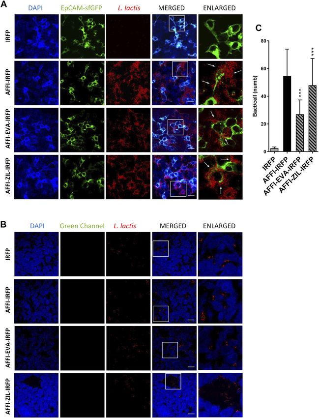
Zahirović et al.                                                                                                                 Engineered Bacteria With Dual Functionality

  FIGURE 8 | The engineered L. lactis specifically adhere to HER2-overexpressing HEK293 cells. Representative confocal microscopy images of adhesion of ZHER
  co-expressing L. lactis to HEK293 cells transfected with HER2 (A) or to nontransfected HEK293 cells (B). Images were quantified by determining the number of adhered
  bacteria per HEK293 cell in 10 representative micrographs for each experimental condition, using ImageJ (C). L. lactis strains tested: L. lactis-ZHER-EVA-IRFP (L. lactis
  co-expressing ZHER, EVA, and IRFP); L. lactis-ZHER-ZIL-IRFP (L. lactis co-expressing ZHER, ZIL, and IRFP); L. lactis-ZHER-IRFP (L. lactis co-expressing ZHER
  and IRFP; positive control); and L. lactis-IRFP (L. lactis expressing only IRFP; negative control). DAPI, DAPI channel showing HEK293 cell nuclei; HEK293-HER2-
  mEmerald, green fluorescence channel showing mEmerald-labeled HER2 overexpressed on the surfaces of the HEK293 cells; L. lactis, red fluorescence channel
  showing L. lactis. Enlarged images show attachment of L. lactis to transfected HEK293 with characteristic adhesion pattern (arrows). White squares indicate enlarged
  sections of the images. Scale bars, 20 μm.

with the bacteria mostly attached to the cell edges (Figure 8A,                            the adhesion was noticed for L. lactis-ZHER-EVA-IRFP and
arrows). Quantification showed that on average 6 L. lactis-                                 L. lactis-ZHER-ZIL-IRFP, respectively. ZHER co-expressing
ZHER-EVA-IRFP, 3 L. lactis-ZHER-ZIL-IRFP, 14 L. lactis-                                    bacteria showed no reactivity towards nontransfected
ZHER-IRFP (positive control), and 1 L. lactis-IRFP (negative                               HEK293 cells, with no more than 1 bacterium adhering per
control) adhered per HER2-bearing HEK293 cell (Figure 8C).                                 nontransfected HEK293 cell (Figure 8B). These data confirm
Compared with positive control, a 57% and a 79% reduction of                               that the displayed HER2-targeting tumor antigen binder

Frontiers in Bioengineering and Biotechnology | www.frontiersin.org                   13                                          January 2022 | Volume 10 | Article 822823
Zahirović et al.                                                                                        Engineered Bacteria With Dual Functionality

ZHER effectively and specifically promotes adherence of L.                  cells. To assess the removal of the cytokines involved in cancer
lactis to the transfected HER2-bearing HEK293 cells.                       inflammatory processes by the engineered bacteria, we
However, the adhesion level was significantly lower for the                 established cell models that mimic certain elements of the
HER2-targeting L. lactis strains compared with the EpCAM-                  inflammatory milieu within the tumor microenvironment.
targeting L. lactis strains.                                               Under inflammatory conditions, cancer cells are a source of
                                                                           IL-8 in the tumor microenvironment. We used the Caco-2 and
                                                                           HT-29 cell lines as a model of CRC cells. To simulate
DISCUSSION                                                                 inflammatory conditions, the Caco-2 and HT-29 CRC cells
                                                                           were primed with IL-1β, a potent inducer of IL-8 secretion.
Tumorigenesis in CRC is driven by soluble mediators,                       The optimal dose of IL-1β required to induce maximal IL-8
including the pro-inflammatory cytokines IL-6 and IL-8,                     secretion in Caco-2 cells was determined to be 25 ng/ml
which orchestrate the many-fold cellular activities that                   (Škrlec et al., 2017). Autocrine secretion of IL-8 can
underlie inflammation. Consequently, modulation of these                    activate intrinsic mechanism of tumor cells to promote
cytokines might be a promising therapeutic strategy for cancer             their escape from stress-induced apoptosis (Doll et al.,
treatments. Here, we engineered L. lactis to serve as a vector             2010). Cancer-cell-derived IL-8 can enhance tumor growth,
for targeted delivery of cytokine-binding proteins to tumor.               invasion, angiogenesis, metastasis, and resistance to therapy
The L. lactis bacteria were constructed by displaying a tumor              (Doll et al., 2010). In breast cancer, it has been shown that
antigen-recognizing moiety and a cytokine-binding moiety on                expression of IL-8 can be modulated by the manipulation of
their surface. These engineered dual functionalized bacteria               EpCAM expression in vitro (Sankpal et al., 2013). Here, we
were shown to selectively target HEK293 cells that                         show that cell-released IL-8 can be effectively captured and
overexpress tumor antigens. Moreover, the engineered                       removed by the engineered bacteria both in the presence and
bacteria removed a high proportion of the IL-8 from the                    absence of the producer cells. IL-8 removal was substantial,
supernatants of immunostimulated Caco-2 and HT-29                          whereby L. lactis co-expressing EVA and tumor antigen
colon adenocarcinoma cell lines, and depleted IL-6 from                    binders removed 65–69% of IL-8 from Caco-2 supernatant
the supernatants of immunostimulated THP-1 and U-937                       and 71–100% of IL-8 from supernatant of HT-29 cells.
monocyte-like cells.                                                           In addition to cancer-cell-derived mediators, pro-
   L. lactis with dual functionality was developed by                      inflammatory cytokines produced by immune cells
simultaneously displaying a combination of a tumor                         (i.e., primarily monocytes and macrophages) also drive
antigen-recognizing protein and a cytokine-binding protein                 inflammation in the CRC microenvironment (Hao et al.,
on the bacterial surface using a dual promoter plasmid                     2012). Monocytes are attracted to tumor tissue by
(pNZDual). IL-8–binding evasin (designated as EVA) and                     chemokines such as IL-8, which is one of the major
IL-6–binding affibody (designated as ZIL) were used to                      chemoattractants in CRC (Ning et al., 2011). In the tumor
achieve cytokine removal, while EpCAM-binding affitin                       microenvironment, monocytes respond to paracrine stimuli
(designated as AFFI) and HER2-binding affibody                              from cancer cells by secreting soluble mediators, most notably
(designated as ZHER) served for tumor antigen targeting.                   IL-6, and these participate in tumor growth, invasion,
   After confirming the co-expression of the fusion proteins                intravasation, and metastasis (Soncin et al., 2018). For the
in all dual plasmid carrying L. lactis strains, their cytokine             analysis of IL-6 removal by the engineered bacteria, we used
binding properties were investigated in relation to the                    the immunostimulated THP-1 human acute monocytic
bacterial cell number using recombinant cytokines. The                     leukemia cell line and the U937 histiocytic lymphoma cell
engineered bacteria removed more than 90% of                               line. PMA-differentiated THP-1 and U-937 cells were used as
recombinant IL-6 from the solution independent of                          cell models of macrophages. It is known that the production of
bacterial cell numbers tested, whereas they removed                        pro-inflammatory cytokines increases when monocytes
between 25 and 81% of recombinant IL-8 in a dose-                          differentiate into macrophages, which suggests that they
dependent manner. In agreement with previous study                         are more likely to induce inflammation than monocytes
(Škrlec et al., 2017), the highest IL-8 removal was achieved               (Soncin et al., 2018). After incubation of the engineered
by 6 × 10⁹ CFU/ml bacterial cells, which falls within the range            bacteria with stimulated cells or their conditioned medium,
of therapeutic doses in current probiotic preparations for                 high levels of IL-6 were removed (>90%) by ZIL co-expressing
human use. Although typical doses of probiotics vary by                    L. lactis in both the undifferentiated and differentiated
product, most clinical studies have examined doses from 1                  monocyte-like cell lines. The removal of IL-6 from cell
to 20 billion CFU per day, with higher doses generally                     culture supernatants by the dual functionalized bacteria
associated with better therapeutic outcomes (i.e., >10                     was comparable to that by its single binder counterpart (L.
billion CFU per day in adults) (Kligler and Cohrssen,                      lactis that expressed only ZIL). This confirms that IL-6
2008). Therefore, we chose 6 × 10⁹ CFU/ml for testing                      removal by the engineered bacteria was not affected by the
removal of IL-8 and IL-6 from the cell culture supernatants.               presence of tumor antigen binders on their surface. Notably,
   The inflammatory cycle in cancer is driven by the pro-                   IL-6 removal by the ZIL co-expressing bacteria was more
inflammatory cytokines that are released in the tumor                       pronounced than IL-8 removal by the EVA co-expressing
microenvironment by cancer cells, in concert with immune                   bacteria, even though both binders have similar affinity

Frontiers in Bioengineering and Biotechnology | www.frontiersin.org   14                                 January 2022 | Volume 10 | Article 822823
Zahirović et al. Engineered Bacteria With Dual Functionality
constants (Kd ~500 pM for ZIL-6 (31), Kd ~430 pM for EVA adhered to nontransfected cells. Hence, by displaying
(Deruaz et al., 2008)). This finding highlights that in addition tumor antigen binding proteins on their surface, L. lactis
to the affinities, the interactions of the ligand-displaying with strong cell adhesion properties were developed from a
bacteria with the target depend on the extent of ligand nonadhesive L. lactis strain. This means that the adhesion is
display, its surface accessibility and/or spatial orientation. achieved via the interaction between the tumor antigen ligand
Overall, the cytokine removal by the engineered bacteria on the bacteria and the receptor on the cell surface, rather
was high and effective across a range of cytokine than via the nonspecific interactions between the
concentrations (107–1,040 pg/ml). Thus, the cytokine peptidoglycans on the bacteria and the cell surface.
binders expressed on L. lactis remained biologically active In general, the microscopy revealed that all of the L. lactis
and bound the IL-8 and IL-6 in the media of four different strains tested were attached to the cell edges. Such an
cell lines. adhesion pattern appears to be characteristic of L. lactis,
Previous studies have shown that neutralization of IL-8 using and is consistent with previous reports of dense clusters of
monoclonal antibodies or small-interfering RNAs is not sufficient L. lactis IBB477 strain interacting with mucus secreted by
to block signaling of other chemokines in the tumor HT29-MTX cells (Radziwill-Bienkowska et al., 2017).
microenvironment (Ning and Lenz, 2012). Moreover, Similarly, previous studies have shown that Lactobacillus
disruption of a single cytokine might trigger compensatory strains also preferentially adhere to cell edges, with the
mechanisms that maintain pro-tumorigenic inflammation and exception of highly adhesive Lb. plantarum C9S2, which
immunosuppression (Jones et al., 2016). These studies suggest adhered throughout the monolayer of the NCM460 normal
that the agent designed to attenuate the effect of a single cytokine human colon mucosal epithelial cell line (Garcia-Gonzalez
involved in cancer inflammation is not sufficient for a complete et al., 2018).
clinical effect. Here, we have shown that multiple cytokines can be Overall, we have demonstrated here that engineered
targeted by engineered L. lactis. The developed bacteria can be bacteria can completely remove or markedly reduce the
further tested for their ability to achieve additive or synergistic concentrations of IL-8 and IL-6 in the media of four
effects as a means to overcome cytokine redundancy. different cell lines. Furthermore, these engineered bacteria
Bacteria have a natural propensity to colonize tumor, and exerted tumor antigen targeting in cell cultures, whereby
although they have been administered safely in clinical trials for strong and highly specific adhesion to HEK293 cells
cancer treatment (Toso et al., 2002), efficient tumor colonization overexpressing the tumor antigens EpCAM or HER2 was
requires administration of high bacterial number into the achieved, particularly by the EpCAM-targeting L. lactis
bloodstream and therefore has a narrow therapeutic index. strains. When dual functionalized bacteria were compared
The oral administration route is a path to selectively deliver with L. lactis strains that display only cytokine binders, the
bacteria to the CRC tumor microenvironment while minimizing EVA co-expressing dual L. lactis strains removed less IL-8,
systemic exposure. Oral delivery of nonpathogenic probiotic whereas the ZIL co-expressing dual L. lactis strains removed
strains equipped with tumor antigen ligands for specific tumor the same amount of the IL-6 and no effect of tumor antigen
targeting might lead to preferential accumulation of bacteria in binders was observed. As expected, the adhesion of dual L.
the CRC as it allows bacteria to closely interact with tumor tissue, lactis strains was lower (about a 10% to a 80% reduction)
thus increasing drug availability in close proximity to responsive compared to the L. lactis displaying the tumor antigen
cells and limiting dilution into the luminal fluid. binders only. Of the four L. lactis strains tested, L. lactis-
The interactions between the engineered bacteria and AFFI-ZIL-IRFP showed the highest cytokine removal and
HEK293 cells that overexpress tumor antigens EpCAM or adhesion efficiency, thus showing the greatest potential for
HER-2 were investigated by fluorescent staining and confocal further exploration of cytokine-targeted therapeutic
microscopy. We found that the engineered bacteria selectively strategies against cancer.
adhered to the transfected cells, with a high level of adhesion
observed for AFFI co-expressing bacteria, and a moderate
level of adhesion for ZHER co-expressing bacteria. The CONCLUSION
ligand-receptor interaction is generally influenced by the
affinity of the ligand for the receptor, the expression and In this study, we have developed a set of new L. lactis strains
surface accessibility of the receptor, and the rate of receptor with dual functionality by displaying two distinct bioactive
internalization upon ligand binding. The difference in relative moieties on the bacterial surface. The surface display of
adhesion efficiency between AFFI and ZHER expressing EpCAM- or HER2-binding proteins enabled selective
strains probably reflects the lower surface accessibility of targeting of the cells overexpressing tumor antigens, while
HER2 receptor compared to EpCAM rather than the the attachment of the IL-8- or IL-6-binding proteins to the L.
affinity, considering that ZHER has a higher affinity lactis surface provided the bacteria with cytokine removal
constant (Kd = 22 pM) for HER2 than AFFI for EpCAM capacity. To the best of our knowledge, this is the first report
(Kd = 110 pM). The interactions were highly specific as of recombinant L. lactis equipped with ligands for tumor
nontargeted bacteria (i.e., L. lactis expressing only IRFP antigen targeting and cytokine neutralization. By using other
intracellularly) showed no adhesion activity towards therapeutic and/or diagnostic domains that bear unique
transfected HEK-293 cells, and neither L. lactis strain functionalities, this strategy can be readily applied to
Frontiers in Bioengineering and Biotechnology | www.frontiersin.org 15 January 2022 | Volume 10 | Article 822823You can also read