Functional siRNA Delivery by Extracellular Vesicle-Liposome Hybrid Nanoparticles
←
→
Page content transcription
If your browser does not render page correctly, please read the page content below
RESEARCH ARTICLE
 www.advhealthmat.de

Functional siRNA Delivery by Extracellular
Vesicle–Liposome Hybrid Nanoparticles
Martijn J. W. Evers, Simonides I. van de Wakker, Ellis M. de Groot, Olivier G. de Jong,
Jerney J. J. Gitz-François, Cor S. Seinen, Joost P. G. Sluijter, Raymond M. Schiffelers,
and Pieter Vader*

 inhibited in a sequence-specific manner.
The therapeutic use of RNA interference is limited by the inability of siRNA This process is mediated by short interfer-
molecules to reach their site of action, the cytosol of target cells. Lipid ing RNA (siRNA) molecules.[1,2] The abil-
nanoparticles, including liposomes, are commonly employed as siRNA carrier ity of RNAi to specifically inhibit trans-
systems to overcome this hurdle, although their widespread use remains lation of (pathological) proteins makes it
 a powerful therapeutic agent applicable in
limited due to a lack of delivery efficiency. More recently, nature’s own carriers
 various areas of disease.[3] However, effec-
of RNA, extracellular vesicles (EVs), are increasingly being considered as tive delivery of siRNA molecules is lim-
alternative siRNA delivery vehicles due to their intrinsic properties. However, ited as unmodified siRNA molecules are
they are difficult to load with exogenous cargo. Here, EV–liposome hybrid instable, immunogenic, and cannot reach
nanoparticles (hybrids) are prepared and evaluated as an alternative delivery their site of action, i.e. the cytosol of tar-
 get cells.[4–7] In order to protect and deliver
system combining properties of both liposomes and EVs. It is shown that
 siRNA into target cells, several RNA deliv-
hybrids are spherical particles encapsulating siRNA, contain EV-surface ery systems have been developed, includ-
makers, and functionally deliver siRNA to different cell types. The functional ing metabolically stable GalNAc-conjugates
behavior of hybrids, in terms of cellular uptake, toxicity, and gene-silencing and lipid-based delivery systems.[3] How-
efficacy, is altered as compared to liposomes and varies among recipient cell ever, these systems have limitations since
types. Moreover, hybrids produced with cardiac progenitor cell (CPC) their tissue distribution and cellular up-
 take is mainly limited to specific sub-
derived-EVs retain functional properties attributed to CPC-EVs such as
 sets of cells in the spleen and liver while
activation of endothelial signaling and migration. To conclude, hybrids only 1–2% of the delivered siRNA reaches
combine benefits of both synthetic and biological drug delivery systems and the cytosol. In addition, the lipids used
might serve as future therapeutic carriers of siRNA. in the formulation of lipid-based delivery
 systems can also show hepatotoxicity.[8–12]
 The delivery of RNA molecules by
 naturally occurring RNA carriers, called
 extracellular vesicles (EVs), is an alternative to current delivery
1. Introduction
 methods. EVs are small, lipid membrane vesicles secreted by
RNA interference (RNAi) is a naturally occurring process a wide variety of cells, and contain biologically active complex
through which messenger RNA (mRNA) translation is molecules such as RNA, proteins, lipids, and sugars.[13,14] EVs

M. J. W. Evers, E. M. de Groot, O. G. de Jong, J. J. J. Gitz-François, S. I. van de Wakker, J. P. G. Sluijter, P. Vader
C. S. Seinen, R. M. Schiffelers, P. Vader Department of Cardiology
CDL Research Laboratory of Experimental Cardiology
University Medical Center Utrecht University Medical Center Utrecht
Utrecht 3584 CX, The Netherlands Utrecht 3584 CX, The Netherlands
E-mail: pvader@umcutrecht.nl O. G. de Jong
 Department of Pharmaceutics
 Utrecht Institute for Pharmaceutical Sciences (UIPS)
 Faculty of Science
 The ORCID identification number(s) for the author(s) of this article Utrecht University
 can be found under https://doi.org/10.1002/adhm.202101202 Utrecht 3584 CG, The Netherlands
© 2021 The Authors. Advanced Healthcare Materials published by J. P. G. Sluijter
Wiley-VCH GmbH. This is an open access article under the terms of the Regenerative Medicine Centre
Creative Commons Attribution-NonCommercial License, which permits UMC Utrecht
use, distribution and reproduction in any medium, provided the original University Utrecht
work is properly cited and is not used for commercial purposes. Utrecht 3584 CT, The Netherlands
DOI: 10.1002/adhm.202101202

Adv. Healthcare Mater. 2021, 2101202 2101202 (1 of 13) © 2021 The Authors. Advanced Healthcare Materials published by Wiley-VCH GmbH
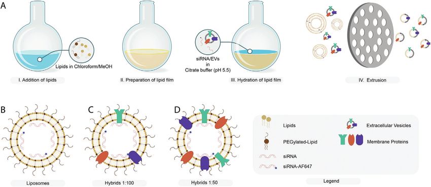
www.advancedsciencenews.com www.advhealthmat.de

comprise a heterogeneous group of vesicles of different intra- 2. Results
cellular origins. At least two different subtypes can be classified
based on their cellular biogenesis: exosomes and ectosomes, the 2.1. Hybrids Carry Physicochemical Features of Both Liposomes
latter also being referred to as microvesicles. and EVs
 Exosomes (30–100 nm) originate in the endosomal pathway
where inward budding of the endosomal membrane results First, SKOV3 EVs were isolated from conditioned medium of
in the formation of intraluminal vesicles which upon release SKOV3 cells via an established size-exclusion chromatography
are referred to as exosomes. Ectosomes (50–1000 nm) are re- protocol.[32] The protein composition of the isolated EVs was then
leased by the cell via direct pinching of the plasma membrane analyzed by western blot to verify the enrichment of specific EV-
at the cell surface. EVs carry different RNA molecules such marker proteins as compared to cell lysate. To this end, we an-
as mRNA and miRNA which can be functionally transferred alyzed expression of the transmembrane proteins CD81, CD63,
to recipient cells and have been suggested to play important and CD9 and luminal proteins Alix and TSG101. As a negative
roles in (patho)physiological processes.[15–17] It is also possible control, expression of endoplasmic reticulum protein Calnexin
to load non-naturally occurring RNA molecules, such as siRNA was analyzed. CD63, CD81, CD9, and Alix were enriched in EVs
and sgRNA, in EVs to be functionally transferred to a recipient as compared to cell lysate (Figure 1A). Expression of TSG101 and
cell.[18–20] Actin in EVs was comparable to that in cell lysate while Calnexin
 As nature’s own carriers of RNA, EVs might be an attractive was clearly negatively enriched. EV purity, as determined by the
alternative carrier system for therapeutic RNA as they have mul- number of particles per μg protein was found to be consistent
tiple potential benefits over current delivery vehicles in terms among isolations (Figure S1, Supporting Information).[33] Mean
of delivery efficacy, intrinsic specific cell targeting properties, and mode EV size was determined by nanoparticle tracking anal-
and toxicity/immunogenicity.[19–26] Interestingly, apart from the ysis (NTA) and found to be 100 and 75 nm, respectively. EV size,
possible benefits for RNA delivery directly, EVs may, as intrin- as determined by dynamic light scattering (DLS), was slightly
sically biologically active entities, induce additional regenera- higher at 150 nm with a polydispersity index (PDI) of ≈0.2 (Fig-
tive or therapeutic effects such as induction of cell prolifera- ure 1B–D). The surface charge (zeta potential) of EVs was nega-
tion, neovascularization, immunomodulation, and prevention of tive, −18 mV, as measured by laser Doppler electrophoresis (Fig-
cell death.[27] Opposite to the beneficial effects of EVs, some ure 1E). Cryo-electron microscopy revealed the typical spherical,
risks might be associated with the use of EVs, which origi- unilamellar morphology of EVs (Figure 1F). All together, these
nate from tumor cells as they have been implicated in cancer analyses confirmed successful isolation of EVs from conditioned
metastasis.[21] medium of SKOV3 cells.[14]
 Although EVs bear great potential as RNA delivery vehicles, These EVs were then used for the production of hybrids
their clinical development is hampered by a low loading effi- encapsulating siRNA via lipid film hydration and subse-
ciency of exogenous RNA molecules.[19,28,29] Multiple methods quent extrusion. Liposomes and hybrids were prepared with
have been developed to achieve RNA loading into EVs either via DLin-MC3-DMA:1,2-dipalmitoyl-sn-glycero-3-phosphocholine
loading during vesicle formation or after vesicle isolation.[30,31] (DPPC):cholesterol:18:1 Biotinyl PE:DMG-PEG in a molar ratio
However, for most methods, reported loading capacities are still of 0.3:0.3:0.355:0.015:0.03 and processed to generate a lipid film.
several orders of magnitude lower compared to that of syn- This lipid film was hydrated with siRNA to form siRNA loaded
thetic delivery systems.[31] Therefore, an alternative approach liposomes (Figure 2A,B). For preparation of hybrids, SKOV3
for active loading of RNA therapeutics in EVs is required to EVs were added at the hydration step at two different ratios of
capitalize on the beneficial properties of EVs as drug delivery EV-protein to total synthetic lipid (w/w), 1:100 and 1:50, and
vehicle. subsequently extruded to produce hybrids (Figure 2C,D).
 Here, we propose a biomimetic approach to generate semisyn- The particles were then analyzed for their physicochemical
thetic hybrid nanoparticles based on EVs and liposomes termed properties to evaluate the influence of increasing numbers of EVs
EV–liposome hybrid nanoparticles (hybrids), thereby combining in the formulation on size, PDI, zeta potential, siRNA encapsula-
the beneficial properties of both liposomes and EVs in a single tion efficiency, and particle morphology (Figure 3). The size and
carrier of siRNA. To this end, we combined SKOV3 EVs and li- PDI of liposomes and hybrids were analyzed by DLS and NTA.
posomes to produce hybrids by lipid-film hydration followed by The average size was close to 150 nm for all formulations as mea-
extrusion. We physicochemically characterized the particles and sured by DLS and ≈ 100 nm as measured by NTA (Figure 3A,E).
analyzed the incorporation of EV-associated membrane proteins PDI seemed to increase slightly as the amount of EV material in
in the hybrids via an antibody-based bead capture assay. Then, the formulation was increased. However, this increase was not
uptake, gene-silencing efficacy, and toxicity of the hybrids were statistically significant (Figure 3B). We did observe that the zeta
evaluated and compared to that of liposomes in multiple cell- potential slightly decreased for hybrids as compared to liposomes
lines. Finally, we used EVs derived from cardiac progenitor cells but no difference was found between hybrids incorporating EVs
(CPCs) to generate hybrid nanoparticles and assessed whether at a ratio of 1:100 or 1:50 (Figure 3C). The decreased surface
the functional regenerative properties of CPC EVs were retained. charge of hybrids could be explained by the incorporation of the
The data show that we successfully produced hybrid nanoparti- negatively charged EV membrane into the newly formed hybrid
cles, which functionally deliver RNA and retain functional prop- nanoparticle leading to a decrease in zeta potential. The encap-
erties attributed to EVs. sulation efficiency of siRNA reduced with increasing amounts of

Adv. Healthcare Mater. 2021, 2101202 2101202 (2 of 13) © 2021 The Authors. Advanced Healthcare Materials published by Wiley-VCH GmbH
www.advancedsciencenews.com www.advhealthmat.de

Figure 1. Physicochemical characterization of SKOV3 EVs. A) Western blot analysis of EV protein markers (Alix, TSG101, CD81, CD9, and CD63) and
EV-negative markers (Calnexin) in SKOV3 cell lysate (CL) and SKOV3 EVs (EV). B) Size distribution of EVs as determined by NTA. C) Average diameter of
EVs as determined by dynamic light scattering. D) Polydispersity index of EVs as measured by dynamic light scattering. E) Surface charge (zeta potential)
of EVs as measured by laser Doppler electrophoresis. F) Cryo-electron microscopy image of EVs isolated from SKOV3 cells. Data are shown as mean ±
SD (n = 3, technical replicates).

Figure 2. Production of liposomes and hybrids. A) Schematic illustration of hybrid production via thin-film hydration and extrusion. B–D) Schematic
illustration of liposomes and hybrids encapsulating a mixture of fluorescent and nonfluorescent siRNA. Hybrids are produced at different protein-to-lipid
ratios (w/w): C) 1:100 and D) 1:50.

Adv. Healthcare Mater. 2021, 2101202 2101202 (3 of 13) © 2021 The Authors. Advanced Healthcare Materials published by Wiley-VCH GmbH
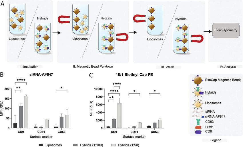
www.advancedsciencenews.com www.advhealthmat.de

Figure 3. Physicochemical characterization of liposomes and hybrids. A) Nanoparticle size as determined by DLS. B) Polydispersity index of nanoparticles
as determined by DLS. C) Zeta potential of nanoparticles as determined by laser Doppler electrophoresis. D) RNA encapsulation efficiency of liposomes
and hybrids. E) Nanoparticle size as determine by NTA. Nanoparticle morphology as determined by cryogenic electron microscopy of F) liposomes,
G) hybrids (1:100), and H) hybrids (1:50). Mean + SD are displayed. n = 8–10 (biological replicates), one-way ANOVA with Tukey’s post-test, ns = not
significant, ** p < 0.01, *** p < 0.001, and **** p < 0.0001.

EVs in the formulation and decreased from ≈80% for liposomes Next, we investigated whether the incorporation of synthetic
to only 50% for hybrids (1:50) (Figure 3D). Furthermore, we lipids in hybrids could also be detected. We prepared lipo-
quantified the overall yield of each production process in terms somes and hybrids containing 0.2 mol% 18:1 Biotinyl Cap PE
of siRNA and cholesterol and found that while the yield for and performed a bead-pulldown with additional staining using
liposomes was ≈50% of both siRNA and cholesterol, the yield streptavidin-PE. We found that 18:1 Biotinyl Cap PE was also
was slightly decreased for both hybrid formulations. Here, the successfully incorporated in the hybrids given the clear increase
influence of EV cholesterol content on the overall amount of in PE-signal for hybrids. Moreover, this experiment confirmed
cholesterol in the formulation was limited (0.6% and 1.2% in that nonspecific binding of liposomes to the beads is very limited
the 1:100 and 1:50 hybrid formulations, respectively) as EVs given the low PE-signal for the liposome sample (Figure 4C). In
contained only ±0.13 μg cholesterol per μg protein (Figure S2, addition, the observed trend in PE signal corresponds to that of
Supporting Information). Cryo-electron microscopy revealed AF647-siRNA where the signal on CD9 beads is slightly higher
that the morphology of the nanoparticles was spherical and compared to CD63 and CD81. All together, these results indicate
that all formulations consisted of unilamellar nanoparticles that we successfully produced EV–liposome hybrid nanoparticles
(Figure 3F–H). carrying EV surface proteins and synthetic lipids while simulta-
 In order to verify successful hybrid formation, we next evalu- neously complexing siRNA.
ated the presence of siRNA and synthetic lipids in liposomes and
hybrids captured using magnetic beads coated with EV-enriched
targets, including CD9, CD81, or CD63. We hypothesized that 2.2. Cellular Uptake of Hybrids Is Dependent on the
only after formation of EV–liposome hybrids, synthetic lipids EV-to-Liposome Ratio and Differs per Cell Type
and siRNA (AF647 labeled) could be detected on the beads (Fig-
ure 4A). For all beads, a clear increase in siRNA-AF647 signal was Next, we evaluated the cellular internalization efficiency of lipo-
observed for hybrid samples as compared to liposomes, which somes and hybrids as this is an important first step in the cytoso-
shows that only hybrids, but not liposomes, contain tetraspanins lic delivery of siRNA. We incubated three different cell types—
that can be captured the beads (Figure 4B). We also observed that SKOV3, HEK293T, and U87-MG—for 4 h with liposomes and hy-
an increase in the number of EVs used in the formulation re- brids (1:100 and 1:50) and analyzed siRNA-AF647 uptake by flow
sulted in a higher siRNA-AF647 signal. cytometry (Figure 5). Uptake of hybrid (1:100) nanoparticles in

Adv. Healthcare Mater. 2021, 2101202 2101202 (4 of 13) © 2021 The Authors. Advanced Healthcare Materials published by Wiley-VCH GmbH
www.advancedsciencenews.com www.advhealthmat.de

Figure 4. Bead capture analysis of siRNA-AF647 and 18:1 Biotinyl Cap PE in liposomes and hybrids on beads targeting CD9, CD63, or CD81. A) Schematic
illustration of bead-capture assay. B) Flow cytometric analysis of siRNA-AF647 on ExoCap beads. Nanoparticles were incubated with beads targeting a
single epitope, CD9, CD63, or CD81, washed and analyzed. C) Flow cytometric analysis of 18:1 Biotinyl Cap PE on ExoCap beads. Data are representative
of three independent experiments and expressed as mean ± SD (n = 3, technical replicates), one-way ANOVA with Tukey’s post-hoc test, * p < 0.05, ** p
< 0.01, and **** p < 0.0001.

Figure 5. Cellular uptake of liposomes, hybrids (1:100), and hybrids (1:50) in A) SKOV3-dluc, B) HEK293T-dluc, and C) U87-MG-dluc. Cells were in-
cubated for 4 h at 37 °C at a concentration of 25 × nm siRNA and cellular uptake was measured by flow cytometry. Data are representative of three
independent experiments and expressed as mean ± SD (n = 3, technical replicates), one-way ANOVA with Tukey’s post-hoc test, * p < 0.05, *** p < 0.001,
and **** p < 0.0001.

Adv. Healthcare Mater. 2021, 2101202 2101202 (5 of 13) © 2021 The Authors. Advanced Healthcare Materials published by Wiley-VCH GmbH
www.advancedsciencenews.com www.advhealthmat.de

Figure 6. Cell viability of different cell types incubated with liposomes and hybrids as determined by an MTS assay. Cells were incubated with liposomes
and hybrids at concentrations ranging from 0.1 to nm siRNA and cell-viability was analyzed after 48 h. A) SKOV3-dluc. B) HEK293T-dluc. C) U87-MG-dluc.
Data are representative of three independent experiments and expressed as mean ± SD (n = 2–3, technical replicates), one-way ANOVA with Tukey’s
post-hoc test, * p < 0.05 and ** p < 0.01.

Figure 7. Gene silencing activity in different cell types treated with liposomes and hybrid nanoparticles encapsulating siRNA. Different cell types were
incubated with liposomes and hybrids at concentrations ranging from 0.1 to 50 nm siRNA and gene-silencing was analyzed after 48 h by measurement
of luciferase expression. Data are plotted as the normalized ratio of firefly to renilla luciferase expression. A) SKOV3-dluc. B) HEK293T-dluc. C) U87-MG-
dluc. Data are representative of three independent experiments and expressed as mean ± SD (n = 3, technical replicates), one-way ANOVA with Tukey’s
post-hoc test, * p < 0.05, ** p < 0.01, and *** p < 0.001.

all three cell types was decreased as compared to liposomes. This hybrids as compared to liposomes. A difference in the effect on
effect was found to be statistically significant in HEK293T and cell viability between liposomes and hybrids was not observed in
U87-MG cells. Interestingly, when more EV components were in- HEK293T and U87-MG cells. In HEK293T cells, a small dose-
corporated in hybrids (1:50), cellular uptake increased again, but dependent decrease in cell viability was seen for all nanoparticles
only at statistically significant levels in HEK293T and U87 cells. with no differences between liposomes and hybrids. In U87-MG,
Almost no nanoparticle uptake was seen at 4 °C which confirmed administration of liposomes and hybrids did not affect the cell vi-
the effects seen are a result of active uptake processes (Figure S3, ability (Figure 6).
Supporting Information). The differences in cellular uptake im- Another possible advantageous functional characteristic of
plicate that the cellular internalization of hybrids varies per cell EVs as compared to liposomes could be an improved siRNA de-
type and that the uptake is affected by the amount of SKOV3 EVs livery efficiency.[19,20] Therefore, we evaluated the gene-silencing
incorporated in the hybrid formulation. efficacy of hybrids as compared to liposomes using a luciferase
 reporter assay. Liposomes or hybrids encapsulating siRNA tar-
2.3. Hybrids Show Limited Toxicity and Functionally Deliver geting firefly luciferase or a nonspecific control siRNA were ad-
siRNA to Multiple Cell Types ministered to SKOV3, HEK293T, and U87-MG (Figure 7; Figure
 S4, Supporting Information). A clear dose-dependent decrease
For successful application of liposomes and hybrids in RNA de- in firefly luciferase was observed in all cell lines. In SKOV3 and
livery, particles must be biocompatible and nontoxic. Therefore, HEK293T cells, the gene-silencing effect of hybrids (1:100 and
we analyzed the toxicity of the nanoparticles using a cell viabil- 1:50) was lower as compared to liposomes. In contrast, in U87-
ity assay. In SKOV3 cells, liposomes showed a dose-dependent MG cells, gene-silencing efficacy of hybrids (1:100 and 1:50) was
decrease in cell viability whereas this effect was not observed for similar to that of liposomes despite a lower uptake efficiency,
hybrids (1:100 and 1:50) indicating increased biocompatibility of which may point toward more efficient cytosolic siRNA delivery.

Adv. Healthcare Mater. 2021, 2101202 2101202 (6 of 13) © 2021 The Authors. Advanced Healthcare Materials published by Wiley-VCH GmbH
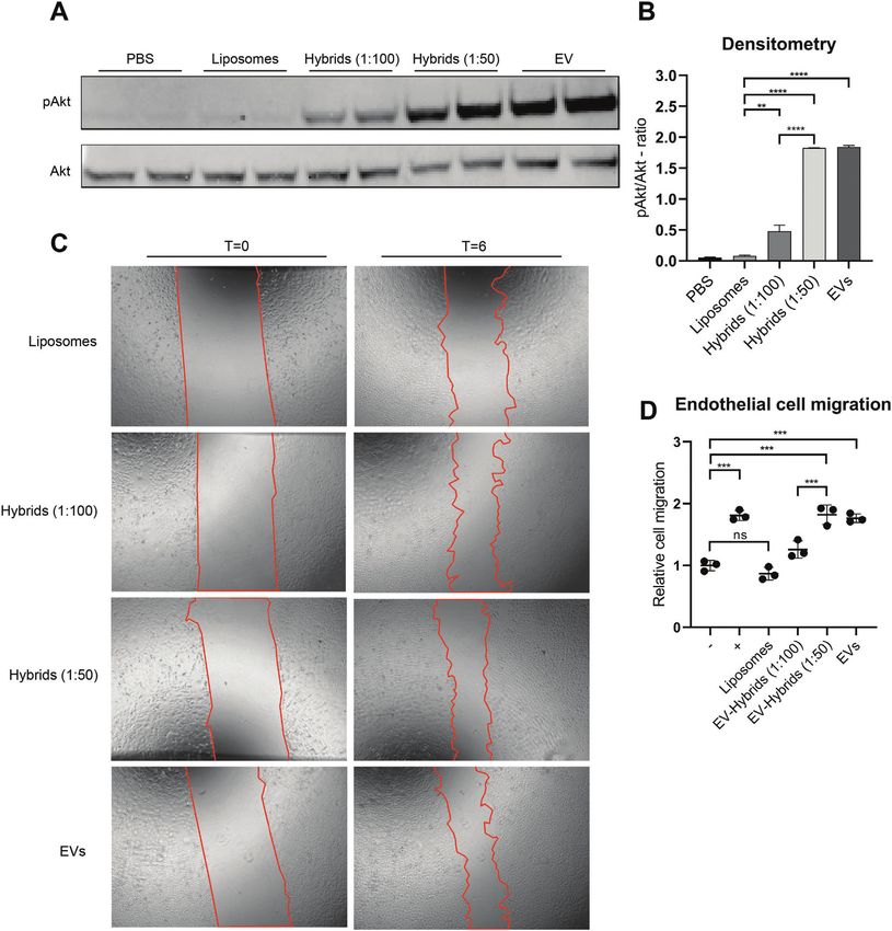
www.advancedsciencenews.com www.advhealthmat.de

There was no difference in gene-silencing efficacy between dif- Here, we produced EV–liposome hybrid nanoparticles, which
ferent EV–liposome hybrids (1:100 and 1:50). This indicates that are nanosized siRNA carriers formed through the merging of
under these conditions, gene-silencing efficacy was not critically EVs and liposomes, via thin-film hydration and extrusion. The
dependent on the EV-protein to lipid ratio. All together, these data anticipated benefits originate from the combination of liposome
show that hybrids are able to functionally deliver siRNA to differ- related properties such as high RNA loading capacity and EV re-
ent cell types, although in these experiments, the potency is re- lated properties such as increased delivery efficacy, cell targeting
duced in SKOV3 and HEK293T cells as compared to liposomes. properties, and possible tissue regenerative properties.
 The thin-film hydration and extrusion method has already
 been previously described to generate different hybrids based
2.4. Hybrids Based on Cardiac Progenitor Cell EVs Retain on the combination of EVs and liposomes.[39,40] For instance,
Functional Regenerative Properties Jhan et al. hydrated a lipid film in PBS and subsequently added
 3T3- or A549-EVs followed by sonication and sequential extru-
Finally, we investigated whether hybrids preserved the biological sion through 400, 200, and 100 nm membrane pores.[39] Rayma-
activity of EVs. To this end, we generated hybrids with CPC EVs. jhi et al. hydrated the lipid film in the presence of J774A.1-EVs,
CPC EVs have been shown to activate endothelial signaling path- sonicated and subsequently extruded the lipid mixture through
ways and migration, and activate in vivo angiogenesis.[34–36] The membrane filters with 400 nm followed by 200 nm pores.[40] The
physicochemical properties were analyzed in a similar manner as use of mechanical extrusion to generate hybrids is not limited
for SKOV3 EV derived hybrids (Figure S5, Supporting Informa- to EVs and lipid-based particles as it has also been used to in-
tion). Liposomes and hybrids had a size of ≈150 nm at a PDI of corporate cellular membranes, such as leukocyte membranes, in
≈0.2. Again, the surface charge of hybrids was lower compared liposomes.[41–43] More recently, extrusion has also been used to
to that of liposomes. surface coat gold nanoparticles with the membrane of EVs.[44]
 We then evaluated the functional capabilities of liposomes and An important variable in the generation of hybrids is the
CPC derived hybrids in two functional assays: an Akt phospho- amount of EVs incorporated in the formulation. A useful met-
rylation assay and a scratch wound healing assay. ric to describe the amount of EVs incorporated in the formula-
 Akt is an important factor in signaling pathways involved in tion is the (EV) protein-to-(liposomal) lipid ratio. In literature,
proliferation, angiogenesis, differentiation, adhesion, migration, ratios can be found ranging from 1:5 to 1:1000 (protein/lipid
and cell survival, and its phosphorylation is an indicator of func- (w/w)).[39–43] For instance, to incorporate the membrane of leuko-
tional CPC EV delivery.[37,38] HMEC-1 cells were serum-starved cytes into phosphatidylcholine liposomes, ratios varying from
and subsequently incubated for 30 min with hybrids (1:100 and 1:100 to 1:300 (protein/lipid; w/w) were used, whereas for the
1:50) and EVs as well as PBS and liposomes as negative con- generation of “macrophage derived hybrid exosomes,” Rayama-
trols. After treatment, cells were lysed and the ratio of phospho- jhi et al. used a ratio of 1:5 (protein/lipid (w/w)).[40,41] A poten-
rylated Akt/Akt was analyzed by western blot analysis. PBS and tial drawback of this metric is that proteins can also be contam-
liposomes did not induce phosphorylation of Akt, whereas this inants of EV isolations, which can vary from batch-to-batch and
was observed for hybrids (1:100 and 1:50) and EVs in a dose- therefore potentially has implications for reproducibility. To ac-
dependent matter (Figure 8A). When analyzed using densitome- count for this, we carefully monitored the number of particles
try, hybrids (1:100 and 1:50) and EVs induced significantly more per μg protein, which was found to be highly consistent among
phosphorylation of Akt as compared to liposomes (Figure 8B). different EV isolations. Here, we generated hybrids by hydration
 Second, we performed a scratch wound healing assay using of a lipid film with EVs at protein-to-lipid ratios of 1:100–1:50
a confluent monolayer of HMEC-1 cells. Samples were normal- (w/w) and subsequent extrusion through membranes with pores
ized based on particle counts as measured by NTA and a total of 1000 nm followed by 100 and then 50 nm. As the majority of
dose of 2 × 10¹² particles for liposomes and hybrids was added as EVs has a size below 100 nm, based on our observation in NTA
well as 3 × 10¹⁰ particles for EVs, which served as positive con- analysis where we observed a size mode value of 75 nm, a 50
trol. The closing of the scratch was then analyzed after 6 h. Hy- nm membrane was chosen as smallest membrane. A possible
brids stimulated closure of the scratch to a larger extent than lipo- limitation of the aforementioned studies regarding EV-based hy-
somes and hybrids (1:50) further increased closure of the wound brid nanoparticles is that samples were extruded through pores
as compared to hybrids (1:100) (Figure 8C,D). This indicates a around or above the median size of EVs, which not necessarily
dose-dependent effect of the amount of CPC EVs used in the for- results in deformation of the EV and subsequent reformation in
mulation on wound closure. a hybrid nanoparticle. In this study, we did take this into account
 These results are in good agreement with the endothelial sig- and extruded EVs together with synthetic liposomes through a
naling assays and indicate that hybrids produced using CPC EVs membrane with pore sizes of 50 nm.
stimulate wound closure and induce phosphorylation of Akt. We analyzed the yield of the production process of this for-
 mulation in terms of siRNA and synthetic lipid yield, which
3. Discussion was found to be maximally 50% of the input siRNA and choles-
 terol. This could potentially be explained by the formation of
The delivery of RNAi therapeutics is challenging given the un- siRNA/ionizable lipid aggregates, which are lost during the
favorable characteristics of siRNA as a drug molecule. siRNA extrusion process. Although this effect has been shown to be
molecules are unstable in circulation, immunogenic, and are un- overcome by the addition of 40% ethanol (v/v) combined with
able due to their molecular properties, to cross cellular mem- a rise in temperature to 65 °C, we decided not to change the
branes to reach their cytosolic target site. production process since higher temperatures and ethanol

Adv. Healthcare Mater. 2021, 2101202 2101202 (7 of 13) © 2021 The Authors. Advanced Healthcare Materials published by Wiley-VCH GmbH
www.advancedsciencenews.com www.advhealthmat.de

Figure 8. Endothelial signaling assay and scratch wound closing assay of HMEC-1 cells treated with liposomes and hybrids. A) Representative western
blot analysis of phosphorylated Akt and Akt expression levels in HMEC-1 cells treated with liposomes, hybrids, and EVs. Liposomes and hybrids were
administered at a total particle dose of 2 × 10¹² and EVs at a total particle dose of 3 × 10¹⁰ . B) Quantification of Akt and pAkt expression levels obtained
via western blot analysis using densitometry expressed as pAkt/Akt-ratio. C) Representative images of scratch wound healing assay before (t = 0) and
after (t = 6 h) incubation with liposomes, hybrids (1:100), hybrids (1:50), and EVs. Liposomes and hybrids were administered at a total particle dose of
2 × 10¹² and EVs at a total particle dose of 3 × 10¹⁰ . D) Cell migration of HMEC-1 expressed relative to the negative control. Incubation of HMEC-1 cells
with hybrids (1:50 and 1:100) and EVs increases wound closure as compared to liposomes. Data are expressed as mean + SD, one-way ANOVA with
post-hoc test, ns = not significant, ** p < 0.01, *** p < 0.001, and **** p < 0.0001.

Adv. Healthcare Mater. 2021, 2101202 2101202 (8 of 13) © 2021 The Authors. Advanced Healthcare Materials published by Wiley-VCH GmbH
www.advancedsciencenews.com www.advhealthmat.de

content could potentially detrimentally affect the EV membrane control making it more difficult to assess the value of the abso-
proteins.[45] When we looked at the influence of EV incorpora- lute toxicity values. However, based on the results we could still
tion in the formulation on several particle characteristics such assess relative differences between the three different nanoparti-
as size, PDI, and zeta potential, we observed that increasing the cle types within the same experiments, which have shown to be
amount of SKOV3 EVs incorporated in the formulation resulted reproducible.
in an increase in PDI, although this effect was not significant. Liposomes and hybrids both functionally delivered siRNA in
For hybrids generated with CPC EVs, an increase in PDI was not a dose-dependent manner in three different cell types although
observed. The surface charge, i.e., zeta potential, was decreased the potency of hybrids was reduced in SKOV3 and HEK293T,
in hybrids, which can most likely be attributed to the incorpo- but not in U87. Again, this is an intriguing observation as it im-
ration of negatively charged EV membrane components. We plies that effects are cell type dependent. A possible explanation
did observe that RNA encapsulation efficiency in hybrids was for the decrease in gene-silencing in HEK293T might be the de-
decreased. This may be the results of competition for the electro- creased uptake. In contrast, the combination of decreased cellular
static interaction with the ionizable lipid by negatively charged uptake and comparable gene-silencing efficacy of liposomes and
EV components such as RNA or negatively charged lipids or hybrids in U87-MG might suggest different intracellular traffick-
proteins. We also evaluated the morphology of the liposomes ing resulting in more efficient escape of siRNA from the endo-
and hybrids. The extrusion process had no apparent detrimental lysosomal pathway for hybrids and is an interesting area to fur-
effects on the morphology of hybrids as they appeared to be ther explore.
spherical, unilamellar membrane enclosed particles, which Our observation that hybrids generated with SKOV3 EVs did
are comparable to liposomes and EVs. We also confirmed that not have a positive effect on gene-silencing efficiency differs
hybrid particles (containing both siRNA and synthetic lipid) from others. Coating of polyethyleneimine-based siRNA particles
were captured by beads coated with antibodies against several with SKOV3 EVs resulted in increased potency in terms of gene-
distinctive EV marker proteins such as CD9, CD63, and CD81 in- silencing efficacy of the EV-modified particle compared to the un-
dicating that surface topology of EVs is at least partly transferred coated particle.[57] This apparent discrepancy may be a result of
to the hybrids. Nevertheless, it remains challenging to assess multiple different causes, including the production method and
the efficiency of this hybridization process and fully exclude the the resulting hybrid composition.
possibility of intact EVs being present in the formulation. Several groups have applied the concept of extracellular
 Next, we quantitatively compared multiple functional charac- vesicle–liposome hybrids to create nanoparticles for tumor-
teristics, including cellular uptake, gene-silencing efficacy, and targeted drug delivery.[39,40,58] Here, we have shown that a similar
cell viability, of liposomes and extracellular vesicle–liposome hy- approach can be used to convey tissue regenerative properties of
brids. The cellular uptake of nanoparticles via endocytosis is CPC EVs to synthetic nanoparticles via the creation of extracellu-
influenced by many variables such as size, charge, and the lar vesicle–liposome hybrids. CPC EVs possess the ability to ac-
biomolecular corona.[46–49] It is known that uptake rate and route tivate endothelial signaling and cell migration in HMEC-1.[34–36]
can vary between different lipid systems and EVs.[47,50–52] As the We observed that hybrids also activated endothelial signaling and
surface charge and membrane surface of hybrids differs from cell migration whereas liposomes did not. This demonstrates that
that of liposomes, we investigated the uptake efficiency of lipo- hybrids produced via thin-film hydration and extrusion can be
somes and hybrids. In HEK293T and U87-MG, we observed a loaded with siRNA and retain functional properties of EVs. This
decrease in uptake for hybrids (1:100) as compared to liposomes. implicates that hybrids potentially can be used as an efficient
At higher concentrations of EVs in the formulation (1:50), cellu- RNA drug delivery system while bearing intrinsic EV function-
lar uptake increased again. The latter observation suggests that ality at the same time. For instance, this can be of relevance in
the uptake mechanism is different for hybrids as compared to the salvage of myocardial tissue upon infarction where CPC EVs
liposomes, changing from predominantly liposome-dictated to have shown to reduce scar size and improve ventricular function
mainly EV-dictated. This may be relevant for cell-targeting pur- after permanent coronary occlusion.[59] Similarly, intracardiac de-
poses, as EVs may have intrinsic capacity to target specific cells livery of a synthetic miRNA mimic of hsa-miR-590-3p via a lipid-
or tissues.[21,24,53] Furthermore, this may affect endocytic routing based system resulted in reduced infarct size and improved car-
and intracellular nanoparticle trafficking, which in turn may in- diac output.[60] Given the results presented in this paper, hybrids
fluence delivery efficiency. might have the potential to combine both treatments in a single
 An important drawback of the usage of liposomes or other lipid particle.
nanoparticles for RNA delivery is the dose/dose-regimen related
hepatotoxicity, which might be related to innate immune system 4. Conclusion
activation.[8,54] In contrast to synthetic systems, EVs are generally
considered to have low immunogenicity and are less toxic as ob- Currently, much is still unknown about how EV composition af-
served in several preclinical studies.[55,56] Here, we observed a de- fects functionality and confers EVs with a potent RNA delivery
crease in in vitro toxicity of hybrids as compared to liposomes in capability. As long as such pivotal data are missing, the produc-
SKOV3 cells, which could be the results of EV component incor- tion of EV–liposome hybrids, which fully reflect the functional
poration in hybrids. The effect was only observed in SKOV3 cells capabilities of EVs, remains challenging. The results presented
suggesting cell-specific effects. These data should be interpreted here show that the production of hybrids via thin film hydration
with care as in vitro to in vivo translation of cell viability data for and subsequent extrusion results in hybrid particles with EV-like
lipid-based drug delivery systems is unclear. Moreover, the MTS- surface topology encapsulating siRNA, which can be functionally
assay as performed in this manuscript lacked an assay positive delivered. The incorporation of EV membrane components leads

Adv. Healthcare Mater. 2021, 2101202 2101202 (9 of 13) © 2021 The Authors. Advanced Healthcare Materials published by Wiley-VCH GmbH
www.advancedsciencenews.com www.advhealthmat.de

to functional differences. Depending on the cell type, uptake is Cell Culture and Isolation of SKOV3-EVs and CPC-EVs: For SKOV3-EV
altered, toxicity of hybrids as compared to liposomes is reduced, production, SKOV3 cells were seeded at an appropriate density and cul-
and gene-silencing effects are retained. Moreover, we also show tured for 48–72 h to a confluence of 80–90% after which the medium was
 replaced and cells were cultured for another 24 h in Opti-Mem supple-
that intrinsic functionalities of CPC EVs such as the ability to ac-
 mented with Glutamax, 100 U mL−1 penicillin, and 100 U mL−1 strepto-
tivate endothelial signaling pathways and stimulate migration of mycin. Conditioned medium was harvested after 24 h and spun down for 5
HMEC are retained in hybrids. Thus, hybrid nanoparticles could min at 300 × g and for 15 min at 2000 × g to remove cells and cell debris, re-
combine the functional characteristics of both liposomes and EVs spectively. The supernatant was filtered through a 0.45 × 10⁻⁶ m PES bottle
and serve as a “best of both worlds” particle for therapeutic deliv- top filter and concentrated to a volume of 15 mL by tangential flow filtra-
ery of siRNA. tion (TFF) using Vivaflow 50R hydrosart casettes, with a membrane cutoff
 of 100 kDa. This concentrate was then further reduced to a volume of ≈5
 mL using 100 kDa Amicon Ultra-15 Centrifugal filter (Merck) and loaded
 on a HiPrep 16/60 Sephacryl S-400 HR column (GE Healthcare, Uppsala,
 Sweden) connected to an ÄKTA Start system (GE Healtcare) containing an
5. Experimental Section UV280 flow cell. For CPC-EVs, the procedure was slightly different. CPCs
 Materials: Cholesterol was obtained from Sigma-Aldrich (Saint-Louis, were seeded at an appropriate density and when a confluency of 80–100%
USA), DPPC from Lipoid GmbH (Ludwigshafen, Germany), DMG-PEG was reached, cells were washed with PBS and medium was replaced for
from NOF Corporation (Tokyo, Japan), and 18:1 Biotinyl Cap PE from basal MEM199. The supernatant was collected after 24 h and centrifuged
Avanti Polar Lipids (Alabama, USA). DLin-MC3-DMA was synthesized in- for 15 min at 2000 × g to remove cells and cell debris and the supernatant
house according to a published protocol.[61] All oligonucleotides were or- was filtered through a 0.45 × 10⁻⁶ m PES bottle top filter. Subsequently,
dered at Integrated DNA Technologies (Iowa, USA). siRNA molecules were the filtrate was concentrated by TFF using a minimate TFF capsule with
ordered as individual strands and annealed for 5 min at 97 °C. The se- a membrane cutoff of 100 kDa. Then, EVs were isolated by size exclusion
quences used can be found in Table S1 in the Supporting Information. chromatography following the same procedure as described for SKOV3-
 Generation of Cells Stably Expressing Firefly and Renilla Luciferase: For EVs. After SEC, the fractions containing EVs were pooled, filtered through
the generation of stable dual luciferase cell lines, the PGK-FFluc-SV40- a 0.45 × 10⁻⁶ m syringe filter, and concentrated using 100 kDa Amicon
Rluc-NeoR_fusion cassette from the pmirGLO Dual-Luciferase miRNA Ultra-15 Centrifugal filter (Merck). Then, the buffer was exchanged to 250
Target Expression Vector (Promega, Leiden, NL) was isolated and trans- × 10−3 m citrate buffer (pH 5.5) and the sample was again concentrated
ferred to a pHAGE2 lentiviral vector. First, pHAGE2-EF1a-IRES-NeoR- using Amicon Spin Filters with a membrane cutoff of 100 kDa. The pro-
WPRE was restricted with SpeI and XbaI restriction enzymes (New Eng- tein concentration was determined via micro-BCA protein determination
land Biolabs, Ipswich, MA, USA) and religated to remove the EF1a pro- kit (Thermo Scientific). EVs were stored at 4 °C until further use. EVs were
moter. Then, the PGK-FFluc-SV40-Rluc-NeoR_fusion cassette was isolated used to prepare hybrids within 72 h after isolation.
from the pmirGLO plasmid using BglII and BstBI restriction enzymes Preparation and Analysis of Liposomes and EV–Liposome Hybrids (Hy-
(New England Biolabs, Ipswich, MA, USA) and ligated into the newly brids): Lipid were dissolved in a mixture of chloroform/methanol (9/1;
formed pHAGE2-IRES-NeoR-WPRE vector digested with BamHI and ClaI v/v) and added to a round bottom flask at a molar ratio of 30:30:35.5:1.5:3
restriction enzymes (New England Biolabs, Ipswich, MA, USA), gener- (DLin-MC3-DMA:DPPC:cholesterol:18:1 Biotinyl Cap PE:DMG-PEG). The
ating a pHAGE2-PGK-FFluc-SV40-Rluc-NeoR_fusion-WPRE plasmid. All organic solvent was evaporated using a RotoVap (Büchi Labortechnik,
ligations were performed using a Quick Ligation Kit (all New England Flawil, Switzerland) at 60 °C and the resulting lipid film was dried under a
Biolabs, Ipswich, MA, USA) and ligation products were subsequently flow of nitrogen for ≈20 min. For the liposomes, the lipid film was hydrated
transformed into One Shot Stbl3 chemically competent Escherichia coli using a mixture of siRNA targeting firefly luciferase and fluorescently la-
(ThermoFisher Scientific, Waltham, MA, USA). For lentiviral production, beled siRNA targeting firefly luciferase in a ratio of 1:1 (siRNA Luc:siRNA
HEK293T cells were transfected overnight with psPAX2, pMD2.G, and Luc-AF488 or AF647) dissolved in 250 × 10−3 m citrate buffer (pH 5.5)
pHAGE2-PGK-FFluc-SV40-Rluc-NeoR_fusion-WPRE plasmids at a 1:1:2 for 1 h at 45 °C. After hydration, the suspension was kept at 45 °C and ex-
ratio using Lipofectamine 2000 (ThermoFisher Scientific, Waltham, MA, truded five times through a polycarbonate filter of 1.0 μm, then five times
USA) according to the manufacturers’ protocol. After 18 h, the culture through 0.1 μm, and finally five times through a polycarbonate filter of 0.05
medium was replaced, and lentiviral supernatant was collected after 48 μm using an Avanti Hand Extruder (Avanti Polar Lipids). Subsequently, the
h. Lentiviral supernatant was cleared from any remaining cells by a 5 min liposomes were dialyzed overnight at 4 °C against an excess of PBS using
1000 × g centrifugation step and subsequent 0.45 μm syringe filter filtra- Slide-A-Lyzer G2 Dialysis Casette with a membrane cutoff of 100 kDa to
tion, and stored at −80 °C until further use. Cells were transduced with change the pH to 7.4 and to remove unencapsulated siRNA. Hybrids were
lentiviral supernatants overnight in the presence of 8 μg mL−1 polybrene produced in a similar fashion, but in this case, the lipid film was hydrated
(ThermoFisher Scientific, Waltham, MA, USA). Starting 24 h after lentivi- using a mixture of siRNA and extracellular vesicles. Extracellular vesicles
ral transduction, cells were cultured with 1000 μg mL−1 G418 for 5 days, were added at different ratios of vesicle protein to total lipid: 1:100 and
after which they were cultured at a 500–1000 μg mL−1 G418, depending 1:50 (protein/total lipid; w/w).
on the cell line, until further use. Transduced cells are referred to by the Characterization of EVs, Liposomes, and Hybrids: Particle size of lipo-
affix -dluc. somes and hybrids was measured via DLS on a Zetasizer Nano S (Malvern
 General Cell Culture: SKOV3, SKOV3-dluc, HEK293T-dluc, and U87- Panalytical, Malvern, UK). Samples were diluted with Dulbecco’s PBS
MG-dluc were cultured in Dulbecco’s modified Eagle Medium (Gibco) (DPBS) to an appropriate concentration and measured in triplicate. Lipo-
supplemented with 10% fetal bovine serum (FBS) (Gibco, Corning). somes, hybrid, and EV size was also measured using NTA on a NanoSight
SKOV3-dluc and HEK293T-dluc were cultured in the presence of 1000 NS500 (Malvern, Panalytical, Malvern, UK). For NTA, samples were di-
μg mL−1 G418 (BioIVT) whereas U87-MG-dluc was cultured in the pres- luted in DPBS to an appropriate particle concentration and loaded in the
ence of 500 μg mL−1 G418. HMEC-1 was cultured in MCDB-131 medium sample chamber. Camera level 16 was selected and sample was measured
supplemented with 10% FBS (Gibco), 2 × 10⁻³ m l-glutamine (Gibco), three times for 30 s and subsequently analyzed using Nanosight NTA 3.4
10 ng mL−1 rhEGF (Peprotech), and 50 × 10−9 m hydrocortisone (Sigma) software at a sensitivity level of 5.
in flasks/plates coated with 0.1% gelatin (Sigma). CPCs were cultured Particle surface potential was measured by laser Doppler electrophore-
in MEM 199 + Earle’s Salts and l-glutamine (Gibco), which was sup- sis on a Zetasizer Nano Z (Malvern Panalytical, Malvern, UK). Samples
plemented with 22% EGM-2 medium (Lonza), 10% FBS, and 1% MEM were diluted in 0.1× DPBS and sample was measured for 20 runs in trip-
NEAA nucleic acids (Gibco). All cells were cultured at 37 °C and 5% CO2 licate.
in the presence of 100 U mL−1 penicillin and 100 U mL−1 streptomycin The RNA concentration was determined based on the fluorescence
(Gibco). emitted by the fluorescently labeled siRNA. Samples were diluted 1:1 in

Adv. Healthcare Mater. 2021, 2101202 2101202 (10 of 13) © 2021 The Authors. Advanced Healthcare Materials published by Wiley-VCH GmbH
www.advancedsciencenews.com www.advhealthmat.de

2% TX-100 in PBS. A calibration curve of fluorescent siRNA was prepared incorporated in the lipid film of both the liposomes and the hybrids. The
in the same medium. Sample fluorescence was measured on a Spec- samples were incubated with ExoCap beads as described above. After the
tramax ID3 (Molecular Devices, San Jose, California, US) at an excita- three initial washing steps, samples were incubated with PE-Streptavidin
tion/emission wavelength of 490/530 nm or 620/665 nm for siRNA-AF488 for 20 min and then washed three times. Samples were then suspended in
or siRNA-AF647, respectively. Concentrations were determined based on 200 μL PBSA and measured by flow cytometry on a BD LSRFortessa (BD,
a reference calibration curve. Franklin Lakes, NJ, US). Flow cytometry data analysis was performed using
 The cholesterol concentration was determined using the LabAssay FlowJo v10 software.
Cholesterol kit (DAKO, JP) in PBS or in the presence of 50% (v/v) iso- Analysis of Cellular Uptake by Flow Cytometry: For the measurement
propanol. Sample concentration was determined using a reference cali- of liposomal and hybrid cellular uptake, flow cytometry was used. Cells
bration curve. Absorbance was measured at 600 nm on a Spectramax ID3 were seeded at an appropriate density in a 48-well plate. SKOV3-dluc was
(Molecular Devices, San Jose, California, US). seeded at 40 000 cells per well 24 h prior to the assay, HEK293T-dluc was
 The encapsulation efficiency of siRNA was calculated by the following seeded at 20 000 cells per well 72 h prior to the assay, and U87-MG-dluc
formula cells were seeded at 40 000 cells per well 24 h prior to the assay. Then, cells
 were incubated with different nanoparticles at a total siRNA concentration
 Encapsulation Efficiency (%) of 25 × 10−9 m per well. As a vehicle control, an equal volume of PBS was
 used. Cells were incubated for 4 h and then cellular uptake was analyzed
 [siRNA]
 after dialysis (100 kDa) by flow cytometry. Cells were washed with PBS, trypsinized, taken up in full
 [cholesterol]
 = × 100 (1) medium, and transferred to a 96 U-Bottom well plate (Greiner). Cells were
 [siRNA]
 [cholesterol]
 before dialysis (100 kDa) then washed with an acid wash (0.5 m NaCL, 0.2 m acetic acid), PBS, and
 taken up in 2% PBSA for analysis on a LSRFortessa (BD, Franklin Lakes,
 Cryogenic Transmission Electron Microscopy: 7 μL of liposomes or hy- NJ, US). For each experiment, cellular uptake was expressed as ratio of the
brid suspension were added to freshly glow-discharged quantifoils and in- uptake of the liposome sample. As a control, uptake was measured at 4 °C.
cubated for at least 10 min in a humidified environment and then vitrified To this end, cells were cooled 30 min prior to incubation in a fridge at 4 °C,
using a FEI Mark IV Vitrobot (FEI, Hillsboro OR, USA). After vitrification, and samples were added and incubated for 4 h at 4 °C. After incubation,
samples were stored in liquid nitrogen until imaging. Samples were im- cells were kept on ice and washed with ice-cold PBS before trypsinization
aged on an FEI Tecnai G2 20 TWIN 200 kV transmission electron micro- and further work-up as described earlier.
scope. Vitrified quantifoils were loaded in a Gatan 70° tilt cryo-transfer sys- Gene-Silencing and Cell Viability: The gene-silencing efficacy of lipo-
tem, which was precooled using liquid nitrogen and inserted in the micro- somes and hybrids was assessed in multiple cell-lines: SKOV3-dluc, U87-
scope. Samples were imaged at a magnification of 29 000× and samples MG-dluc, and HEK293T-dluc. All cells expressed a dual luciferase con-
images were acquired by the bottom mounted FEI High-Sensitive 4k × 4k struct containing both firefly and renilla luciferase under G418 selection.
Eagle camera. SKOV3-dluc and HEK293T-dluc cells were seeded in a 96-well plate at a
 Western Blotting: Protein concentration was determined via a micro- density of 5000 cells per well 48 h prior to transfection or 10 000 cells per
BCA assay and ≈10 μg protein was used per sample. Samples were mixed well 24 h prior to transfection. U87-MG-dluc were seeded at a density of
with 4× sample buffer (40% v/v glycerol, 8% w/v SDS, 8% v/v bromophe- 5000 cells per well 24 h prior to transfection. Samples were added at con-
nol blue, in 0.25 m Tris-HCL) with or without dithiothreitol (DTT) for re- centrations ranging from 0 × 10−9 to 50 × 10−9 m siRNA. As a positive
duced or nonreduced conditions, respectively. Samples were heated to control, cells were transfected using Lipofectamine RNAiMAX according
95 °C for 5 min and separated on a 4–12 Bis-Tris polyacrylamide gel to the manufacturer’s instructions. Luciferase activity was assessed after
(Thermo Scientific). Proteins were then electrotransferred to immobilon- another 48 h of culture. Luciferase activity was measured using the Stop
FLR polyvinylidene difluoride (PVDF) membranes and blocked with 50% & Glo System (Promega, Leiden, NL) according to the manufacturer’s in-
v/v Odyssey Blocking Buffer (LI-COR Biosciences) in Tris buffered saline structions. In short, medium was aspirated and replaced by 50 μL of fresh
(TBS). All immune-labeling was performed with 50% v/v Odyssey Block- medium. 50 μL of Glo substrate was added and cells were incubated for
ing Buffer in TBS containing 0.1% Tween 20 (TBS-T). Primary antibod- 10 min. After 10 min, 100 μL of lysate was transferred to a white 96-well
ies were used overnight at 4 °C and included mouse anti-CD63 (Abcam, plate and firefly luciferase activity was measured. Then, 50 μL of Stop & Glo
MEM-259; 1:1000), Mouse Anti-CD81 (Santa Cruz, SC-166029; 1:500), rab- buffer was added and, after an incubation of 10 min, renilla luciferase ac-
bit anti-TSG101 (Abcam, ab30871, 1:1000), mouse anti-Alix (Thermo Sci- tivity was measured. Both firefly luciferase and renilla luciferase activities
entific, 3A9, 1:1000), mouse-anti- -actin (Cell Signaling Technology, clone were measured on a Spectramax ID3 (Molecular Devices, San Jose, CA,
8H10D10, 1:1000), rat anti-Calnexin (Tebu-Bio, N3C2, 1:1000), rabbit anti- USA) at an integration time of 1000 ms. For data analysis, firefly luciferase
AKT (Cell Signaling Technology, 9272S, 1:1000), rabbit anti-pAKT (Cell activity was normalized based on renilla luciferase activity and expressed
Signaling Technology, 4060S, 1:1000), and mouse anti- -actin (Sigma, as percentage of the blank—0 × 10−9 m siRNA—sample.
A5441,1:1000). Cell viability was measured using CellTiter 96 AQueous MTS Reagent
 Secondary antibodies included Alexa Fluor 680-conjugated antirab- Powder according to the manufacturer’s instructions. As a negative con-
bit antibodies (LI-COR Biosciences, A-21076, 1:7500–1:10 000), Alexa trol, MTS medium was added to wells, which did not contain any cells
Fluor 680-conjugated antimouse antibodies (LI-COR Biosciences, A- and this background value was subtracted from sample values. Samples
21057; 1:7500–1:10 000), IRDye 800CW antimouse antibodies (LI-COR were normalized to untreated, blank cells, whose value was set at 100%.
Biosciences, 926-32212, 1:7500–1:10 000), and IRDye 800CW antirabbit Absorbance was measured at 490 nm using a Spectramax ID3 (Molecular
antibodies (LI-COR Biosciences, 926-32211, 1:7500–1:10 000). Imaging Devices).
was performed on an Odyssey Infrared Imager (LI-COR Biosciences, Leus- Scratch Migration Assay: For the migration assay, HMEC-1 cells were
den, The Netherlands) at 700 and 800 nm. seeded in a 48-well plate at a density of 90 000 cells per well 48 h prior
 Proof of Hybridization: Analysis Using ExoCap CD9/CD81/CD63 Beads: to the assay. A scratch was made by hand using a pipet tip and the de-
Samples were incubated overnight at 4 °C with 0.75 μL ExoCap beads (JSR tached cells were washed away with MCDB-131 medium without any sup-
Life Sciences, Tokyo, Japan) in a total volume of 50 μL 2% BSA in PBS plementation. Subsequently, the cells were incubated in the basal MCDB-
(PBSA). Samples were normalized based on siRNA concentration. Sam- 131 medium with different samples in triplicate for 6 h. PBS was used as
ples were incubated separately with three different beads: CD9, CD81, and a negative control. At t = 0 h and t = 6 h, two pictures per well were made
CD63. After incubation, beads were captured on a magnetic plate and with the EVOS microscope (Life Technologies). The closing of the scratch
washed three times with PBSA. Successful bead pulldown was analyzed was measured by image analysis using Image J software. The mean width
by measurement of siRNA-AF647 using flow cytometry. of each scratch of t = 0 h was subtracted by the mean width at t = 6 h to
 Incorporation of a synthetic lipid was also analyzed. To this end, 1.5% determine the migrated area. The relative wound closure was calculated
18:1 Biotinyl Cap PE (Avanti Polar Lipids, Alblaster, Alabama, USA) was as compared to the negative control.

Adv. Healthcare Mater. 2021, 2101202 2101202 (11 of 13) © 2021 The Authors. Advanced Healthcare Materials published by Wiley-VCH GmbH
www.advancedsciencenews.com www.advhealthmat.de

 Endothelial Signaling Activation Assay: For the endothelial signaling ac- Received: June 18, 2021
tivation assay, HMEC-1 cells were used to measure phosphorylation of Revised: July 30, 2021
AKT after incubation with liposomes, hybrids, and EVs. HMEC-1 cells were Published online:
seeded in a 48-well plate at a concentration of 90 000 cells per well and
incubated for 48 h. Then, the medium was replaced with basal medium
(MCDB-131 medium without any supplementation), and the cells were
starved for 3 h in the basal medium. After 3 h, samples were added to
the wells and PBS was used as vehicle control. After 30 min, the medium [1] S. M. Elbashir, J. Harborth, W. Lendeckel, A. Yalcin, K. Weber, T.
was aspirated and the wells were washed with PBS. To lyse the cells, 100 Tuschl, Nature 2001, 411, 494.
μL complete lysis-M buffer (Roche, Basel, Switzerland) including protease [2] A. Fire, S. Xu, M. K. Montgomery, S. A. Kostas, S. E. Driver, C. C.
inhibitors (Roche) and phosphatase inhibitors (Roche) was added and in- Mello, Nature 1998, 391, 806.
cubated for 5 min on ice. Every well was scraped and the lysate was trans- [3] R. L. Setten, J. J. Rossi, S. Han, Nat. Rev. Drug Discovery 2019, 18, 421.
ferred to an Eppendorf tube. Samples were vortexed and centrifuged for
 [4] D. V. Morrissey, K. Blanchard, L. Shaw, K. Jensen, J. A. Lockridge, B.
15 min at 12 000 × g at 4 °C. Expression of AKT and phosphorylated AKT
 Dickinson, J. A. McSwiggen, C. Vargeese, K. Bowman, C. S. Shaffer,
(pAKT) was analyzed by western blotting as described above in the “West-
 B. A. Polisky, S. Zinnen, Hepatology 2005, 41, 1349.
ern Blotting” section. Protein concentration of samples was measured by
Pierce BCA Protein Assay Kit and samples were normalized based on pro- [5] W. J. Kleinschmidt, L. F. Ellis, R. M. Van Fbank, E. B. Murphy, Nature
tein concentration. 1968, 220, 167.
 Statistical Analysis: Data were presented as mean ± SD, unless other- [6] K. A. Whitehead, J. E. Dahlman, R. S. Langer, D. G. Anderson, Annu.
wise stated. Differences in terms of particle characteristics and functional- Rev. Chem. Biomol. Eng. 2011, 2, 77.
ity between liposomes, hybrids (1:100), and hybrids (1:50) were analyzed [7] S. F. Dowdy, Nat. Biotechnol. 2017, 35, 222.
by one-way ANOVA with Tukey’s post-hoc test. An outcome was consid- [8] S. Sabnis, E. S. Kumarasinghe, T. Salerno, C. Mihai, T. Ketova, J. J.
ered statistically significant if a p-value of ≤0.05 was obtained. Statistical Senn, A. Lynn, A. Bulychev, I. McFadyen, J. Chan, Ö. Almarsson, M.
analysis was performed using GraphPad Prism v8.3 software (GraphPad G. Stanton, K. E. Benenato, Mol. Ther. 2018, 26, 1509.
Software, San Diego, CA, USA). [9] C. D. Sago, B. R. Krupczak, M. P. Lokugamage, Z. Gan, J. E. Dahlman,
 Cell. Mol. Bioeng. 2019, 12, 389.
 [10] N. Yamamoto, Y. Sato, T. Munakata, M. Kakuni, C. Tateno, T. Sanada,
Supporting Information Y. Hirata, S. Murakami, Y. Tanaka, K. Chayama, H. Hatakeyama, M.
 Hyodo, H. Harashima, M. Kohara, J. Hepatol. 2016, 64, 547.
Supporting Information is available from the Wiley Online Library or from [11] Y. Sato, H. Hatakeyama, M. Hyodo, H. Harashima, Mol. Ther. 2016,
the author. 24, 788.
 [12] B. L. Mui, Y. K. Tam, M. Jayaraman, S. M. Ansell, X. Du, Y. Y. C. Tam,
 P. J. Lin, S. Chen, J. K. Narayanannair, K. G. Rajeev, M. Manoharan,
Acknowledgements A. Akinc, M. A. Maier, P. Cullis, T. D. Madden, M. J. Hope, Mol. Ther.–
 Nucleic Acids 2013, 2, e139.
The work of M.J.W.E., R.M.S., and P.V. was supported by the European
 [13] M. Yáñez-Mó, J. Extracell. Vesicles 2015, 4, 27066.
Union’s Horizon 2020 Research and Innovation program in the project B-
SMART (to P.V. and R.M.S.) under grant agreement no. 721058. O.G.d.J. [14] C. Théry, K. W. Witwer, E. Aikawa, M. J. Alcaraz, J. D. Anderson, R.
was supported by a VENI Fellowship (VI.Veni.192.174) from the Dutch Andriantsitohaina, A. Antoniou, T. Arab, F. Archer, G. K. Atkin-Smith,
Research Council (NWO). S.I.v.d.W. was supported by the Van Herk Fel- D. C. Ayre, J.-M. Bach, D. Bachurski, H. Baharvand, L. Balaj, S. Bal-
lowship. This work was also supported by the Project EVICARE (No. dacchino, N. N. Bauer, A. A. Baxter, M. Bebawy, C. Beckham, A.
725229) of the European Research Council (ERC) to J.P.G.S., PPS grant Bedina Zavec, A. Benmoussa, A. C. Berardi, P. Bergese, E. Bielska,
(No. 2018B014) to J.P.G.S./P.V., the Dutch Ministry of Economic Affairs, C. Blenkiron, S. Bobis-Wozowicz, E. Boilard, W. Boireau, A. Bongio-
Agriculture and Innovation and the Netherlands CardioVascular Research vanni, et al., J. Extracell. Vesicles 2018, 7, 1535750.
Initiative (CVON): the Dutch Heart Foundation to J.P.G.S., Dutch Feder- [15] J. Skog, T. Wurdinger, S. van Rijn, D. Meijer, L. Gainche, M. Sena-
ations of University Medical Centers, the Netherlands Organization for Esteves, W. T. Curry Jr., R. S. Carter, A. M. Krichevsky, X. O. Breakefield,
Health Research and Development, and the Royal Netherlands Academy Nat. Cell Biol. 2008, 10, 1470.
of Sciences. P.V. acknowledges support from the Dutch Heart Foundation [16] D. M. Pegtel, K. Cosmopoulos, D. A. Thorley-Lawson, M. A. J. van
(Dr. E. Dekker Senior Scientist grant, no. 2019T049). Eijndhoven, E. S. Hopmans, J. L. Lindenberg, T. D. de Gruijl, T. Wur-
 dinger, J. M. Middeldorp, Proc. Natl. Acad. Sci. USA 2010, 107, 6328.
 [17] H. Valadi, K. Ekström, A. Bossios, M. Sjöstrand, J. J. Lee, J. O. Lötvall,
Conflict of Interest Nat. Cell Biol. 2007, 9, 654.
 [18] O. G. de Jong, D. E. Murphy, I. Mäger, E. Willms, A. Garcia-Guerra,
R.M.S. is the CSO and shareholder of EXCYTEX B.V., a company devoted J. J. Gitz-Francois, J. Lefferts, D. Gupta, S. C. Steenbeek, J. van Rhee-
to the development of extracellular vesicle research tools. P.V. serves on
 nen, S. El Andaloussi, R. M. Schiffelers, M. J. A. Wood, P. Vader, Nat.
the scientific advisory board of Evox Therapeutics.
 Commun. 2020, 11, 1113.
 [19] R. Reshke, J. A. Taylor, A. Savard, H. Guo, L. H. Rhym, P. S. Kowalski,
 M. T. Trung, C. Campbell, W. Little, D. G. Anderson, D. Gibbings, Nat.
Data Availability Statement Biomed. Eng. 2020, 4, 52.
The data that support the findings of this study are available from the cor- [20] D. E. Murphy, O. G. de Jong, M. J. W. Evers, M. Nurazizah, R. M.
responding author upon reasonable request. Schiffelers, P. Vader, Nano Lett. 2021, 21, 1888.
 [21] A. Hoshino, B. Costa-Silva, T. L. Shen, G. Rodrigues, A. Hashimoto,
 M. Tesic Mark, H. Molina, S. Kohsaka, A. Di Giannatale, S. Ceder, S.
Keywords Singh, C. Williams, N. Soplop, K. Uryu, L. Pharmer, T. King, L. Boj-
 mar, A. E. Davies, Y. Ararso, T. Zhang, H. Zhang, J. Hernandez, J. M.
drug delivery, exosomes, extracellular vesicles, liposomes, nucleic acids, Weiss, V. D. Dumont-Cole, K. Kramer, L. H. Wexler, A. Narendran, G.
siRNA K. Schwartz, J. H. Healey, P. Sandstrom, et al., Nature 2015, 527, 329.

Adv. Healthcare Mater. 2021, 2101202 2101202 (12 of 13) © 2021 The Authors. Advanced Healthcare Materials published by Wiley-VCH GmbH
You can also read