G protein-coupled receptor-receptor interactions give integrative dynamics to intercellular communication
←
→
Page content transcription
If your browser does not render page correctly, please read the page content below
Rev. Neurosci. 2018; 29(7): 703–726

Diego Guidolin*, Manuela Marcoli, Cinzia Tortorella, Guido Maura and Luigi F. Agnati

G protein-coupled receptor-receptor interactions
give integrative dynamics to intercellular
communication
https://doi.org/10.1515/revneuro-2017-0087
Received October 23, 2017; accepted January 1, 2018; previously
                                                                       Introduction
published online February 21, 2018
                                                                       In his essay ‘Evolution and Tinkering’ published 40 years
Abstract: The proposal of receptor-receptor interactions               ago, Jacob (1977) proposed to describe the process of evo-
(RRIs) in the early 1980s broadened the view on the role               lution with the concept of ‘tinkering’, stating that the
of G protein-coupled receptors (GPCR) in the dynamics                  natural selection’s creative force is evident in its ability to
of the intercellular communication. RRIs, indeed, allow                recombine old material into novelties. In other words, new
GPCR to operate not only as monomers but also as recep-                biological structures emerge from previously unseen asso-
tor complexes, in which the integration of the incoming                ciations of already available material. Modern neurosci-
signals depends on the number, spatial arrangement, and                ence provided several examples of this concept. Anderson
order of activation of the protomers forming the complex.              (2007, 2010), for instance, has put forward the interesting
The main biochemical mechanisms controlling the func-                  proposal of the creative reuse of existing neural compo-
tional interplay of GPCR in the receptor complexes are                 nents, a process that likely played a significant role in the
direct allosteric interactions between protomer domains.               evolutionary development of cognition. A particularly
The formation of these macromolecular assemblies has                   interesting example of tinkering, however, may be found
several physiologic implications in terms of the modula-               in nerve cells at the molecular level.
tion of the signaling pathways and interaction with other                   The G protein-coupled receptor (GPCR) superfamily
membrane proteins. It also impacts on the emerging field               represents the largest family of integral membrane recep-
of connectomics, as it contributes to set and tune the syn-            tors, contributing to all physiologic processes in mammals
aptic strength. Furthermore, recent evidence suggests that             and representing the most common target of drugs (Wise
the transfer of GPCR and GPCR complexes between cells                  et al., 2002; Lefkowitz, 2007). The GPCR family involves
via the exosome pathway could enable the target cells                  about 800 human receptors, organized into five subfami-
to recognize/decode transmitters and/or modulators for                 lies, namely classes A (the largest group), B, C, frizzled,
which they did not express the pertinent receptors. Thus,              and adhesion (Foord et al., 2002). It is well known from
this process may also open the possibility of a new type of            in vitro and in vivo experiments that GPCR monomers can
redeployment of neural circuits. The fundamental aspects               recognize/decode signals. In this respect, worth mention-
of GPCR complex formation and function are the focus of                ing are studies in which the monomeric entities of three
the present review article.                                            class A GPCRs (namely rhodopsin, β2-adrenergic, and
                                                                       μ-opioid receptors) trapped into nanodiscs were able to
Keywords: allosteric interaction; exosomes; G protein-
                                                                       signal as monomers (Bayburt et al., 2007; Whorton et al.,
coupled receptors; oligomerization; receptor-receptor
                                                                       2007; Kuszak et al., 2009). Furthermore, signaling from
interactions.
                                                                       GPCR monomers is characterized by an intrinsic plastic-
                                                                       ity, as GPCR activation can result in different patterns of
*Corresponding author: Diego Guidolin, Department of
                                                                       signal transduction, such as G protein and/or arrestin
Neuroscience, University of Padova, via Gabelli 65, I-35121 Padova,
Italy, e-mail: diego.guidolin@unipd.it                                 pathways (Zidar et al., 2009). The concept of biased GPCR
Manuela Marcoli and Guido Maura: Department of Pharmacy and            agonism, meaning functional selectivity, was developed
Center of Excellence for Biomedical Research, University of Genova,    by Kenakin (2011). The agonist stabilization of distinct
I-16126 Genova, Italy                                                  active states in receptor conformation was suggested to
Cinzia Tortorella: Department of Neuroscience, University of Padova,
                                                                       be the mechanism involved in producing the activation of
via Gabelli 65, I-35121 Padova, Italy
                                                                       discrete signal transduction pathways by GPCR.
Luigi F. Agnati: Department of Biomedical Sciences, University of
Modena and Reggio Emilia, I-41121 Modena, Italy; and Department             In the 1980s, however, by means of in vitro and in vivo
of Neuroscience, Karolinska Institutet, S-17177 Stockholm, Sweden      experiments, Agnati, Fuxe, and their coworkers gave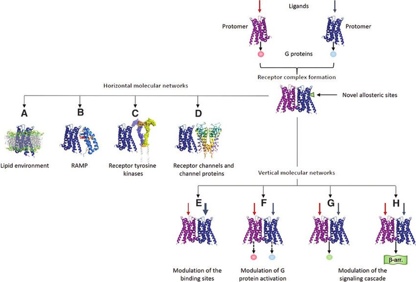
704 D. Guidolin et al.: GPCR-receptor interactions give integrative dynamics to intercellular communication
indirect biochemical and functional evidence that GPCRs of class A GPCR complexes in native systems (Bouvier and
could also establish structural receptor-receptor interac- Hebert, 2014). Furthermore, in view of the fact that the
tions (RRIs; Agnati et al., 1980, 1983; Fuxe et al., 1983). three above-mentioned class A GPCRs shown to be func-
The term RRI emphasized the existence of an interaction tional as monomers also exist as dimers or higher-order
requiring a direct physical contact between the involved oligomers (see below), the existence of class A GPCR func-
receptor proteins leading to the formation of multimeric tional oligomers cannot be excluded (see Franco et al.,
assemblies of receptors (dimers or high-order oligomers) 2016, for a recent discussion of the topic). In this respect,
at the cell membrane, operating as integrative input units of interest are studies showing that class A receptors
of membrane-associated molecular circuits (see Kenakin appear to exist in a monomer-dimer equilibrium, where
et al., 2010). The concept of GPCR oligomerization was class A GPCR dimers are often transient as seen from their
later confirmed in 1998–1999 by studies reporting that half-lives determined from the rate of association and dis-
two nonfunctional class C GPCR monomers, GABAB1 and sociation (Gurevich and Gurevich, 2008). This may help
GABAB2, assembled in a signaling heterodimer (Marshall explain opposing views on the role of class A GPCR mono-
et al., 1999a). In the years that followed, several groups mers versus dimers (Chabre and le Maire, 2005).
provided direct evidence for the existence of receptor com- The amount of RRIs identified so far is very high and
plexes formed by GPCR (Fuxe et al., 1998; Bockaert and their number is continuously increasing (see Farran, 2017,
Pin, 1999; Marshall et al., 1999b; Xie et al., 1999; Franco for a recent review). They are mostly stored in the GPCR
et al., 2000; Lee et al., 2000; Overton and Blumer, 2000; Oligomerization Knowledge Base (http://www.gpcr-okb.
Zeng and Wess, 2000; Angers et al., 2001; Dean et al., org; Khelashvili et al., 2010) and, for what it concerns the
2001; Kenakin, 2002; Waldhoer et al., 2005). heteromers, in the GPCR-HetNet (http://www.iiia.csic.
The amount of data supporting the existence of GPCR es/∼ismel/GPCR-Nets/index.html; Borroto-Escuela et al.,
heteromers showed a huge increase as far as biophysical 2014) containing more than 500 entries.
techniques capable of detecting the spatial proximity of The basic molecular mechanism leading to the for-
protein molecules were developed and became widespread mation of these receptor assemblies are allosteric inter-
(Bai, 2004; Guidolin et al., 2015). Biological methods actions (see Changeux, 2013), and as recently outlined
for identifying GPCR oligomers in cells and tissues or in by Changeux and Christopoulos (2016), the cooperativity
recombinant mammalian expression systems presently that emerges in the actions of orthosteric and allosteric
include energy transfer-based methods [fluorescence res- ligands of the GPCR forming the complex provides the
onance energy transfer (FRET) and bioluminescence reso- cell decoding apparatus with sophisticated dynamics in
nance energy transfer (BRET); Fernandez-Dueñas et al., terms of modulation of recognition and signaling. Thus,
2012], bimolecular luminescence or fluorescence comple- for an assembly of multiple receptors, the term ‘receptor
mentation (Gandia et al., 2008), total internal reflection mosaic’ (RM) was also introduced (Agnati et al., 1982) to
fluorescence microscopy (Hern et al., 2010), fluorescence better indicate the ‘integrated output’ of such an input
correlation spectroscopy (Chen et al., 2003), analysis of unit, stressing the concept that the emergent properties of
colocalization in immunohistochemical preparations the receptor assemblage depend on the location and the
(Agnati et al., 2005a), coimmunoprecipitation (Skieterska order of activation of the participating receptors (Agnati
et al., 2013), assays based on bivalent ligands (Yekkirala et al., 2007) as well as on the type of allosteric interactions
et al., 2013), and in situ proximity ligation assays (Trifilieff (entropic and/or enthalpic) within such an integrative
et al., 2011). complex (Fuxe et al., 2009; Agnati et al., 2010a). The
It is now well accepted that class C GPCRs form consti- assessment of RRI, therefore, provided a broadened view
tutive homomers or heteromers (Kniazeff et al., 2011) and on the role of GPCR in the dynamics of synaptic function,
some evidence exists suggesting that also class B GPCRs indicating that they can operate not only as monomers
could oligomerize (see Ng and Chow, 2015). but also as integrated units.
The oligomerization process in class A GPCRs is a This finding led to the suggestion (Fuxe et al., 2013,
debated question (see Milligan, 2009), especially for what 2014a; Gomes et al., 2013) that RRI could open new targets
it concerns its occurrence in living tissues, as no single for drug development and allow new strategies of treat-
presently available experimental approach can lead to ment. This aspect is presently the subject of intense
a conclusive demonstration of GPCR complexes in vivo research (see Guidolin et al., 2015; Borroto-Escuela et al.,
(Lambert and Javitch, 2014). However, the overall avail- 2017; Farran, 2017, for recent reviews). In recent years,
able evidence (obtained through multiple approaches such an effort allowed the characterization of a panel of
with consistent results) strongly supports the possibility receptor complexes representing possible targets for the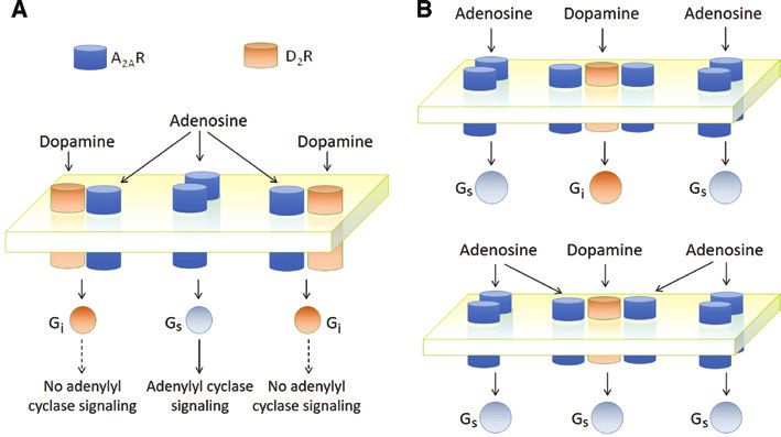
D. Guidolin et al.: GPCR-receptor interactions give integrative dynamics to intercellular communication   705

treatment of pathologic conditions, such as Parkinson’s             of the TM region (Palczewski et al., 2000; Katritch et al.,
disease (Fuxe et al., 2015), schizophrenia and depression           2012), which also harbors a number of kinks elicited by
(Fuxe et al., 2013; Sahlholm et al., 2017), neuropathic pain        Pro residues, segregating the receptor into ligand binding
(Bushlin et al., 2012), addiction (Gomes et al., 2013), and         and receptor signaling ‘modules’ (Latek et al., 2012). As
food intake disorders (Kern et al., 2012). On this basis,           shown by crystallographic studies, the overall structure of
novel strategies for drug treatment have also been pro-             GPCR proteins is highly conserved, but significant diversi-
posed. Interestingly, such protocols when compared to               ties can be observed in the loop regions and in the pitch
the traditional ones often appear able to reduce collateral         and orientation of individual TM in the helical bundle (Lu
effects (Le Naour et al., 2014). Of particular interest were        and Wu, 2016). Such a quite high plasticity of the GPCR
recent advances leading to the development of receptor              structure is likely a consequence of the presence of intrin-
complex-specific ligands (Bhushan et al., 2004; Daniels             sically disordered segments that do not fold into a stable
et al., 2005) that could lead to the identification of new          secondary structure (Agnati et al., 2008; Venkatakrishnan
tools for pharmacologic intervention.                               et al., 2014). Computational and structural studies have
     The formation of receptor complexes, however, has              revealed that GPCRs harbor disordered segments in the
also an impact on neurophysiology (Farran, 2017), espe-             extracellular N-terminus and large disordered areas in the
cially for what it concerns the emerging field of ‘connec-          cytosolic region, mainly in the intracellular C-terminus
tomics’ (see Guidolin et al., 2017, for a recent review), as        and in the ICLs, particularly ICL3 (Agnati et al., 2008;
it allows an integration of the incoming signals already            Guidolin et al., 2011a; Venkatakrishnan et al., 2014). These
at the plasma membrane level and can significantly con-             results have been recently supported by Tovo-Rodrigues
tribute to set and tune the efficiency of the connections           et al. (2014) who provided a detailed analysis of disordered
between cells and, in particular, the synaptic strength.            domains in 75 GPCRs involved in synaptic transmission
     The fundamentals of GPCR complex formation and                 using computational tools for the sequence-based predic-
the functional roles these structures can play at the syn-          tion of intrinsically disordered regions within a protein.
aptic level in terms of the modulation of signaling path-           As, in many cases, disordered segments assume a stable
ways [also called vertical molecular networks (VMNs);               folding following the binding with some partner, these
Agnati et al., 2005b] and interaction with other mem-               unstructured sequences are particularly suited for inter-
brane proteins [i.e. in the so-called horizontal molecular          action. Interestingly, some common interaction partners
networks (HMNs); Agnati et al., 2005b] will be the focus            of GPCRs, such as GPCR kinases (GRK), have the possibil-
of the present review article. When available, molecular            ity to couple to disordered regions of the receptor compris-
and bioinformatics models concerning the structure and              ing ICL3 and the C-terminal tail (Boguth et al., 2010; Elgeti
function of GPCR complexes will be briefly summarized               et al., 2013). The presence of these highly flexible linkers
and discussed.                                                      also facilitates conformational changes allowing large
                                                                    movements of the TM domains (Rasmussen et al., 2011),
                                                                    making possible a diversity of TM interactions.

Structural biology of receptor                                           Structural plasticity and malleability, however, are
                                                                    crucial not only for conformational fluctuations and
complexes                                                           intrareceptor interactions, but they are also of paramount
                                                                    importance to establish allosteric RRIs, allowing the for-
GPCRs have a complex structure that occupies a volume               mation of receptor complexes.
of about 3–4 nm by side (Zoffmann et al., 2007) and ranges
three different microenvironments (extracellular space,
membrane lipid bilayer, and cytoplasm). From a global               RRI as allosteric interactions
structural point of view, it is possible to distinguish seven
α-helixes piercing the entire plasma membrane (trans-               It has been known for a quite long time that receptors can
membrane domains, TM), which are interconnected via                 functionally interact by sharing signaling pathways or by
an extracellular loop (ECL) and an intracellular loop (ICL).        mechanisms of transactivation (Luttrel et al., 1999; Köse,
The extracellular region (comprising the N terminus of              2017). This formally fits the definition of RRI in a func-
the protein) is characterized by a high structural diversity        tional sense, although the involved proteins may never
allowing the recognition of a wide spectrum of ligands.             physically come into contact with each other (see Prezeau
Interhelical bonds and hydrophobic interactions between             et al., 2010, for a detailed analysis). What we are here
highly conserved residues in GPCR provide the stability             discussing, on the contrary, are RRIs requiring a direct
706 D. Guidolin et al.: GPCR-receptor interactions give integrative dynamics to intercellular communication
physical contact between the involved receptors leading light of biophysical investigations (Fotiadis et al., 2003;
to the formation of receptor complexes at the cell mem- Tateyama et al., 2004; Hern et al., 2010), the ‘domain
brane. The definition of ‘physical contact’, however, can contact’ is presently considered as the main mechanism
be debated as proteins have varying degrees of associa- of GPCR association. Regardless of the type of geometry
tion. A specific international consensus workshop in 2010 assumed for the association, the specific interacting resi-
(see Kenakin et al., 2010) provided a definition that will dues that form the interaction interface represent a signifi-
be adopted here: ‘Receptor-receptor interactions: when cant target of current research on GPCR oligomerization
the binding of a ligand to the orthosteric or allosteric sites (Skrabanek et al., 2007). In fact, the nature of the interac-
of one receptor causes, via direct allosteric interactions, a tion interface not only specifies which GPCR can exhibit
change in the ligand recognition, decoding and trafficking significant interactions but also influences the models for
processes of another receptor’. potential allosteric interactions between partners.
Allostery (Tsai et al., 2009; Tsai and Nussinov, 2014;
Liu and Nussinov, 2016) is a mode of long-distance com-
munication between distal sites in proteins, in which the Interaction interfaces
energy released as a consequence of conformational or
dynamic changes at one site can travel along specific path- The research in this field benefited of a combined use
ways within the protein structure to other sites, chang- of bioinformatics methods to predict the amino acid
ing their conformational or dynamic properties (Liu and sequences involved in the interaction interfaces and
Nussinov, 2017). Computational methods directly relat- experimental work.
ing protein structural dynamics to information exchange Several bioinformatics methods have been devised
between functional sites have also been devised (Lenaerts to predict the interfaces available to a given GPCR for RRI
et al., 2008). Because allostery involves changes in protein (Filizola and Weinstein, 2005; Guidolin et al., 2011a). They
conformation, the ability of a protein to take on new con- can, in principle, be categorized into three broad classes
formations is related to the ability of the protein to be according to the type of strategy followed to perform the
allosterically modulated. Therefore, a protein with an analysis (Simpson et al., 2010):
already rigid structure is less inclined to be allosterically –– The first type of approach is based on the identifi-
modulated than a protein with a high degree of intrinsic cation of protein regions exhibiting some property
disorder. In this respect, molecular dynamics studies sug- (potentially relevant for the interaction with other
gested that signaling proteins, such as GPCR, are ideal proteins) that can be deduced simply by the analy-
candidates to be allosterically modulated (Liu et al., sis of the primary structure (i.e. the amino acid
2006a; Hilser and Thompson, 2007). Thus, when two sequence). Using sequence features, for instance, sev-
protomers establish direct RRI, the energy released fol- eral methods have been developed to classify whether
lowing a perturbation event at one site of a protomer can any given residue belongs to protein segments poten-
pass over the receptor interface into the other protomer tially relevant for protein-protein interaction, such as
(Agnati et al., 2010b; Fuxe et al., 2012) to change its con- intrinsically disordered regions (Ferron et al., 2006).
formation and functional features. Extensive reviews on Agnati et al. (2008) introduced a ‘disorder index’ as
allostery at GPCR homomers and heteromers, with a clear the weighted average of the results provided by 10
discussion of the topic, have been provided by Kenakin predictors, covering a wide spectrum of the strate-
and Miller (2010) and by Smith and Milligan (2010). gies to identify disordered regions in proteins using
As far as the modes of association of GPCR mono- their primary structure. The results suggested ICL3
mers into oligomers are concerned, two modes have been and C-terminal domains as potential sites of inter-
proposed (Gouldson et al., 2000). One of them is called action interfaces for A1 and A2A adenosine receptors,
‘domain contact’ dimerization, corresponding to the whereas, in A2B and A3 subtypes, only the C-terminal
interaction of the molecular surfaces at specific binding domain exhibited a significant score. The analysis of
interfaces, without largely changing the conformation of disordered domains, however, should be integrated
the monomer structure. The other one, termed ‘domain with other methods, as it does not help for the identi-
swapping’ dimerization, is a mechanism in which a sub- fication of putative interaction sites located in the TM
structure (or domain) of a monomer is exchanged with helices. Thus, an approach based on the evaluation
the corresponding substructure (or domain) of the other of the aggregation propensity of natural amino acids
monomer. Thus, a large conformational change of a (Sànchez de Groot et al., 2005) was also proposed
monomer structure is required for this mechanism. In the (Agnati et al., 2009a), allowing the identification ofD. Guidolin et al.: GPCR-receptor interactions give integrative dynamics to intercellular communication 707 protein regions that are especially relevant for pro- homodimer was very recently provided (Altwaijri tein aggregation (‘hot spots’) by simply analyzing the et al., 2017) using a new coarse-grained approach to amino acid sequence. When applied to the analysis molecular dynamics simulations, specifically devel- of GPCRs, such as adenosine A2A, dopamine D2, can- oped for identifying helix-helix interactions in GPCR. nabinoid CB1, and glutamate mGlu5, this approach predicted ‘hot spots’ in specific regions of TM4–TM6. Results obtained from computational predictions and Other sequence-based computational methods that concerning a panel of receptor complexes are summa- have been used include variants of the ‘evolutionary rized in Table 1. These analyses, however, only provide trace’ method, ‘level entropy’ and ‘sequence space suggestions that should be confirmed by experimental automation’ methods, and ‘correlated mutation anal- data. Recent advances in experimental methods have ysis’ (see Filizola and Weinstein, 2005; Vohra et al., equipped researchers with a repertoire of tools to get 2007; Guidolin et al., 2011a, for reviews). According details about the interaction interfaces. The last years, to a meta-analysis reported by Filizola and Weinstein for instance, have seen a significant advancement in crys- (2005), most of the identified residues are within tallization techniques with important consequences for TM4–TM6, further suggesting a specific role for these the analysis of GPCR and an increase of the number of three helices in the dimerization/oligomerization experimentally assessed structures (Grisshammer, 2017). interfaces of GPCR. Additional experimental tools encompass atomic force –– A second type of approach is based on the analysis of microscopy (Liang et al., 2003; Agnati et al., 2010b) and the three-dimensional structure (as obtained by experi- novel super-resolution imaging approaches, such as pho- mental investigation or by homology modeling) of the toactivated localization microscopy (PALM; Jonas et al., protein under scrutiny to identify the possible surfaces 2016), far-ultraviolet circular dichroism spectroscopy, and of interaction with other proteins. Of particular interest sodium dodecyl sulfate-polyacrylamide gel electropho- in this field are recent developments in protein-protein resis using synthetic peptides corresponding to different docking software that lead to the prediction of the pos- TMs (Thevenin and Lazarova, 2008). Woods et al. (Ciruela sible binding sites between two molecules, allowing et al., 2004; Woods et al., 2005) using mass spectrom- the formation of a stable complex (Simpson et al., 2010; etry in combination with collision-induced dissociation Kaczor et al., 2015; Soni and Madhusudhan, 2017). experiments investigated intracellular domains (namely Examples of this approach include the study of the ICL3 and C terminus), demonstrating strong electrostatic lutropin receptor dimerization (Fanelli, 2007), stud- interactions in heteromers. Results from experimental ies aimed at characterizing the interaction interface in investigations are reported in Table 1. As shown, they serotonin (5-HT)4 (Bestel, 2005; Soulier et al., 2007) and were, in general, supportive of the predictions provided rhodopsin complexes (Han et al., 2009), and the gen- by bioinformatics. Both computational and experimen- eration of models for the heterodimeric mGluR2-5-HT2A tal methods suggest that GPCR structures are capable of complex (Bruno et al., 2009) and for the dopamine D1- interacting via multiple interfaces, but some domains D2 receptor dimer (Agnati et al., 2016). were observed more often than others. TM4–TM6 and –– Molecular dynamics and coarse-grained simulations ICL3, for instance, were reported as the main interfaces are one of the most versatile and widely applied com- in a quite large number of GPCR complexes. For what it putational techniques for the study of membrane concerns the possible involvement of ECLs in the interac- proteins (Almeida et al., 2017), as they consider the tion, it was demonstrated for some class A GPCR (Huang tertiary structure of the studied proteins and can et al., 2013), and evidence was provided for interaction by implement an energy landscape to estimate the disulfide bridges between extracellular domains in some molecular interactions, also accounting for the role class C GPCR (Kniazeff et al., 2011). of the lipid microenvironment. Such methods have An interesting finding from computational and exper- also been used to study of GPCR dimerization and oli- imental studies on GPCR oligomerization interfaces is the gomerization (Fanelli et al., 2013; Jonas et al., 2015; presence at the interface of specific motifs that appear Altwaijri et al., 2017). Examples include the study of of particular importance for the allosteric interaction. As rhodopsin dimer (Filizola et al., 2006) and simula- demonstrated by Woods and colleagues (Woods, 2004; tions of vasopressin receptor oligomerization (Witt Woods and Ferré, 2005), the electrostatic interactions et al., 2008) and of the mGluR2-5-HT2A heterodimeric between intracellular domains occur between a negatively complex (Bruno et al., 2009). A structural char- charged serine-phosphate-containing intracellular motif acterization of the human A2A adenosine receptor of one receptor and a positively charged arginine-rich
708            D. Guidolin et al.: GPCR-receptor interactions give integrative dynamics to intercellular communication

Table 1: Interaction interfaces: computational predictions and experimental findings.

GPCR complex                    Predicted                References                      Experimental             References

Homodimers
 Adenosine A2A-A2A              TM1,2,3–TM1,2,3          Fanelli and Felline, 2011       TM1,3–TM5,6              Liu et al., 2012
                                TM1–TM1
 β1-β1-Adrenergic               TM1–TM1                  Mondal et al., 2013             TM1,H8–TM1,H8            Huang et al., 2013
                                TM5–TM5                                                  TM4,5–TM4,5              Cordomì et al., 2015
 β2-β2-Adrenergic               TM6–TM6                  Mondal et al., 2013             TM6–TM6                  Hebert et al., 1996
                                TM4,5–TM4,5
                                TM1–TM1                  Prasanna et al., 2014
                                H8–H8                    Ghosh et al., 2014
 Chemokine CXCR4-CXCR4          TM3–TM4,5                Rodriguez and Gutierrez-de-     TM3–TM4                  Wu et al., 2010
                                                         Teran, 2012
                                TM5–TM5                                                  TM5–TM6
 Dopamine D2-D2                 TM4–TM4                  Nemoto and Toh, 2005            TM4–TM4                  Guo et al., 2003
 δ-δ Opioid                     TM4–TM4                  Johnston et al., 2011           TM4–TM4                  Johnston et al., 2011
                                TM4,5–TM4,5
 κ-κ Opioid                     TM1–TM2                  Kaczor et al., 2013             TM1,2,H8–TM1,2,H8        Johnston and Filizola, 2014
 μ-μ Opioid                     TM1,2,H8–TM1,2,H8        Marino et al., 2016             TM1,2,H8–TM1,2,H8        Manglik et al., 2012
                                TM5–TM5                                                  TM5,6–TM5,6
                                TM1,2,H8–TM5
 Serotonin 5-HT1A-5-HT1A        TM4,5–TM4,5              Gorinski et al., 2012           TM4,5–TM4,5              Gorinski et al., 2012
Heterodimers
 A2A-D2                         TM5,6,ICL3–TM3,4         Canals et al., 2003             TM5,6,ICL3–TM3,4         Canals et al., 2003
                                TM4,5–TM3,4,5
                                TM4,5–TM4,5              Borroto-Escuela et al.,         TM4,5–TM4,5              Borroto-Escuela et al., 2010
                                                         2010
                                H8–ICL3                  Agnati et al., 2008             H8–ICL3                  Ciruela et al., 2004
                                                                                                                  Woods et al., 2005
 D1-D2                          TM4,5–TM1,2              Agnati et al., 2016             H8–ICL3                  Hasbi et al., 2014
                                H8–ICL3
Trimers
  A2A-CB1-D2                    TM4–TM4                  Agnati et al., 2010c            TM4–TM4                  Navarro et al., 2010
                                TM6–TM6                                                  TM5–TM5
                                H8–ICL3                                                  H8–ICL3

H8, C-terminal amphipathic helix 8.

motif of a second receptor. Once established, they possess               Most of them are leucine-rich motifs. Another minor type
a covalent-like stability and likely represent the main                  of triplets contains charged amino acids and the electro-
mechanism for heteromer assemblage. More recently, it                    static interaction between triplets may guide-and-clasp
has been reported that a highly conserved small-XXX-small                protein-protein interactions (Borroto-Escuela et al., 2011;
motif found in TM1 of the fungal GPCR Mam2 promotes                      Fuxe et al., 2014a).
TM1 self-association (Lock et al., 2014). Small-XXX-small
motifs are motifs of two residues (typically glycine, but
also alanine or serine) separated by three amino acids                   Quaternary structure of GPCR complexes
in the polypeptide chain, thus physically placing them
on the same face of an α-helix. Colocation of these two                  The basic structure generated by the interaction of GPCR
small residues results in a ‘groove’ that allows two helices             is the dimeric structure (Figure 1A), in which pairs of
to interlock via many favorable van der Waals contacts,                  protein molecules (protomers) associate. Homodimers
thereby promoting helix-helix interactions (Jenei et al.,                are pairs of the same protomer, whereas heterodimers
2009). Based on a bioinformatics approach, Tarakanov                     are formed from distinct GPCR. The number of described
and Fuxe (2010) have deduced a set of triplet homologies                 homodimers and heterodimers in both cellular systems
that may be responsible for RRIs. Such amino acid triplets               and native tissues is at present very high (see Fuxe et al.,
resulted mainly located at the receptor-receptor interface.              2015; Guidolin et al., 2015; Borroto-Escuela et al., 2017;
D. Guidolin et al.: GPCR-receptor interactions give integrative dynamics to intercellular communication 709
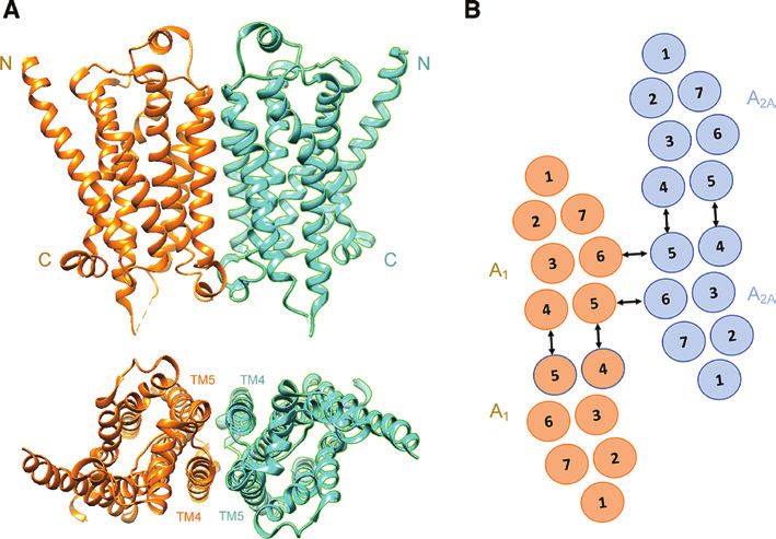
Figure 1: Examples of quaternary structures formed by GPCR.
(A) Monomers of the β1-adrenergic receptor (PDB code: 4GPO) as obtained by X-ray crystallography (Huang et al., 2013), arranged to form
a β1-β1 homodimer as suggested by Cordomì et al. (2015). Top, the N and C termini are indicated; bottom, a top view (from the extracellular
surface) is provided with indication of the interacting TM4/TM5 domains. (B) Schematic representation of the heterotetramer formed by A1
and A2A adenosine receptors as proposed by Navarro et al. (2016). Arrows indicate the TM4–TM5 interface mediating homodimerization and
the TM5–TM6 interface exploited for heterodimerization. These interfaces give a rhombus-shaped receptor complex organization.
Farran, 2017, for reviews), and Table 1 only provides some et al., 2016). Tetrameric assemblies of β2-adrenergic
example. However, the evidence that a GPCR can exploit receptors were demonstrated by the group of Kobilka
multiple interaction interfaces can significantly influence to occur spontaneously following reconstitution into
the architecture of the resulting receptor complexes in at phospholipid vesicles (Fung et al., 2009), suggest-
least two aspects: ing that β2-adrenergic receptor oligomerization is an
–– The first concerns the stoichiometry of the complex intrinsic property of the receptor. A possible heterote-
(i.e. the number of component subunits) opening trameric structure has been recently proposed for the
the possibility that oligomeric assemblies of different complexes formed by adenosine A1 and A2A receptors
order could be formed (Agnati et al., 2010a). Navarro (Navarro et al., 2016), in which a TM4–TM5 interface
et al. (2010) were among the first to advance evidence mediates homodimerization and a TM5–TM6 interface
for the role of interaction interfaces between protom- is exploited for heterodimerization (Figure 1B). Some
ers in orchestrating the quaternary structure of het- evidence also exists of higher-order GPCR oligomers.
eromers. This study was focused on adenosine A2A, For instance, combined BRET/FRET and complemen-
dopamine D2, and cannabinoid CB1 receptors. Each of tation studies have revealed that the assemblage of
them exhibited two intracellular domains that inter- dopamine D2 receptors by symmetrical interfaces at
acted in a specific manner with intracellular domains TM4 and TM1 can lead to a complex composed of at
of the other two protomers through electrostatic least four protomers in the plasma membrane of liv-
interactions, leading to the assembly of dimers (A2A- ing mammalian cells (Guo et al., 2008). Moreover,
D2, A2A-CB1, and CB1-D2) but also to the formation of a based on an analysis of PALM data, it has been pro-
A2A-D2-CB1 heterotrimer. Trimeric receptor complexes posed that direct RRI could lead to the formation
have been indeed identified (Gandia et al., 2008). of high-order oligomers (tetramers, octamers, and
They include the A2A-D2-mGlu5 (Cabello et al., 2009) larger-sized complexes) depending on the specific
heteroreceptor complex, the muscarinic M2 homo- membrane microenvironment (Scarselli et al., 2016).
trimer (Park and Wells, 2004), the dynamic Gal1-5- –– The existence of multiple interaction interfaces opens
HT1A-GPR39 heterotrimer (Tena-Campos et al., 2015), the possibility that the assemblage of a given set of
and the putative Gal1-Gal2-5-HT1A heterotrimer (Millón receptor molecules to form a complex could occur710 D. Guidolin et al.: GPCR-receptor interactions give integrative dynamics to intercellular communication
in a number of different geometrical arrangements orthosteric pockets, in receptor structures, allosteric
(Agnati et al., 2009a) depending on a number of con- binding sites can be located in various regions of the mole-
ditions including not only the physical properties of cule (Bartuzi et al., 2017). For class A GPCR, in most cases,
the interacting proteins (surface charge, hydropho- allosteric binding sites are located in the same region as
bicity, etc.) but also the microenvironment surround- the orthosteric one (i.e. within the seven TM), whereas,
ing the interacting partners (i.e. the energy landscape; in class C GPCR, the two sites are usually well separated
Frauenfelder et al., 1991). As better discussed in the (see Wu et al., 2014). When a receptor complex forms, the
next section, the topological arrangement of the allosteric binding sites on single monomers may undergo
receptor complex can influence its functional behav- structural and functional changes (see Shivnaraine et al.,
ior. An interesting experimental evidence of this con- 2016). Of significant interest, however, is the possibility
cept was recently provided by Jonas et al. (2015) using that, when the complex forms, the quaternary structure
a super-resolution imaging approach. The study was could display novel specific allosteric sites suitable for
focused on two functionally defined mutant luteiniz- the binding of some modulator. Thus, ligands could also
ing hormone receptors, which only function via inter- exist specific to the receptor complex as such (see Fuxe
molecular cooperation with favored oligomeric over et al., 2010). Studies on the effect of homocysteine (Agnati
dimeric formation. PD-PALM imaging of trimers and et al., 2006, 2008) on the A2A-D2 heterodimer provided
tetramers showed that monomers interconnected by a first example of the possible existence of allosteric mod-
complex helix interfaces can assume a variety of dis- ulators of a receptor complex. In Chinese hamster ovary
tinct spatial arrangements that also differ from each cells stably cotransfected with adenosine A2A and dopa-
other in terms of signal sensitivity and strength. mine D2 receptors, homocysteine selectively decreased the
ability of D2 receptor stimulation to internalize the recep-
A further aspect of substantial interest has been high- tor complexes. Mass spectrometric analysis showed that,
lighted by studies using single-GPCR imaging (see by means of an arginine-thiol electrostatic interaction,
Sungkaworn et al., 2013) in living cells. They revealed homocysteine forms noncovalent complexes with the two
the kinetics of complex formation, indicating that GPCR arginine-rich epitopes of the third ICL of the D2 receptor,
can form either stable or transient complexes at the cell one of them being involved in the receptor heteromeri-
surface depending on the interaction energy (Gurevich zation interface. However, homocysteine was unable to
and Gurevich, 2008). To exist as a stable dimer with a half- prevent or disrupt A2A-D2 receptor heteromerization as
life comparable to that of even short-lived GPCR (2–20 h), demonstrated by FRET experiments. Thus, it likely acts as
a binding energy of at least ~60 kJ/mol is required. This a modulator of the allosteric process of energy transmis-
condition is often fulfilled by class C GPCR, explaining sion between the two protomers.
why they often appear as stable dimers. For what it con-
cerns family A GPCR dimers, they are often transient as
seen from their half-lives. In the case of the neurotensin
NTS1 dimer, a half-life of 340 s has been observed (White
Dynamic behavior of receptor
et al., 2007) and evidence has been found that M1 mus- complexes and the concept of RM
carinic receptor dimers have an estimated half-life of 0.5 s
(Hern et al., 2010). In a study by Calebiro et al. (2013), The existence of these supramolecular assemblies is con-
β1- and β2-adrenoceptors were monitored on the surface sidered of particular importance because it allows the
of living cells and the kinetics of the interactions leading emergence of integrative functions (Agnati et al., 2010b)
to the formation of oligomers was characterized. All these performed by a receptor complex as a whole. In fact,
receptors dynamically formed dimers and high-order oli- owing to allosteric RRI, a configuration change of a given
gomers, with an apparent half-life in the order of 4–6 s. protomer will change the probability of changing the con-
Thus, a dynamic equilibrium condition was established at figuration for the adjacent receptors in the complex and
the cell surface, with constant formation and dissociation the effect will propagate throughout the cluster, leading
of new receptor complexes. The relative amount of the to a complex collective behavior and to an integrated
different stoichiometries was dependent not only on the regulation of multiple effectors (Fuxe et al., 2012). These
subtype of receptor but also on the receptor density. concepts were well illustrated by a simple mathematical
A final relevant aspect of the receptor complex struc- approach to the cooperativity in complexes formed by
ture can be appreciated when the allosteric binding dimers of identical receptors and/or by receptors binding
sites of the monomers are considered. In contrast to the to the same ligand (Agnati et al., 2005c). The model wasD. Guidolin et al.: GPCR-receptor interactions give integrative dynamics to intercellular communication 711
based on a ‘symmetry rule’, which has been proven for results of thermodynamics-based approaches to modeling
hemoglobin (Ackers et al., 1992), and this model main- (Jackson, 2006). In this formulation, each protomer can
tains that a quaternary switching from tense form (the exist in distinct conformational states and makes rapid sto-
‘deoxy’, low-affinity state) to relaxed form (the ‘oxy’, chastic transitions between these states, with Boltzmann-
high-affinity state) occurs whenever heme-site binding weighted probability. The energy of each protomer in the
creates a tetramer with at least one ligated subunit on complex was in turn dependent on three energy terms:
each dimeric half-molecule. When the same basic rule the energy associated with its configuration, the energy
is applied to assemblies formed by homodimers (as evi- associated with external inputs, and the energy due to the
denced for dopamine receptors; Guo et al., 2008), the inte- coupling with adjacent protomers. In the model discussed
grative cooperativity of the complex appeared to depend by Duke et al. (2001), when coupling exists between
not only on the composition (number of dimers) but also neighboring proteins, a conformational spread occurs,
on its spatial organization (respective location of the driving the system to a switch-like, sigmoid response to
dimers) and order of activation (order according to which changes in ligand concentration. A theoretical analysis of
the single receptors are ligated). To investigate in more the role of the spatial arrangement of monomers within
detail the potential complex cooperative behavior of the the complex based on thermodynamical considerations
receptor assemblies, a number of computational models was provided by Agnati et al. (2010c), who showed that,
and computer simulations were proposed (see Guidolin for each given set of binding and interaction constants,
et al., 2011a, for a review). the theoretical saturation curves of trimeric or tetrameric
A first class of models aimed at describing the receptor complexes were dependent on the geometry of
dynamics of receptor complexes was based on methods the receptor complex.
from discrete dynamics (Martelli, 1999). According to this Thus, receptor complexes appear to be endowed with
approach, individual receptors can be assumed to have ‘emergent properties’, that is, with biochemical charac-
two broad classes of conformational states with respect teristics and functions that could not be fully anticipated
to the macromolecular effectors: one active and one inac- by analyzing the characteristics of the single participat-
tive. Owing to RRIs, however, a state change of a given ing receptor monomers. In particular, due to differences
receptor will change the probability of changing the state in topology and rank order of activation, it is possible (at
for the adjacent receptors in the RM and the effect will least from a theoretical standpoint) to have markedly dif-
propagate throughout the cluster, leading to a complex ferent integrative functions for receptor complexes formed
cooperative behavior. On this basis, Boolean net- by the same set of monomers (i.e. same stoichiometry).
works (BN) were proposed as a suitable abstract model To grasp this fundamental aspect of GPCR assemblies,
to explore how complex properties may emerge from the term RM was proposed (Agnati et al., 1982, 2010a) to
systems permeated by deterministic local interactions of identify them. The concept was inspired from the mosaic
many simple components operating in parallel (Agnati as defined in figurative art: namely the process of assem-
et al., 2007). Very common ‘macroscopic’ properties of a bling images by inlaying small pieces of colored stones
receptor system (such as a sigmoidal response curve to (tesserae) according to a structural plan (Agnati et al.,
an extracellular ligand) were captured from a model of 2009b). The term ‘mosaic’ thus describes how a limited
this type (Guidolin et al., 2011b). The response, however, set of building blocks could be arranged into different
was modulated by changes in the topology and/or in the patterns according to distinct designs, resulting in sets of
local interactions between the receptor units forming the elements endowed with differential emergent properties
assembly. Furthermore, the system exhibited a limited depending on their respective interactions. According to a
number of equilibrium configurations or ‘attractors’, metaphor proposed by Kenakin (2009), RM would operate
leading to the hypothesis that such a set of configurations as a sort of molecular ‘microprocessor’, as they are not just
could be interpreted as a form of information storage ‘on-off’ switches but exhibit a high capability to elaborate
(engram) at the level of synapses and intercellular con- information. A straightforward example is likely provided
nections (Agnati et al., 1982, 2004; Guidolin et al., 2007). by RM formed by isoreceptors (i.e. receptors for the same
Thus, the suitable reorganization of receptor complexes ligand), such as, for instance, the D1–D2 (Lee et al., 2004) or
in the postsynaptic membrane has been recently pro- the D1–D3 (Marcellino et al., 2008) isoreceptor complexes.
posed as the molecular basis of learning and memory It has been suggested that an analogy with an electronic
(Fuxe et al., 2014b; Borroto-Escuela et al., 2015). apparatus like the ‘demultiplexer’ (a device taking one
Consistent with the just mentioned characteristics of input signal and selecting one of many-data-output lines
a receptor complex suggested by BN models are also the to send it) could be exploited to describe their functional712 D. Guidolin et al.: GPCR-receptor interactions give integrative dynamics to intercellular communication
Figure 2: Basic operations described in Sherrington’s studies that could be realized at the molecular level by receptor complexes (Guidolin
et al., 2016).
(A) Possible “lateral inhibition” process at microscale level in a system of RM involving A2A and D2 receptors with reciprocal inhibitory activity
(see Guidolin et al., 2015) and A2A homodimers. The activation of D2 receptors by dopamine leads to a reduction in the affinity of A2A recep-
tors in the heterodimers, hence in a sharpening of the Gs-mediated signaling. (B) Schematic representation of the possible implementation
at molecular level of the concept of “fringe”. A low concentration of adenosine can activate only Gs-mediated signaling from A2A homodimers
(top). However, high concentrations of adenosine can induce, via RRI, an inhibition of the D2 receptor in the heterotrimer and its shift from
Gi- to Gs-mediated signaling (bottom).
behavior (Agnati et al., 2016). Furthermore, it has been multiple intracellular phosphorylation pathways involved
proposed (see Figure 2) that the interplay between RM in the regulation of gene expression and diverse biological
may implement at membrane-level basic operations (e.g. responses, such as proliferation and differentiation. Acti-
‘lateral inhibition’) that were described at the level of neu- vated GPCR, however, also interact with cytosolic ligands
ronal networks in Sherrington’s studies (Guidolin et al., (see Premont and Gainetdinov, 2007; Magalhaes et al.,
2016). 2012, for reviews), such as GRK (Premont et al., 1994)
As illustrated in Figure 3, RM operate as special- and arrestins (Lohse et al., 1990). GRK regulate GPCR
ized devices in two complementary contexts (see Agnati desensitization by both phosphorylation-dependent and
et al., 2010a). From one side, they are part of the so-called phosphorylation-independent mechanisms (Dhami and
VMNs, that is, the molecular pathways involved in signal Ferguson, 2006). Arrestins turn off the GPCR response or
recognition and transduction, extending from the extra- adapt the system to a persistent stimulus by coordinat-
cellular space to the cytoplasm and nucleus. On the other ing spatially and temporally the uncoupling of G protein
side, they can also partake HMNs, i.e. networks formed from receptors and by mediating agonist-promoted recep-
by interacting membrane components, regulating infor- tor internalization (Lefkowitz and Shenoy, 2005). In addi-
mational exchange between the extracellular and the tion, arrestins have been demonstrated to scaffold a wide
intracellular environments. This twin role will be the variety of signaling complexes and there is now extensive
focus of the sections that follow. evidence indicating that ligands that interact with GPCR
can selectively activate G protein- versus arrestin-medi-
ated signaling pathways (Reiter and Lefkowitz, 2006).
RM and VMNs When RRI in the plane of the membrane occur, the
formation of RM can lead to several changes in the chain
A wide variety of extracellular ligands (biogenic amines, of events linking ligand recognition to signal transduction
amino acids, ions, lipids, peptides, proteins, sensory from the single protomers. They can be briefly summa-
stimuli, etc.) can be detected by GPCR, and these events rized as follows (see Guidolin et al., 2015; Borroto-Escuela
switch the receptor to an active conformational state that et al., 2017; Farran, 2017, for more specific reviews):
permits its coupling and activation of heterotrimeric GTP- –– Modulation of the binding sites has been reported to
binding proteins (Gαβγ proteins), leading to the regulation of occur in a variety of RM as a consequence of allostericD. Guidolin et al.: GPCR-receptor interactions give integrative dynamics to intercellular communication 713
Figure 3: Receptor complexes appear to be endowed with characteristics and functions that could not be fully derived from the characteris-
tics of the single participating receptor monomers.
When the complex forms, for instance, the quaternary structure could display novel specific allosteric sites (Agnati et al., 2008).
Furthermore, RM are in the center of two complementary networks of interactions: HMN involving (A) the lipid environment (Gahbauer
and Böckmann, 2016), (B) RAMP (Foord and Marshall, 1999), (C) RTK (Borroto-Escuela et al., 2012), and (D) membrane channels (Liu et al.,
2006a; Gamo et al., 2015) and VMN leading to (E) modulation of the binding sites (Fuxe et al., 1998), (F) modulation of G protein activa-
tion (Ferrada et al., 2009), (G) modulation of the signaling cascade, among others, and (H) switching from G protein to β-arrestin signaling
(Rashid et al., 2007; Rozenfeld et al., 2012).
RRI. One of the first examples was the A2A-D2 hetero causing allosteric conformational changes, due to the
dimer, where the binding of the adenosine A2A ago- repulsive effect generated by the negatively charged
nist CGS-21680 lead to a reduction of the affinity of phosphate, thus modulating heteromerization and
the high-affinity dopamine D2 agonist binding site affecting the stability of heteromers’ interactions and
(Fuxe et al., 1998). In this RM, a reciprocal interaction their binding affinity. In this instance, phosphoryla-
between D2 and A2A receptors also exists, as D2 recep- tion is not just an ‘on-off switch’; instead, by weak-
tor can inhibit the A2A-induced increase in cyclic AMP ening the noncovalent bond, heteromerization acts
(cAMP) accumulation via Gi/o at the level of the ade- as the entity that controls the stability of the heter-
nylate cyclase (Kull et al., 1999). As discussed by Woods omer through the activation or inhibition of adenylate
and Jackson (2013), in this heterodimer, the first step cyclase.
driving heteromerization involves the phosphoryla- –– Modulation of G protein activation causes changes
tion of the serine/threonine in an epitope containing in the decoding of signals impinging on protomers.
a casein kinase 1/2-consensus site, and dopaminergic An example is provided by the heterodimer formed by
neurotransmission, through cAMP-dependent pro- dopamine D1 and histamine H3 receptors. In this RM,
tein kinase A (PKA), slows down heteromerization. there is a change in the D1 receptor coupling from the
In addition, the negative charge, acquired by phos- Gs to the Gi protein, to which H3 receptors are already
phorylating a serine/threonine in a PKA consensus coupled. In fact, in the presence of the H3 receptor, D1
site in the arginine-rich epitope, affects the activ- receptors were no longer coupled to Gs and could not
ity of the receptors involved in heteromerization by activate adenylyl cyclase but were coupled to Gi, which714 D. Guidolin et al.: GPCR-receptor interactions give integrative dynamics to intercellular communication
transduced the signal toward the mitogen-activated parameter for receptor function (Mitchell et al., 1990), but
protein kinase pathway (Ferrada et al., 2009). direct cholesterol-receptor interactions have also been
–– Modulation of the signaling cascade occurs when described (Albert et al., 1996). Remarkably, the effect of
the RM recruits a G protein different from those cholesterol on GPCR function is receptor dependent. For
usually associated to the monomers (as in the D1-D2 example, cholesterol modulates agonist binding to oxy-
dimer; Rashid et al., 2007) or when the oligomeriza- tocin receptors (Gimpl and Fahrenholz, 2002), 5-HT recep-
tion process leads to a switch between G protein and tors (Pucadyil and Chattopadhyay, 2004) and μ-opioid
β-arrestin signaling (Rozenfeld et al., 2012), such as, receptors (Qiu et al., 2011), whereas other GPCRs are less
for instance, in the κ-μ and κ-δ opioid heteromers influenced (Oates and Watts, 2011). Stability studies have
(Le Naour et al., 2014). demonstrated the binding of cholesterol molecules to a
conserved motif located between helices 1–4 (Hanson
It has to be emphasized that the role played by RM in VMN et al., 2008). Recently, relevant phospholipids were found
is of particular importance from the pharmacologic point to affect GPCR function. In β2-adrenergic receptors recon-
of view, as it discloses a marked rise of the repertoire of stituted in high-density lipoparticles, for instance, phos-
GPCR recognition and signaling (Guidolin et al., 2015; phatidylgycerol markedly favored agonist binding and
Borroto-Escuela et al., 2017). facilitated receptor activation, whereas phosphatidyletha-
nolamine favored antagonist binding and stabilized the
inactive state of the receptor (Dawaliby et al., 2016). These
RM and HMNs data suggested that phospholipids could act as direct
allosteric modulators of GPCR activity. Lipids, however,
The pattern of interactions between molecules embed- can also be covalently bound to GPCR (see G ahbauer and
ded and/or associated with the cell membrane form the Böckmann, 2016). Due to a post-translational modification
so-called HMNs that can operate as modules carrying out called palmitoylation, the saturated fatty acid palmitic
specialized tasks (Agnati et al., 2010a). A functional and acid (16 carbons) can be added to C-terminal cysteine resi-
structural relationship exists between GPCR and HMN. dues via a thioester-type bond (Chini and Parenti, 2009).
GPCR can interact with several membrane components It was reported that GPCRs can be mono-, bis-, or even tris-
(Gahbauer and Böckmann, 2016) and play an important palmitoylated and that this lipid modification is reversible
role in the formation of HMN, where they can function as well as adjustable, thereby allowing the regulation of
as sophisticated signaling processing centers (Kenakin, GPCR function (Qanbar and Bouvier, 2003).
2007). As far as membrane proteins are concerned, the
In this respect, the first aspect deserving considera- most interesting association with GPCR was identified
tion is the lipid environment. It was shown to influence in a set of three homologous transmembrane proteins
GPCR function and several health disorders during aging that were named receptor activity-modifying membrane
were assigned to changes in the membrane composi- (RAMP) protein (Foord and Marshall, 1999). When asso-
tion that altered GPCR signaling (Alemany et al., 2007). ciated to the calcitonin-like receptor (CLR), they sig-
The preferential localization of GPCR and other compo- nificantly modify its function: the complex RAMP1-CLR
nents involved in signal propagation in dynamic mem- behaves phenotypically as a calcitonin gene-related
brane nanodomains (lipid rafts and caveolae) has been peptide receptor, whereas the association of RAMP2 or
reported in a vast number of studies (Insel et al., 2005; RAMP3 with CLR provides specificity for adrenomedul-
Lingwood and Simons, 2010; Simons and Sampaio, lin (Poyner et al., 2002). Other family B GPCRs have also
2011). These nanodomains are densely packed, dynamic been shown to associate with RAMP. They include para-
membrane areas with increased concentrations of gly- thyroid hormone and glucagon receptors (see Kenakin
cosphingolipids and cholesterol. Caveolae (‘little caves’) and Miller, 2010).
show a similar lipid composition, but they additionally Of special relevance for structural plasticity would
contain the protein caveolin on the inner leaflet of the be the recruitment of receptor tyrosine kinases (RTK) to
bilayer (Insel et al., 2005). One of the most prominent the receptor complexes formed, which might result, for
membrane components, which shows enlarged con- example, in synergistic increases in neurite densities and
centrations in membrane nanodomains, is cholesterol. spines. Processes of transactivation of RTK by GPCR have
It was frequently reported to regulate GPCR signaling been reported (Flajolet et al., 2008; Asimaki et al., 2011; Di
(Oates and Watts, 2011). The effect of cholesterol on the Liberto et al., 2014), generating neuroplasticity in cultured
order and fluidity of the membrane may be an important neurons. Recently, however, the formation (by direct RRI)You can also read