Inhibitory activity of medicinal mushroom Ganoderma lucidum on colorectal cancer by attenuating inflammation
←
→
Page content transcription
If your browser does not render page correctly, please read the page content below
Precision Clinical Medicine, 0(0), 2021, 1–15

                                                                             https://doi.org/10.1093/pcmedi/pbab023
                                                                             Advance Access Publication Date: 28 August 2021
                                                                             Research Article

                                                                                                                                                         Downloaded from https://academic.oup.com/pcm/advance-article/doi/10.1093/pcmedi/pbab023/6359148 by guest on 19 November 2021
RESEARCH ARTICLE

Inhibitory activity of medicinal mushroom
Ganoderma lucidum on colorectal cancer by
attenuating inflammation
Mandy M. Liu1,§ , Tiantian Liu2,§ , Steven Yeung1 , Zhijun Wang3 ,
Bradley Andresen1 , Cyrus Parsa4,5 , Robert Orlando4,5 , Bingsen Zhou6 ,
Wei Wu6 , Xia Li6 , Yilong Zhang6 , Charles Wang2, * and Ying Huang1, *
1
  Department of Pharmaceutical Sciences, College of Pharmacy, Western University of Health Sciences,
Pomona, CA 91766, USA
2
  Center for Genomics & Department of Basic Sciences, School of Medicine, Loma Linda University, Loma
Linda, CA 92350, USA
3
  Department of Pharmaceutical Sciences, College of Pharmacy, Marshall B. Ketchum University, Fullerton, CA
92831, USA
4
  College of Osteopathic Medicine of the Pacific, Western University of Health Sciences, Pomona, CA 91766, USA
5
  Department of Pathology, Beverly Hospital, Montebello, California, CA 90640, USA
6
  Beijing Tong Ren Tang Chinese Medicine Co., Ltd., New Territories, Hong Kong 999077, China
∗
  Correspondence: Charles Wang, chwang@llu.edu; Ying Huang, yhuang@westernu.edu
Charles Wang, http://orcid.org/0000-0001-8861-2121
§
  Mandy M. Liu and Tiantian Liu contributed equally to this work.

Abstract
The medicinal mushroom Ganoderma lucidum (GL, Reishi or Lingzhi) exhibits an inhibitory effect on cancers.
However, the underlying mechanism of the antitumor activity of GL is not fully understood. In this study, we
characterized the gene networks regulated by a commercial product of GL containing a mixture of spores and
fruiting bodies namely “GLSF”, in colorectal carcinoma. We found that in vitro co-administration of GLSF extract
at non-toxic concentrations significantly potentiated growth inhibition and apoptosis induced by paclitaxel in
CT26 and HCT-15 cells. GLSF inhibited NF-κB promoter activity in HEK-293 cells but did not affect the function
of P-glycoprotein in K562/DOX cells. Furthermore, we found that when mice were fed a modified diet contain-
ing GLSF for 1 month prior to the CT26 tumor cell inoculation, GLSF alone or combined with Nab-paclitaxel
markedly suppressed tumor growth and induced apoptosis. RNA-seq analysis of tumor tissues derived from
GLSF-treated mice identified 53 differentially expressed genes compared to normal tissues. Many of the GLSF-
down-regulated genes were involved in NF-κB-regulated inflammation pathways, such as IL-1β, IL-11 and Cox-2.

Received: 2 June 2021; Revised: 16 August 2021; Accepted: 23 August 2021

C The Author(s) 2021. Published by Oxford University Press on behalf of the West China School of Medicine & West China Hospital of Sichuan University.

This is an Open Access article distributed under the terms of the Creative Commons Attribution License (https://creativecommons.org/licenses/by/4.0/),
which permits unrestricted reuse, distribution, and reproduction in any medium, provided the original work is properly cited.

                                                                                                                                                    1
2    Mandy M. Liu et al.

Pathway enrichment analysis suggested that several inflammatory pathways involving leukocyte migration and
adhesion were most affected by the treatment. Upstream analysis predicted activation of multiple tumor sup-
pressors such as α-catenin and TP53 and inhibition of critical inflammatory mediators. “Cancer” was the major
significantly inhibited biological effect of GLSF treatment. These results demonstrate that GLSF can improve
the therapeutic outcome for colorectal cancer through a mechanism involving suppression of NF-κB-regulated
inflammation and carcinogenesis.

Key words: Ganoderma lucidum; colorectal carcinoma; RNA-seq; NF-κB; inflammation

                                                                                                                             Downloaded from https://academic.oup.com/pcm/advance-article/doi/10.1093/pcmedi/pbab023/6359148 by guest on 19 November 2021
Introduction
                                                               cancer prevention, that is prophylaxis. In several reports,
Colorectal cancer is the third most common type of can-        the water extracts of the mycelia culture media of GL
cer and also the third most common cause of cancer-            prevented chemical carcinogen-induced colorectal car-
related death in both men and women in the United              cinogenesis in mice or rats.6–8 The triterpene extract pre-
States.1 Currently, surgery and chemotherapy are the           vented colitis-associated colon carcinogenesis in mice.9
main treatment options for colorectal cancer, depend-          Feeding water extract of GL powder to rats with a high-
ing on the stage, grade, and tumor location at diagnosis.      fat diet reduced fecal secondary bile acids and intestinal
Chemotherapy is used for patients with metastatic col-         bacteria known to be related to colon carcinogenesis.10
orectal cancer as primary therapy, and for patients with       GL polysaccharide consumption reduced 30% of mor-
early-stage colorectal cancer before or after surgery as       tality, specific bacteria and expression of cancer-related
adjuvant therapy. However, chemoresistance is a com-           genes in mice with colon cancer induced by chemi-
mon obstacle that occurs in almost all cases of col-           cal carcinogens.11 Second, GL has shown some clinical
orectal cancer. In addition, because chemotherapy non-         evidence of preventing development of colon cancer or
selectively affects all cells that are active in the cell      modulating some biomarkers related to immunomodu-
growth cycle, it causes toxicity in normal organs such         lation or carcinogenesis. For example, the same mycelia
as the bone marrow, gastrointestinal tract, and hair folli-    extract mentioned above suppressed the number and
cles. Therefore, the common clinical practice is to use a      size of precancerous colorectal adenomas in human sub-
combination of several anticancer agents with different        jects who took the mycelia extract at 1.5 g/day for 12
mechanisms of actions or toxicity profiles to overcome         months.12 In patients with advanced colorectal cancer,
drug resistance and reduce toxic reactions.                    taking oral GL at 5.4 g/day for 12 weeks induced some
   Globally, it has become more and more popular for           changes in immune-related parameters such as IL-1 and
cancer patients to use complementary and alterna-              TNF-α, although the effects were not statistically sig-
tive medicine derived from natural sources during or           nificant.13 Third, GL has shown some antitumor effi-
after the course of conventional anticancer therapies. A       cacy in a few preclinical studies of human colon can-
nationwide survey conducted in Japan revealed a preva-         cer cell lines in vitro. For example, the effect of enzy-
lence of herbal use in 44.6% of cancer patients and in         matically hydrolyzed GL polysaccharide has been exam-
25.5% of non-cancer patients with benign tumors, and           ined in human colon cancer cell lines, indicating inhibi-
the most frequently used herbal products were mush-            tion of apoptosis via up-regulation of BCL-2 associated
rooms.2 One of the most well-known medicinal mush-             X protein (Bax) and down-regulation of COX-2.14 How-
rooms is Ganoderma lucidum (GL), also commonly named           ever, besides reports of the prophylactic efficacy for GL,
as Reishi or Lingzhi, which has been used for centuries in     few studies have reported the therapeutic effects of GL
East Asia to treat a variety of disorders, including inflam-   products as a single therapy or combined with conven-
mation and cancer, without any obvious toxicity.3,4 GL         tional chemotherapies for already diagnosed colorectal
and related products are referred to as ‘The Mushroom          cancer both in vitro and in vivo. Therefore, it is largely
of Immortality’ because of beneficial effects on health        unknown whether colorectal cancer patients could ben-
and longevity. The spores of GL are the reproductive           efit from treatment with GL products.
cells of the fungus, which are ejected from the cap after          The present study aimed to determine the effect of
the fruiting bodies become mature. The major bioactive         GL on colorectal cancer in vitro and in vivo. A com-
components identified in GL spores and fruiting bod-           mercial GL product containing a mixture of spores
ies are polysaccharides, which are known to stimulate          and fruiting bodies, namely “GLSF”, was used. We
the immune system, as well as triterpenes, which may           firstly evaluated whether the GLSF extracts prepared
directly inhibit proliferating cancer cells.5                  in several methods including the use of artificial gas-
   Anticancer activity has been investigated using differ-     trointestinal juice (to simulate in vivo digestion) have
ent parts of the GL mushroom, the spores, fruiting bodies      any direct anticancer effects on cancer cells in cul-
or mycelia (the early harvest), in human and preclinical       ture. As colorectal cancers are commonly resistant
animal models. The evidence related to colorectal cancer       to taxanes, we next evaluated whether artificial gas-
can be summarized into three types of studies. First, GL       trointestinal juice digested GLSF has any chemosen-
has shown efficacy in certain preclinical models of colon      sitizing effect in combination with paclitaxel in vitro.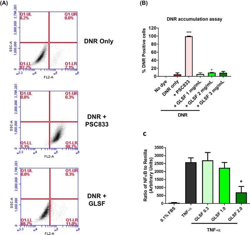
Antitumor activity of Ganoderma lucidum               3

Furthermore, we evaluated whether a GLSF modified diet            ganoderenic Acid C, ganoderic Acid C2, ganoderic Acid G,
was able to inhibit tumor growth in a mouse model bear-           ganoderic Acid B, ganoderenic Acid B, ganoderic Acid A,
ing murine colorectal carcinoma CT26, which is one of             ganoderic Acid H, ganoderenic Acid D, ganoderic Acid D,
the most extensively used syngeneic mouse tumor mod-              ganoderic Acid F, ganoderic Acid DM, ganoderol A, and
els.15 Tumor samples dissected from GLSF-treated and              ergosterol. The individual contents in the extracts were
control diet-treated tumor-bearing mice were analyzed             quantified and used as a fingerprint for GL products. All
by RNA sequencing (RNA-seq). Potential mechanisms of              the extractions and fingerprint analysis were conducted
the anticancer and chemosensitization effect of GLSF              in triplicate.

                                                                                                                                   Downloaded from https://academic.oup.com/pcm/advance-article/doi/10.1093/pcmedi/pbab023/6359148 by guest on 19 November 2021
in colorectal carcinoma were further explored through
bioinformatics analysis.                                          Cell lines and cell culture
                                                                  CT26, HCT-15, HT-29, and HEK-293 cells were purchased
Materials and methods                                             from ATCC (Manassas, VA). K562/DOX cell line (a gift from
Compounds and reagents                                            J.P. Marie, INSERM, E9912, University of Paris, France) was
                                                                  obtained by in vitro passaging of K562 in progressively
Paclitaxel (T7402) purchased from Sigma Aldrich (St
                                                                  increasing doses of daunorubicin.18 These cells were
Louis, MO) was used in vitro. AbraxaneR for injectable
                                                                  maintained in RPMI-1640 or DMEM supplemented with
suspension (Celgene, Summit, NJ) was used in vivo. A sin-
                                                                  10% fetal bovine serum and 1% penicillin-streptomycin.
gle batch of a commercial product manufactured by Bei-
                                                                  Cells were incubated at 37 ◦ C with 5% CO2 /95% air.
jing Tong Ren Tang Chinese Medicine Co. (Hong Kong,
China), named as GLSF, contains a mixture of the spores
and fruiting bodies of GL at a 30:8 ratio, was used               SRB cell proliferation assay
throughout the present study.                                     Plates (96-well) were seeded with 3000 cells per well and
                                                                  the cells were allowed to attach overnight. Cells were
Preparation of GLSF extracts and quantification of                treated with drugs for 72 hours and incubated at 37 ◦ C
the major components                                              in 5% CO2 /95% air. Cell viability was determined using
                                                                  a Sigma sulforhodamine B (SRB) assay according to the
As there is no standardized preparation approach for
                                                                  manufacturer’s protocol.
GL and related products, several reagents were used to
extract GLSF including ethanol, methanol, hot water, as
well as artificial gastric and intestinal (GI) juice. For the     Apoptosis analysis
ethanol/methanol extract, 0.5 g of GLSF was extracted             Flow cytometry was used to assess apoptosis by FITC-
with 12.5 ml solvent in a round bottom flask attached to          labeled annexin-V (Sigma) and propidium iodide, respec-
a reflux condenser at 80 ◦ C for 2 hours. The mixture was         tively, as previously described.19 Briefly, 1 × 10⁵ cells were
centrifuged at 4500 g for 10 minutes. The supernatant             seeded per well in six-well tissue culture plates for 24
was collected, and the pellet was extracted by repeat-            hours. The cells were then incubated with drugs for 72
ing the steps above. The supernatant collected was com-           hours. Cells were collected and washed twice with cold
bined and filtered using Whatman filter paper. The sol-           PBS. Approximately 5 × 10⁵ cells were mixed with bind-
vent was removed using a rotary evaporator.                       ing buffer with or without Annexin V or PI. Fluorescence
   For the hot water extract, 10 g of GLSF was extracted          was detected in fluorescence channels FL1 and FL3. Data
with 200 ml of nanopure water at 85 ◦ C for 15 minutes            acquisition and analysis were performed using Accuri
under stirring. The mixture was then centrifuged for 15           C6 Flow Cytometer (BD Biosciences, Franklin Lakes, NJ).
minutes at 4500 g. The supernatant was filtered using             Analysis was based on acquisition of data from 10 000
Whatman filter paper and filtrate lyophilized. The pow-           cells. Early apoptotic cells are Annexin-V positive and PI-
der obtained was weighed and stored at −20 ◦ C before             negative, whereas late apoptotic cells are both Annexin-
analysis.                                                         V and PI-positive.
   The extracts of GLSF were prepared in the artificial
gastrointestinal juice, which was prepared according to
                                                                  Intracellular daunorubicin (DNR) accumulation
reported methods.16,17 In brief, 5 g of GLSF powder was
                                                                  assay
mixed in 50 ml of artificial gastric juice at 37 ◦ C with shak-
ing for 1 hour, then 50 ml of artificial intestinal juice was     The capacity of a compound to inhibit P-glycoprotein-
added and incubated at 37 ◦ C with shaking for an addi-           mediated efflux from K562/DOX cells, was measured by
tional 5 hours. The mixture was centrifuged at room tem-          flow cytometry as described previously.20 Briefly, the cell
perature for 15 minutes at 4500 g, and the supernatant            pellet was resuspended in culture media at a concentra-
was collected. The extract was then neutralized to pH 7.0         tion of 6 000 000 cells/ml. Aliquots (50 μl) of the cell sus-
using 0.2 M NaOH, filtered using Whatman filter paper,            pension were transferred to tubes containing 1.95 ml of
lyophilized, and stored at −20 ◦ C.                               media in the presence or absence of test drugs in media
   The major active components were determined using              and 5 μM of DNR for 50 minutes at 37 ◦ C. After cen-
a validated HPLC-DAD method (unpublished), which was              trifugation and removal of supernatant, cold PBS was
able to quantify 13 major components in GLSF including            added to each tube, and cell suspension was transferred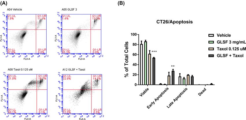
4    Mandy M. Liu et al.

to FACS tubes, which were placed on ice until analysis.        RNA extraction, library preparation, and
Flow cytometry was performed with an Accuri C6 Flow            next-generation sequencing
Cytometer. PSC833 (10 μM) served as a positive control.
                                                               Tumors from the rodent diet-based drug regimen study
                                                               were dissected, snap frozen, and stored at −80 ◦ C. The
Luciferase reporter gene assay
                                                               total RNA was extracted from the tumor tissues using
Dual luciferase assay for NF-κB has been described in          TRIzol reagent (Invitrogen, Grand Island, NY) and a
previous work.21 In brief, HEK-293 cells were transfected      RNeasy Mini Kit (Qiagen, Germantown, MD). The qual-
with pRL-TK-Luc Renilla luciferase (Promega, Madison,          ity of RNA samples was determined with an Agilent 2100

                                                                                                                               Downloaded from https://academic.oup.com/pcm/advance-article/doi/10.1093/pcmedi/pbab023/6359148 by guest on 19 November 2021
WI) and pGL4.22-NF-κB (Promega) plasmids at a ratio            Bioanalyzer. A total of eight RNA samples (four sam-
of 1:30 using FuGENE HD Transfection Reagent (Roche            ples per treatment, two groups) were sent to Fulgent
Applied Science, Indianapolis, IN). Twenty-four hours          Genetics (Temple City, CA) for library preparation and
later the cells were exposed to TNF-α (10 ng/ml) and           sequencing. Briefly, the library was constructed using
incubated in fresh growth medium containing drugs for          the NEBNext  R Poly(A) mRNA Magnetic Isolation Mod-

an additional 5 hours. Cell lysates were analyzed by the       ule + NEBNext   R UltraII Directional RNA Library Prep

dual luciferase reporter gene assay (Promega) with Renilla     Kit (New England Biolabs, Ipswich, MA). The RNA-seq
luciferase serving as a normalization factor.                  libraries were sequenced using an Illumina HiSeq 4000
                                                               with 150 bp, paired-end, at a minimum depth of total 60
Rodent diet preparation                                        million reads per sample.

For in vivo studies, 1.25% of GLSF powder was incorpo-
rated into mouse diets for oral administration. The modi-
fied animal diet was ordered through Newco Distributors,       Computational RNA-seq data analysis
Inc (Rancho Cucamonga, CA). The formulation included
                                                               Raw data were converted into fastq files by Illu-
98.45% Laboratory Rodent Diet #5001, 1.25% GLSF, and
                                                               mina bcl2fastq2 v2.20. Read quality was assessed using
0.3% color dye. The calculation of the drug was based on
                                                               FastQC. The sequence reads were mapped to mouse ref-
an average daily intake of 4 g per mouse. Food consump-
                                                               erence genome GRCm38 using the Edico Genome Dra-
tion for each group was monitored during the study and
                                                               gen aligner with default settings. Duplicate reads, as
it was confirmed that the dose of GLSF was ∼2.0 g/kg.
                                                               marked by the Dragen aligner, were removed before cov-
                                                               erage analysis. The aligned bam files were processed by
Animal experiment                                              HTSeq for gene quantification. Only genes with counts
All animal studies were carried out in strict accordance       per million (CPM) values above 0.5 in at least two sam-
with the recommendations in the Guide for the Care             ples were included in the differential expression anal-
and Use of Laboratory Animals of the National Institutes       ysis. Differentially expressed genes were identified by
of Health and approved by the Western University of            R package EdgeR (version 3.24.3). Briefly, read counts
Health Sciences Institutional Animal Care and Use Com-         were fitted into negative binomial distribution. The dif-
mittees. A pilot study was conducted using five male and       ferential analysis was carried out using quasi-likelihood
six female BALB/c mice. CT26 cells (3 × 10⁶ ) in 100 μl        F-test. Genes with P values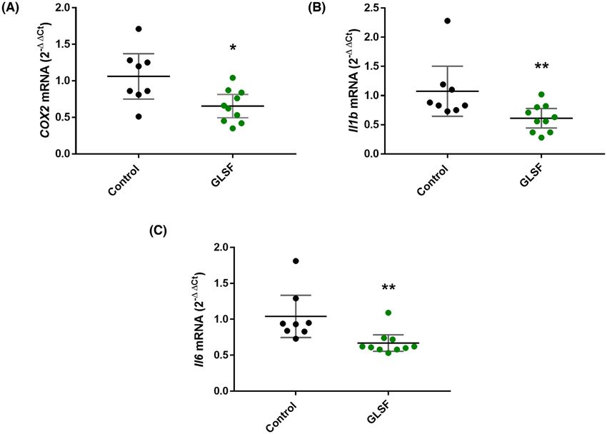
Antitumor activity of Ganoderma lucidum                    5

                                                                                                                                                   Downloaded from https://academic.oup.com/pcm/advance-article/doi/10.1093/pcmedi/pbab023/6359148 by guest on 19 November 2021
Figure 1. Effects of GLSF gastrointestinal (GI) juice extract on the growth inhibitory effects of paclitaxel in colorectal cancer cells. (A) SRB
cytotoxicity assay was used to determine the effects of paclitaxel, GLSF, alone or in combination in the mouse colon cancer CT26 cells. (B) The
cytotoxicity of paclitaxel, GLSF, alone or in combination in human colon cancer HCT-15 cells. (C) The effects of paclitaxel, GLSF alone or in
combination in human colon cancer HT-29 cells. ∗ : P < 0.05, ∗∗∗ : P < 0.001, comparing groups treated with paclitaxel alone with other groups,
as determined by t test.

Results                                                                   markers for GLSF. A HPLC-DAD fingerprint method has
                                                                          been developed22 and was used to quantify these mark-
Identification of optimal extraction methods for
                                                                          ers in the extract of GLSF. The contents of 13 com-
GLSF
                                                                          pounds are shown in Supplementary Table S2. Among
As there is no standardized preparation approach for GL                   the 13 assessed compounds, only ganoderol A was
and related products, we tried several methods for prepa-                 not detectable; the other 12 compounds were identi-
ration of GLSF extracts, including the use of ethanol,                    fied with substantial amounts. The inter-batch varia-
methanol, hot water, as well as artificial gastrointesti-                 tion was below 16.5% for all these compounds. Although
nal juice. To compare the pharmacologic activity of these                 the chemical markers mainly consisted of triterpenes
extracts, we examined their ability to inhibit cancer cell                (polysaccharides not included), the results indicated that
growth in vitro. The extracts were examined on a panel                    at least for selected active components GLSF was effi-
of cancerous and non-cancerous cell lines derived from                    ciently and consistently extracted.
different tissues of origin (PC3, MDA-MB-231, A375, H460,
HT-29, HepG2, NCI-N87 and NHDF) using a sulforho-
damine B (SRB) colorimetric assay. Comparison of the                      Effects of GLSF extract on chemosensitivity of
cytotoxicity data for the four extracts in melanoma A375                  colorectal cancer
or breast cancer MDA-MB-231 indicated that the gas-                       Paclitaxel (taxol) exhibited a modest inhibitory effect on
trointestinal juice and hot water extracts exhibited rel-                 CT26 cells (IC50 was 0.45 ± 0.02 μM), while GLSF did
atively higher potencies of cancer cell growth inhibition                 not cause cytotoxicity in CT26 at concentration up to
(Supplementary Figs. S1 and S2). The viability data and                   3 mg/ml. However, the cells that were co-treated with
IC50 values in these cell lines for gastrointestinal juice                taxol (0.125 μM) and GLSF (0.3 mg/ml) showed signifi-
and hot water extracts are summarized in Supplemen-                       cantly increased cytotoxic effects compared to the taxol
tary Table S1. Additionally, our previous study showed                    treatment alone (P < 0.05) (Fig. 1a). We observed a dose-
higher contents of triterpenes from GLSF in the gas-                      dependent chemosensitizing effect of GLSF, where a
trointestinal juice extract compared to the chloroform                    higher concentration (3.0 mg/ml) showed an increased
extract.22 As the gastrointestinal juice extract showed                   chemosensitizing effect compared to GLSF at 0.3 mg/ml
the highest potency on the majority of cancer cell lines, it              (P < 0.001). Similar effects were observed for combined
was selected for chemical fingerprint analysis and exam-                  treatment of GLSF (0.3 or 3.0 mg/ml) with a higher con-
ined for in vitro anticancer effects.                                     centration of taxol (0.5 μM). To confirm the nature of the
                                                                          interaction between GLSF and taxol, combination anal-
                                                                          yses were performed with the Combination Index (CI)
Chemical fingerprint analysis of gastrointestinal
                                                                          method,23,24 using drug combinations at various ratios.
juice extract of GLSF by HPLC
                                                                          A synergistic interaction was produced (CI = 0.04) when
Based on a literature search for components of GL                         taxol 0.125 μM was combined with GLSF at 3.0 mg/ml
products, 13 compounds were selected as chemical                          (CI values < 1 indicate synergistic activity). To confirm
6 Mandy M. Liu et al.
Downloaded from https://academic.oup.com/pcm/advance-article/doi/10.1093/pcmedi/pbab023/6359148 by guest on 19 November 2021
Figure 2. Effects of GLSF GI juice extract on apoptosis induced by paclitaxel in mouse colorectal cancer CT26 cells. (A) Cell apoptosis was assessed
by Annexin V/PI double staining which was detected by flow cytometry FACS analysis; Annexin-V-FITC staining in y axis (FL1) and PI in x axis
(FL3). (B) The Annexin V/PI assay was performed three times and the average percentage of viable, early apoptosis, late apoptosis, and necrosis
populations of cells were plotted. ∗∗ : P < 0.01, ∗∗∗ : P < 0.001, compared with vehicle control group, as determined by t test.
these results in human colorectal cancer cell lines, SRB of viable cells (P < 0.001) (53.7 ± 1.6% of viable cells in
assays were performed on human colorectal cancer HCT- control) and increase in the number of early apoptotic
15 and HT-29 cells, treated with taxol and GLSF alone cells (P < 0.01). This result is consistent with those data
or in combination. The IC50 in HCT-15 treated with taxol obtained by SRB in CT26 cells (Fig. 1a), indicating that
was 0.40 ± 0.002 μM and similar to the CT26 cells, GLSF the co-treatment with non-toxic concentration of GLSF
did not cause any cytotoxicity in HCT-15 cells up to increased the anticancer potency of taxol via enhancing
3 mg/ml concentration. The cells that were co-treated apoptosis in CT26 cells.
with taxol (0.25 μM) and GLSF (0.11–3 mg/ml) showed a
dose-dependent increase in cytotoxic effects compared
to the taxol treatment alone (P < 0.05 for GLSF 0.11
mg/ml; P < 0.001 for GLSF 1.0 and 3.0 mg/ml) (Fig. 1b). Effects of GLSF extract on P-glycoprotein (MDR1)
Although HT-29 cells were much more sensitive to taxol activity in K562/DOX cells
(IC50 = 0.0035 ± 0.001 μM) than CT26 and HCT-15 and
GLSF at 3 mg/ml alone was also nontoxic, co-treatment Previous studies have shown that both CT2625 and HCT-
with GLSF and taxol did not demonstrate increased or 1526 express the efflux transporter P-glycoprotein (P-gp
decreased effect on cell viability (Fig. 1c). Therefore, the or MDR1, ABCB1 gene), which is part of the mechanism
chemosensitizing effect for GLSF is variable in different for their intrinsic resistance to taxanes and other P-
cancer cell lines. gp substrate drugs such as daunorubicin (DNR). There-
fore, we examined whether GLSF was able to inhibit
P-gp-mediated drug efflux from the multi-drug resis-
Effects of GLSF extract on taxol-induced tant cell line K562/DOX, which overexpresses P-gp, using
daunorubicin (DNR) as a fluorescent substrate.20 Few
apoptosis in CT26 cells
cells were positive for DNR accumulation without P-
The degree of apoptosis induced by GLSF (3 mg/ml), taxol gp inhibitor (Fig. 3a). Co-treatment with a known P-gp
(0.125 μM), or their combination was evaluated in CT26 inhibitor PSC833 (10 μM) as a positive control greatly
cells. The cells were incubated for 72 hours with com- increased DNR positive cells to nearly 100%. However,
binations of drugs and the level of apoptosis was quan- GLSF (1, 2 and 3 mg/ml) only slightly increased DNR
tified by an Annexin-V binding and PI staining assay. accumulation (Fig. 3b). Although the treatment effect
As shown in Fig. 2, GLSF treatment slightly increased of GLSF (2 mg/ml) was significant (P < 0.05), compared
the viable cells compared to vehicle control (P > 0.05). to the positive control PSC833, the degree of inhibition
Taxol decreased the viable cell population to 60.1 ± 6.2% was negligible. This result suggests that the chemosen-
(P > 0.05). When the cells were exposed to a combina- sitizing activity of GLSF is not attributed to P-gp
tion of GLSF and taxol, there was a significant reduction inhibition.Antitumor activity of Ganoderma lucidum 7
Downloaded from https://academic.oup.com/pcm/advance-article/doi/10.1093/pcmedi/pbab023/6359148 by guest on 19 November 2021
Figure 3. Effects of GLSF GI juice extract on P-glycoprotein activity in K562/DOX cells and NF-κB activity in HEK-293 cells. (A) Flow cytometry
analysis of DNR accumulation in cells incubated with DNR, treated with or without PSC833 (10 μM) or GLSF. Representative flow cytometry data
are shown. (B) Graphs showing percentage of cells that were positive for DNR in cell samples co-treated with GLSF and DNR in comparison
with those co-treated with 10 μM PSC833 and DNR. The data are presented as the mean ± SD from 3–6 independent experiments. ∗ : P < 0.05
compared to the DNR only group. (C) Effects of GLSF on TNF-α-induced NF-κB promoter activity. HEK-293 cells transfected with NF-κB luciferase
reporter and Renilla control reporter were treated with vehicle, TNF-α (10 ng/mL), GLSF (0.3, 1.0, 2.0 mg/mL) combined with TNF-α for 5 hours.
Data are expressed as mean +/- SEM; n = 3–6. ∗ : P < 0.05 as per one-way ANOVA.
Effects of GLSF extract on NF-κB promoter activity various types of cancer27 and conferring chemoresis-
in HEK-293 cells tance to paclitaxel,28 inhibition of NF-κB by GLSF might
partly explain the in vitro chemosensitizing activity and
We next evaluated the effect of GLSF on TNF-α pro-
the in vivo activity described below.
moted NF-κB activation. The HEK-293 cells were trans-
fected with NF-κB-luc firefly luciferase reporter construct
and a plasmid encoding the Renilla luciferase for the dual
Anticancer efficacy of GLSF oral administration to
luciferase assay. The transfected cells were treated with
mice bearing murine colorectal cancer tumor
vehicle (control) or GLSF extracts at 0, 0.3, 1.0, or 2.0
CT26
mg/ml, followed by a 5-hour co-incubation with TNF-α
(10 ng/ml). As expected, TNF-α strongly stimulated NF- To evaluate the anticancer activity of GLSF in vivo, a syn-
κB activity (Fig. 3c). The promoter activity stimulated geneic model of colorectal cancer-bearing BALB/c mice
by TNF-α was inhibited significantly by the GI extract was used. We firstly conducted a pilot study using a small
at 2.0 mg/ml in a dose-dependent manner (Fig. 3c). number of mice with two tumors (CT26) implanted in
As NF-κB plays a role in promoting carcinogenesis of each mouse (n = 2 each gender specific group).8 Mandy M. Liu et al.
Downloaded from https://academic.oup.com/pcm/advance-article/doi/10.1093/pcmedi/pbab023/6359148 by guest on 19 November 2021
Figure 4. Effects of GLSF on tumor growth and apoptosis in a colorectal cancer syngeneic model. (A) The CT26 cells were subcutaneously
implanted into the female BALB/c mice after the mice were fed with the control diet (groups 1 and 3) or diet containing GLSF (groups 2 and 4)
for 4 weeks. Groups 3 and 4 mice were treated with a single dose of abraxane R on Day 13. Tumor volumes were measured on Days 13 and 19
(Day 0 was the day of tumor injection). ∗ : P < 0.05 by Tukey-Kramer Multiple-Comparison test (n = 8 for G1 and G2; n = 4 for G3 and G4). (B)
Representative photos of mice in control and GLSF groups. (C) Tumor tissues were stained with H&E or immunohistochemical staining of Ki-67.
(D) Western blot analysis of PARP, cleaved PARP, and β-actin. Representative gel images are shown. Bar graphs represent the ratios of cleaved
PARP/total PARP. ∗ : P < 0.05 as compared with the control group by t test. PARP, poly (ADP-ribose) polymerase. Control n = 5 tumors, GLSF n = 4
tumors.
The treatment by oral gavage (2.0 g/kg, daily, Monday 3) or modified diet containing GLSF (groups 2 and 4)
to Friday, nine doses in total) was started on the 11th day 1 month before tumor implantation. As GLSF showed
after tumor implantation. Although statistically insignif- antitumor effects in both genders in the pilot study
icant, a trend of treatment effect was observed in tumor and the CT26 tumor was derived from female mice,15
volume in both males and females (Supplementary Fig. only females were included in this experiment. Abrax-
S3): the average tumor volumes in GLSF-treated mice ane treatment was given for groups 3 and 4 to observe
were smaller than in untreated control. In addition, the the effects of combination treatments. Two tumors were
mice did not lose body weight compared to initial weight, implanted in each mouse. Consistent with the pilot
indicating the treatment was not toxic. study, GLSF treatment alone inhibited tumor growth
To explore the potential anticancer mechanism, the (Fig. 4a), although repeated measures ANOVA analysis
effect of GLSF treatment on mouse spleen lympho- did not show a statistically significant difference. At the
cyte proliferation induced by concanavalin A (Con A) or end of the experiment, 100% of the mice had two tumors
lipopolysaccharides (LPS) in vitro was determined using in the control group, while only 63% of mice in the GLSF
the spleen samples obtained from the CT26 tumor- group had two tumors, and two of the eight mice (25%)
bearing mice. Compared with non-tumor-bearing mice, in the GLSF group did not show any tumors (Fig. 4b). The
proliferation of spleen T and B lymphocytes induced by Tukey-Kramer post hoc analysis showed that on Day 19
Con A and LPS were strongly declined in tumor-bearing after tumor inoculation, co-treatment with abraxane and
mice (Supplementary Fig. S3). However, treatment with GLSF significantly suppressed tumor growth compared
GLSF of these mice increased spleen T and B lymphocyte with the control group (Fig. 4a). Furthermore, tumors in
proliferation in CT26-bearing mice, although this was not the control group (group 1) showed a significant increase
statistically significant. These data indicate a potential when comparing tumor size on Day 13 and 19, while
immunomodulatory activity of GLSF, consistent with a groups 2, 3 and 4 did not show time-dependent tumor
previous report.4 size increase, confirming the treatment effect.
To confirm the anticancer activity of GLSF, BALB/c Tumor tissues dissected from the control and GLSF
mice were pre-treated with control diet (groups 1 and groups were subjected to hematoxylin and eosin (H&E)Antitumor activity of Ganoderma lucidum 9
Downloaded from https://academic.oup.com/pcm/advance-article/doi/10.1093/pcmedi/pbab023/6359148 by guest on 19 November 2021
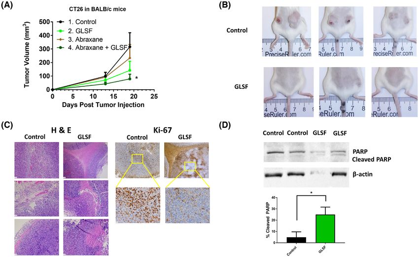
Figure 5. RNA-seq analysis of CT26 syngeneic tumors in mice treated with control diet or GLSF-modified diet. (a) Multi-dimensional scaling plot
of detected genes in GLSF treatment (T, red) and control group (C, green). The distances correspond to leading log2 fold-changes between each
pair of samples. (b) Volcano plots of differentially expressed genes between treatment and control groups. The red dots indicate up- and down-
regulated DEGs with P < 0.05 and absolute log2 FC > 1. (c) Heat map of top 30 significantly differentially expressed genes between treatment (T)
and control groups (C). (d) Genes that are up- and down-regulated in treatment group (compared to control) are displayed within red or green
nodes, respectively. The predicted inhibited biology effects are presented in blue nodes. Blue (predicted to be inhibited) or gray (undetermined
direction) dash lines represent relationships with causal consistency.
staining. The image of H&E sections indicated that tumor RNA-seq profiling of GLSF-treated tumors
tissues from both groups consisted of a morphologically
To explore the mechanism underlying the treatment
similar differentiated adenocarcinoma. However, tumors
effects by GLSF, RNA-seq analysis was performed to com-
in the GLSF group showed a conspicuous increase of
pare the gene expression profiles between tumors iso-
necrosis in all the four tumor samples submitted for H&E
lated from mice subjected to the control diet (n = 4)
staining (representative images are shown in Fig. 4c). Ki-
or GLSF modified diet (n = 4). On average, 40 million
67 staining showed that the tumors in the control group
pair-end reads pairs per sample were generated, with
consisted entirely of one cell type with high prolifera-
all of their uniquely mapping rates above 91%. Over-
tive index, while the tumors in the GLSF group demon-
all, RNA sequencing detected 25 051 genes across eight
strated nonspecific staining of necrotic/apoptotic cells
samples, of which 13 790 genes had a CPM value above
but showed fainter staining of fewer Ki-67 positive viable
0.5 in at least two samples. A multi-dimensional scal-
cells (Fig. 4c).
ing (MDS) plot showed a trend of clustering separation
Western blot analysis was used to examine the
between treated group and control group (Fig. 5a). Out of
expression of the apoptosis marker PARP and cleaved
the 13 790 genes, 53 were identified as significant differ-
PARP in tumor tissue lysates. The expression of cleaved
entially expressed genes (DEGs) between the two groups
PARP was markedly increased in the GLSF treatment
with a absolute log2 fold-change >1 and a P value10 Mandy M. Liu et al.
Table 1. Top 12 up-regulated genes in tumors from mice treated with GLSF compared to control group.
Gene ID Gene name Log FC P value
Itga10 Integrin, alpha 10 1.07 9.34E-05
Zbtb16 Zinc finger and BTB domain containing 16 2.07 2.79E-04
Gm807 Predicted gene 807 1.26 9.47E-04
Lvrn Laeverin 1.17 1.07E-03
Gm6093 Predicted gene 6093 1.63 1.49E-03
Hrct1 Histidine rich carboxyl terminus 1 1.33 2.15E-03
Downloaded from https://academic.oup.com/pcm/advance-article/doi/10.1093/pcmedi/pbab023/6359148 by guest on 19 November 2021
Aldh1a1 Aldehyde dehydrogenase family 1, 1.08 2.59E-03
subfamily A1
Inpp5j Inositol polyphosphate 5-phosphatase J 1.67 3.06E-03
Rcan2 Regulator of calcineurin 2 1.00 3.39E-03
Ifng Interferon gamma 1.09 3.52E-03
Retnla Resistin like alpha 2.52 3.63E-03
Fmo2 Flavin containing monooxygenase 2 1.35 7.23E-03
∗ Log FC (log2 Fold change) larger than 1 indicates higher expression in GLSF-treated tumors.
Table 2. Top 10 down-regulated genes in tumors from mice treated with GLSF compared to control group.
Gene ID Gene name Log FC P value
Il1b Interleukin 1 beta − 1.31 9.57E-06
Il11 Interleukin 11 − 1.09 4.89E-04
4930565N06Rik RIKEN cDNA 4930565N06 gene − 1.06 6.84E-04
Nppb Natriuretic peptide type B − 1.44 7.72E-04
Ptgs2 Prostaglandin-endoperoxide synthase 2 (Cox-2) − 1.11 1.18E-03
Mmp10 Matrix metallopeptidase 10 − 1.51 1.31E-03
Mmp13 Matrix metallopeptidase 13 − 1.16 1.68E-03
Stamos Signal transducing adaptor molecule (SH3 domain and − 1.13 1.82E-03
ITAM motif) 1, opposite strand
Cxcl1 Chemokine (C-X-C motif) ligand 1 − 1.56 2.07E-03
Mmp12 Matrix metallopeptidase 12 − 1.03 4.01E-03
∗ Log FC (log2 Fold change) smaller than −1 indicates lower expression in GLSF-treated tumors.
fold-change lower than −1 indicated that the expression and “Agranulocyte adhesion and diapedesis”, which are
of the gene was decreased by GLSF treatment, whereas known immune/inflammatory pathways.
a log2 fold-change more than 1 suggested an increased Upstream analysis through IPA was used to predict
gene expression in tumor tissues derived from GLSF the upstream regulators potentially causing changes
treatment. A heat map of the top 30 significant DEGs in gene expression and regulation direction based on
was plotted with unsupervised hierarchical clustering the DEGs. Predicted significantly activated and inhib-
(Fig. 5b). A volcano plot was generated with the signifi- ited regulators are listed in Supplementary Table S5 (P
cant DEGs highlighted in red color based on their P val- value < 0.05, |Z-score|>2). The two significantly activated
ues and fold changes (Fig. 5c). It is notable that many upstream regulators were alpha-catenin and TP53, both
of these GLSF down-regulated genes are involved in NF- of which are known tumor suppressors for colon cancer.
κB-regulated inflammation, such as IL-1β (Il1b) and IL-11 On the other hand, the most inhibited upstream regula-
(Il11) (Table 2, Fig. 5b). tors were factors that play critical roles in inflammatory
To explore the possible biological functions linked response and tumorigenesis, including TNFSF12, KRT17,
with these DEGs, ingenuity pathway analysis (IPA) was IL17RA, TNF, and IL17A.
performed to identify canonical pathways, upstream reg- ”Diseases and functions” in IPA was used to predict
ulators, diseases and functions that are associated with affected biology and its regulation direction associated
GLSF treatment. Based on the ratio of the number of DEGs with GLSF treatment. Using a cutoff of P value < 0.05,
in our dataset to the total number of reference genes |Z-score|>2, there was no significantly activated biology
in the specific pathways in the IPA knowledge bases, a effect identified, while the top five significantly inhib-
Fisher’s exact test was used to determine the canonical ited biological effects are presented in Fig. 5d, which
pathways associated with the treatment effect. Using a included “Cancer”, “Epithelia neoplasm”, “Carcinoma”,
cutoff P valueAntitumor activity of Ganoderma lucidum 11
Downloaded from https://academic.oup.com/pcm/advance-article/doi/10.1093/pcmedi/pbab023/6359148 by guest on 19 November 2021
Figure 6. qRT-PCR results from tumor samples of control or GLSF groups for (A) COX-2, (B) IL-1β, and (C) IL-6. β-actin was used as normalization
control and data are expressed as mean with 95% CI. n = 8 tumor samples from control group. n = 10 tumor samples for GLSF. ∗ : P < 0.05; ∗∗ :
P < 0.01 via a Mann-Whitney U approximation without correction for normality.
RT-PCR analysis confirmed that GLSF inhibited GL extracts. In vitro, GLSF alone showed modest cytotoxi-
the expression of selected inflammatory genes, city on colon cancer cells, which is consistent with a pre-
cyclooxygenase-2 (Ptgs2 or Cox-2) (P < 0.05), interleukin- vious study indicating that the direct anticancer effect of
1β (Il1b) (P < 0.01), and interleukin-6 (Il6) (P < 0.01) GL is limited.3 However, when GLSF was combined with
(Fig. 6). taxol, it induced stronger tumor inhibition and apopto-
sis, suggesting that GLSF may be used as a chemosen-
sitizer (Fig. 1a and b). As GLSF is not a P-gp inhibitor
(Fig. 3a and b), its chemosensitizing mechanism remains
to be determined. One possible mechanism is through its
Discussion inhibitory activity against NF-κB signaling, suggested by
The present study examined the in vitro and in vivo anti- the dual luciferase assay (Fig. 3c). Consistently, previous
cancer activity of GLSF, which contains a mixture of bro- studies have shown that triterpene ganoderic acid C1 iso-
ken spores and fruiting bodies of mushroom. As differ- lated from GL inhibited inflamed Crohn’s disease colonic
ent methods of extraction affect the release, bioavailabil- mucosa as a result of blockage of NF-κB.31
ity and pharmacological activity of active components, In vivo, GLSF alone or in combination with abrax-
extracts of GLSF were prepared in ethanol, methanol, ane (nanoparticle albumin bound paclitaxel or nab-
hot water, or artificial gastrointestinal juice and were paclitaxel) induced tumor growth inhibition and apop-
examined in a panel of cell lines. We found that extracts tosis (Fig. 4). The syngeneic tumor model used in the
prepared using gastrointestinal juice had the highest study was murine colorectal carcinoma cell line Colorec-
potency of cell growth inhibition and therefore were used tal Tumor #26 (CT26).15 CT26 cells were derived by expos-
in our in vitro studies. This extraction method is physi- ing BALB/c mice to the chemical carcinogen N-nitroso-
ologically relevant to clinical application. Although the N-methylurethane, resulting in a rapid-growing grade IV
method of extraction has been used for ginseng29 and carcinoma that is easily implanted and readily metasta-
notoginseng (“San-Qi”),30 to our knowledge, this is the sizes.32 As CT26 tumor in the immunocompetent BALB/c
first study using artificial gastrointestinal juice to prepare mice provides a syngeneic model, it is frequently used for12 Mandy M. Liu et al.
developing and testing immunotherapeutic agents.15,33 Another down-regulated gene in the GLSF-treated
Compared with in vitro data, a large in vivo effect suggests group was the inflammatory chemokine Cxcl1, which
that an intact immune system or tumor microenviron- is involved in enhanced metastatic potential of colon
ment may be essential for the pharmacological action of cancer by increasing cell migration, matrix metal-
GLSF. One limitation for these in vivo studies was that a loproteinases (MMP) expression, and epithelial-to-
single dose was used (2.0 g/kg), which was derived from mesenchymal transition and therefore has a negative
published data of GL.34–36 Dose-response studies should prognostic impact to the clinical outcome.49 Probably
be considered in the future. Although taxol or abraxane associated with Cxcl1 down-regulation, three MMP genes
Downloaded from https://academic.oup.com/pcm/advance-article/doi/10.1093/pcmedi/pbab023/6359148 by guest on 19 November 2021
are effective to many types of cancer such as breast or were among the top 20 down-regulated genes: MMP10,
ovarian cancer, their application in colorectal cancer has MMP13, and MMP12. These genes and their products
been limited because of intrinsic resistance.37 Thus, GLSF have been identified as negative prognostic markers
may be used as a chemosensitizer for taxanes. The ratio in colon cancer patients.50,51 GLSF down-regulated
of GLSF versus taxol or abraxane should be optimized Nppb, encoding for natriuretic peptide B, which has
in future studies to identify the optimal combinational been shown to be a key oncogene candidate for colon
regimen. Further studies comparing therapeutic effects tumors and suggested as one of the early biomarkers for
of GLSF in different tumor models and with other anti- prevention in the clinical setting.52
cancer agents should be considered. The GLSF-up-regulated genes have various functions.
To gain mechanistic insight, RNA-seq was performed Although most of these genes have not yet been asso-
to evaluate genome-wide transcriptome changes asso- ciated with colon cancer, they may contribute to GLSF-
ciated with GLSF treatment. Based on the differentially induced anti-cancer activity. Some of them may be tumor
expressed genes (DEGs), expression levels of IL-1β and IL- suppressor genes, for example, Fmo2 (flavin containing
11, genes encoding for cytokines in the tumor microen- monooxygenase 2) plays a role as a tumor suppressor in
vironment that promote colorectal cancer progression, lung cancer.53 The lower level of Inpp5j, which encodes
were decreased by GLSF. These cytokines, also includ- inositol polyphosphate-5-phosphatase J, has been asso-
ing Il-6 which was not picked up by RNA-seq analysis ciated with more aggressive tumors and poorer sur-
because of low CPM values, are produced by myeloid vival of cutaneous squamous cell carcinoma,54 and was
and T-helper interleukin (IL)-17-producing (Th17) cells found to be deficient in oropharyngeal squamous cell
that are accumulated in the tumor microenvironment.38 carcinoma.55 Itga10, a top DEG of the GLSF-up-regulated
Because these cytokines directly or indirectly activate genes, encodes integrin subunit α 10, which binds to col-
neoplastic epithelium, therapies that target their acti- lagen and plays a role in cell adhesion and cell-surface
vation, for example anti-IL-11 therapy, have been pro- mediated signaling. Although the activity of increased
posed to treat colorectal cancer.38 In addition, the GLSF expression of Itga10 by GLSF is unknown, the interac-
down-regulated Ptgs2, encoding for Cox-2, known to tion of integrin and collagen may mediate an anti-tumor
be an inflammatory mediator that promotes colorectal immune response.56
cancer.39 RT-PCR results confirmed that in GLSF-treated The canonical pathway analysis highlighted “Gran-
mice the tumor cell expression of Cox-2, IL-1β, and IL-6 ulocyte adhesion and diapedesis” and “Agranulocyte
was significantly down-regulated at mRNA level (Fig. 6). adhesion and diapedesis” as the most significantly reg-
These results suggest that inflammation is a potential ulated pathways influenced by GLSF treatment. Both
target for treatment of some types of colorectal cancer.40 pathways contain the same set of genes, mainly MMPs,
Consistent with the luciferase assay data (Fig. 3c), these and have been associated with immunity and inflam-
GLSF-down-regulated genes are under the control of NF- mation,57 as well as tumor invasion and metastasis.58
κB transcription factor. Furthermore, CT26 cells harbor These results support the notion that these two path-
the constitutively activating mutant KRAS,15 which has ways were involved in inflammation associated with col-
been shown to trigger the production of several inflam- orectal cancer, as previously reported,59 and may con-
matory mediators including IL-6,41 IL-1β 42 and associ- tain the molecular targets that underlie the anticancer
ated with COX-2.43 Interestingly, NF-κB is activated in effect of GLSF. Based on the DEGs between the treat-
KRAS-mutated cancer and has been suggested as a tar- ment and control groups, IPA predicted the upstream reg-
get to treat KRAS-induced cancer.44,45 In addition, a pre- ulators and their activation states which might result
vious study showed that CT26 cells not only secret IL- in these gene expression changes. Several inflamma-
6, but also the growth of CT26 tumor depends on IL- tory cytokines were at the top of the inhibition list
6.46,47 Thus, these inflammatory factors, many of which such as TNFSF12, TNF, and IL17A. On the other hand,
are under the regulation of NF-κB, are very likely tar- tumor suppressors such as alpha-catenin and TP53 were
gets of a GL-mediated anticancer effect. These results activated (activation z-score > 2) according to the pre-
also suggest that inflammation-associated cancers and diction. This prediction further implied that GLSF may
KRAS-driven tumors might be more likely to respond elicit anti-tumor activity through its anti-inflammatory
to GLSF. As inhibiting KRAS directly is very challenging, effects. More specifically, one possible mechanism is that
approaches to disrupt downstream signaling pathways GLSF treatment resulted in an inhibition of the NF-kB
may be a better approach to treat these types of highly pathway, a shared target of the aforementioned pre-
aggressive cancer.48 dicted upstream regulators, which eventually may leadAntitumor activity of Ganoderma lucidum 13
to tumor suppression. Further studies are required to val- experiments, wrote and reviewed the manuscript. All
idate this hypothesis. Based on the IPA prediction, there authors read and approved the final manuscript. C.W.
was no significantly activated biology effect while the top submitted the manuscript.
five inhibited biology effects were all related to “Cancer”
and displayed the network of tumor suppression induced
by GL treatment (Fig. 5d). These results implied a possi- Acknowledgements
ble mechanism of GL’s anti-tumor effect as alleviating the We would like to thank Lily Kong Lim, A.S.C.P. (H.T.),
immune suppression in colorectal tumor tissue by pro- at the Beverly Hospital for preparing the formalin-fixed
Downloaded from https://academic.oup.com/pcm/advance-article/doi/10.1093/pcmedi/pbab023/6359148 by guest on 19 November 2021
moting the recruitment of anti-tumor immune cells, or paraffin-embedded tissues and H&E staining. The GLSF
by inhibiting immune suppressive cells such as myeloid is available from the Beijing Tong Ren Tang Chinese
derived suppressor cells. This hypothesis align well with Medicine Co., Ltd. This study was funded by a con-
the observation that GLSF alone showed little cytotox- tract grant (YH) from the Beijing Tong Ren Tang Chinese
icity in vitro because of a lack of immune system. It is Medicine Co., Ltd. This study was also partially supported
notable that the in vivo studies used GLSF powder while in by Loma Linda University (LLU) GCAT grant (CW).
vitro assays used extract. Identification of the active com-
ponents that are responsible for these observed effects
requires further investigation. Conflict of interests
In conclusion, this study evaluated the anticancer
effects and possible underlying anticancer mechanism B. Zhou, W. Wu, X. Li and Y. Zhang are employees or con-
of GLSF using both human and murine colorectal can- sultants of the Beijing Tong Ren Tang Chinese Medicine
cer cell lines, and examined GLSF alone or in combi- Co., Ltd. The other author(s) declare no competing inter-
nation with a commonly used chemotherapeutic agent ests. Besides, as an Editorial Board Member of Precision
paclitaxel. In vitro data revealed that although GLSF alone Clinical Medicine, the corresponding author Charles Wang
had little anticancer effect, it had a chemosensitiza- was blinded from reviewing and making decision on this
tion effect in certain colon cancer cells and that NF-κB manuscript.
played a major role in mediating the in vitro anticancer
effect of GLSF. Immunocompetent mice carrying the syn-
References
geneic tumor CT26 were used for investigating the in
vivo anticancer activity of GL. RNA-seq and bioinformat- 1. Siegel RL, Miller KD, Jemal A. Cancer statistics, 2016. CA Can-
ics analysis, for the first time, indicated that GLSF tar- cer J Clin 2016;66:7–30. doi:10.3322/caac.21332.
geted inflammation and carcinogenesis and confirmed 2. Hyodo I, Amano N, Eguchi K, et al. Nationwide sur-
vey on complementary and alternative medicine in
the role of NF-κB as the potential target. Advanced col-
cancer patients in Japan. J Clin Oncol 2005;23:2645–54.
orectal cancer shows inherent resistance to paclitaxel or
doi:10.1200/JCO.2005.04.126.
related chemotherapeutic agents. With the use of GLSF, 3. Sanodiya BS, Thakur GS, Baghel RK, Ganoderma lucidum: a
a non-toxic natural product, the antitumor effects of potent pharmacological macrofungus. Curr Pharm Biotechnol
chemotherapy may be enhanced. GSLF alone also had 2009;10:717–42. doi: 10.2174/138920109789978757.
anticancer activity in vivo through an inhibitory effect 4. Wang PY, Zhu XL, Lin ZB. Antitumor and Immunomod-
against inflammation, NF-kB, and/or KRAS activation. ulatory Effects of Polysaccharides from Broken-Spore
of Ganoderma lucidum. Front Pharmacol 2012;3:135.
doi:10.3389/fphar.2012.00135.
Supplementary data 5. Sliva D. Ganoderma lucidum in cancer research. Leuk Res
2006;30:767–8. doi:10.1016/j.leukres.2005.12.015.
Supplementary data are available at PCMEDI online.
6. Lu H, Kyo E, Uesaka T, et al. Prevention of develop-
ment of N,N’-dimethylhydrazine-induced colon tumors by a
Data and material availability water-soluble extract from cultured medium of Ganoderma
lucidum (Rei-shi) mycelia in male ICR mice. Int J Mol Med
All materials, data, and associated protocols are avail- 2002;9:113–7. doi:https://doi.org/10.3892/ijmm.9.2.113..
able to readers without undue qualifications in material 7. Lu H, Kyo E, Uesaka T, et al. A water-soluble extract from cul-
transfer agreements. The GLSF is available from the Bei- tured medium of Ganoderma lucidum (Rei-shi) mycelia sup-
jing Tong Ren Tang Chinese Medicine Co., Ltd. presses azoxymethane-induction of colon cancers in male
F344 rats. Oncol Rep 2003;10: 375–9.
8. Lu H, Uesaka T, Katoh O, et al. Prevention of the devel-
Author contributions opment of preneoplastic lesions, aberrant crypt foci, by
a water-soluble extract from cultured medium of Gano-
Y.H. conceived and designed the study. M.L. carried
derma lucidum (Rei-shi) mycelia in male F344 rats. Oncol Rep
out most in vitro and in vivo experiments and ana-
2001;8:1341–5. doi: 10.3892/or.8.6.1341.
lyzed the data; T.L. and C.W. analyzed the RNA-seq 9. Sliva D, Loganathan J, Jiang J, et al. Mushroom Ganoderma
data and revised the manuscript; B.A. conducted sta- lucidum prevents colitis-associated carcinogenesis in mice.
tistical analysis; S.Y. and Z.W performed HPLC finger- PLoS One 2012;7:e47873. doi:10.1371/journal.pone.0047873.
print analysis; C.P. and R.O. performed the pathology 10. Yang Y, Nirmagustina DE, Kumrungsee T, et al. Feeding of
analysis; B.Z., W.W., X.L., Y.Z. and Y.H. coordinated the the water extract from Ganoderma lingzhi to rats modulates14 Mandy M. Liu et al.
secondary bile acids, intestinal microflora, mucins, and pro- 27. Hu M, Peluffo G, Chen H, et al. Role of COX-2 in epithelial-
pionate important to colon cancer. Biosci Biotechnol Biochem stromal cell interactions and progression of ductal carci-
2017;81:1796–804. doi:10.1080/09168451.2017.1343117. noma in situ of the breast. Proc Natl Acad Sci 2009;106:3372–7.
11. Luo J, Li T, Xie J, et al. Guar gum different from Gano- doi:0813306106 [pii] 10.1073/pnas.0813306106.
derma lucidum polysaccharide in alleviating colorectal can- 28. Chen X, Sun X, Guan J, et al. Rsf-1 influences the sensitiv-
cer based on omics analysis. Food Funct 2020;11:572–84. ity of non-small cell lung cancer to paclitaxel by regulating
doi:10.1039/c9fo02786f. NF-kappaB pathway and its downstream proteins. Cell Phys-
12. Oka S, Tanaka S, Yoshida S, et al. A water-soluble extract from iol Biochem 2017;44:2322–36. doi:10.1159/000486116.
culture medium of Ganoderma lucidum mycelia suppresses 29. Rimar S, Lee-Mengel M, Gillis CN Pulmonary protective and
Downloaded from https://academic.oup.com/pcm/advance-article/doi/10.1093/pcmedi/pbab023/6359148 by guest on 19 November 2021
the development of colorectal adenomas. Hiroshima J Med Sci vasodilator effects of a standardized Panax ginseng prepa-
2010;59:1–6. ration following artificial gastric digestion. Pulm Pharmacol
13. Chen X, Hu ZP, Yang XX, et al. Monitoring of immune 1996;9:205–9. doi: 10.1006/pulp.1996.0025.
responses to a herbal immuno-modulator in patients with 30. Wang JR, Yau LF, Zhang R, et al. Transformation of gin-
advanced colorectal cancer. Int Immunopharmacol 2006;6:499– senosides from notoginseng by artificial gastric juice can
508. doi:10.1016/j.intimp.2005.08.026. increase cytotoxicity toward cancer cells. J Agric Food Chem
14. Bai JH, Xu J, Zhao J, et al. Ganoderma lucidum polysaccha- 2014;62:2558–73. doi:10.1021/jf405482s.
ride enzymatic hydrolysate suppresses the growth of human 31. Liu C, Dunkin D, Lai J, et al. Anti-inflammatory
colon cancer cells via inducing apoptosis. Cell Transplant effects of ganoderma lucidum triterpenoid in human
2020;29: 963689720931435. doi:10.1177/0963689720931435. crohn’s disease associated with downregulation of NF-
15. Castle JC, Loewer M, Boegel S, et al. Immunomic, genomic kappaB signaling. Inflamm Bowel Dis 2015;21:1918–25.
and transcriptomic characterization of CT26 colorectal carci- doi:10.1097/MIB.0000000000000439.
noma. BMC Genomics 2014;15:190. doi:10.1186/1471-2164-15- 32. Griswold DP, Corbett TH. A colon tumor model for anticancer
190. agent evaluation. Cancer 1975;36:2441–4. doi: 10.1002/1097-
16. Chrubasik S, Sporer F, Dillmann-Marschner R, et al. Physico- 0142(197512)36:63.0.co;2-p.
chemical properties of harpagoside and its in vitro release 33. Lechner MG, Karimi SS, Barry-Holson K, et al. Immunogenic-
from Harpagophytum procumbens extract tablets. Phy- ity of murine solid tumor models as a defining feature of in
tomedicine 2000;6:469–73. doi:10.1016/S0944-7113(00)80076-8. vivo behavior and response to immunotherapy. J Immunother
17. Panda V, Khambat P, Kundnani K, et al. Evaluation of antacid 2013;36:477–89. doi:10.1097/01.cji.0000436722.46675.4a.
activity of garcinia indica fruit rind by a modified artificial 34. Wang S, Wang X, Lin L, et al. Inhibitory effect of Lucid Gan-
stomach model. Bull Environ Pharmacol Life Sci 2013;2:38–42. oderma spore on human hepatocarcinoma cell line HepG2
18. Marie JP, Faussat-Suberville AM, Zhou D, et al. Daunorubicin and growth of transplanted tumor in nude mice. World
uptake by leukemic cells: correlations with treatment out- Chinese J Digestol 2008;16:1114–8. doi:10.3969/j.issn.1009-
come and mdr1 expression. Leukemia 1993;7:825–31. 3079.2008.10.017.
19. Liu M, Ravula R, Wang Z, et al. Traditional Chinese medicinal 35. Sun L, Zhao M, Huang Y, Influence of ganodema lucidum
formula Si-Wu-Tang prevents oxidative damage by activat- spore on dendritic cells from mice bone marrow and its anti-
ing Nrf2-mediated detoxifying/antioxidant genes. Cell Biosci tumor effect. (In Chinese).Shanxi Med J 2006;35:698–700.
2014;4:8. doi:10.1186/2045-3701-4-8. 36. Huang G, Zhong W. Inhibition of mouse transplanted hep-
20. Wang EJ, Casciano CN, Clement RP, et al. In vitro flow atoma growth and telomerase activities by germination-
cytometry method to quantitatively assess inhibitors of P- activating sporoderm-broken ganoderma spores (GSGS). (In
glycoprotein. Drug Metab Dispos 2000;28:522–8. Chinese). China Medical Engineering 2007;15:1–8.
21. Huang KM, Liang S, Yeung S, et al. Topically applied carvedilol 37. Wang Y, Zhang C, Zhang S, et al. Kanglaite sensitizes
attenuates solar ultraviolet radiation induced skin carcino- colorectal cancer cells to Taxol via NF-kappaBeta inhibi-
genesis. Cancer Prev Res 2017;10:598–606. doi:10.1158/1940- tion and connexin 43 upregulation. Sci Rep 2017;7:1280.
6207.CAPR-17-0132. doi:10.1038/s41598-017-01480-2.
22. Yeung S, Chen Q, Yu Y, et al. Quality evaluation of 38. Ernst M, Putoczki TL. Targeting IL-11 signaling in colon can-
commercial products of Ganoderma lucidum made cer. Oncotarget 2013;4:1860–1. doi:10.18632/oncotarget.1410.
from its fruiting body and spore. Acta Chromatogr 2021. 39. Janakiram NB, Rao CV. The role of inflammation in colon
doi:10.1556/1326.2020.00825. cancer. Adv Exp Med Biol 2014;816:25–52. doi:10.1007/978-3-
23. Chou TC, Talalay P. Quantitative analysis of dose-effect rela- 0348-0837-8 2.
tionships: the combined effects of multiple drugs or enzyme 40. Ray AL, Berggren KL, Restrepo Cruz S, et al. Inhibition of
inhibitors. Adv Enzyme Regul 1984;22:27–55. doi:10.1016/0065- MK2 suppresses IL-1beta, IL-6, and TNF-alpha-dependent
2571(84)90007-4. colorectal cancer growth. Int J Cancer 2018;142:1702–11.
24. Chou TC, Motzer RJ, Tong Y, et al. Computerized quanti- doi:10.1002/ijc.31191.
tation of synergism and antagonism of taxol, topotecan, 41. Ancrile B, Lim KH, Counter CM. Oncogenic Ras-induced
and cisplatin against human teratocarcinoma cell growth: a secretion of IL6 is required for tumorigenesis. Genes Dev
rational approach to clinical protocol design. J Natl Cancer Inst 2007;21:1714–9. doi:10.1101/gad.1549407.
1994;86:1517–24. doi: 10.1093/jnci/86.20.1517. 42. Beaupre DM, Talpaz M, Marini FC, 3rd, et al. Autocrine
25. Lin F, Hoogendijk L, Buil L, et al. Sildenafil is not a useful interleukin-1beta production in leukemia: evidence for
modulator of ABCB1 and ABCG2 mediated drug resistance the involvement of mutated RAS. Cancer Res 1999;59:
in vivo. Eur J Cancer 2013;49:2059–64. doi:10.1016/j.ejca.2012. 2971–80.
12.028. 43. Pan Y, Jiang Y, Tan L, et al. Deletion of cyclooxygenase-
26. Naito M, Matsuba Y, Sato S, et al. MS-209, a quinoline-type 2 inhibits K-ras-induced lung carcinogenesis. Oncotarget
reversal agent, potentiates antitumor efficacy of docetaxel in 2015;6:38816–26. doi:10.18632/oncotarget.5558.
multidrug-resistant solid tumor xenograft models. Clin Can- 44. Mizumoto Y, Kyo S, Kiyono T, et al. Activation of NF-kappaB is
cer Res 2002;8:582–8. a novel target of KRAS-induced endometrial carcinogenesis.You can also read