Chlamydia pneumoniae can infect the central nervous system via the olfactory and trigeminal nerves and contributes to Alzheimer's disease risk
←
→
Page content transcription
If your browser does not render page correctly, please read the page content below
www.nature.com/scientificreports
OPEN Chlamydia pneumoniae can
infect the central nervous system
via the olfactory and trigeminal
nerves and contributes
to Alzheimer’s disease risk
Anu Chacko1,5, Ali Delbaz1,5, Heidi Walkden1, Souptik Basu1, Charles W. Armitage2,
Tanja Eindorf1, Logan K. Trim3, Edith Miller1, Nicholas P. West1, James A. St John1,4,6,
Kenneth W. Beagley3,6 & Jenny A. K. Ekberg1,4,6*
Chlamydia pneumoniae is a respiratory tract pathogen but can also infect the central nervous system
(CNS). Recently, the link between C. pneumoniae CNS infection and late-onset dementia has become
increasingly evident. In mice, CNS infection has been shown to occur weeks to months after intranasal
inoculation. By isolating live C. pneumoniae from tissues and using immunohistochemistry, we show
that C. pneumoniae can infect the olfactory and trigeminal nerves, olfactory bulb and brain within
72 h in mice. C. pneumoniae infection also resulted in dysregulation of key pathways involved in
Alzheimer’s disease pathogenesis at 7 and 28 days after inoculation. Interestingly, amyloid beta
accumulations were also detected adjacent to the C. pneumoniae inclusions in the olfactory system.
Furthermore, injury to the nasal epithelium resulted in increased peripheral nerve and olfactory bulb
infection, but did not alter general CNS infection. In vitro, C. pneumoniae was able to infect peripheral
nerve and CNS glia. In summary, the nerves extending between the nasal cavity and the brain
constitute invasion paths by which C. pneumoniae can rapidly invade the CNS likely by surviving in glia
and leading to Aβ deposition.
Chlamydia pneumoniae is a gram-negative respiratory pathogen, responsible for causing 5–20% of community-
acquired pneumonia1,2. C. pneumoniae primarily infects the pulmonary and nasal mucosa, but has in recent
years been linked to diseases distinct from the respiratory tract, such as atherosclerosis/coronary d isease3,
asthma4, inflammatory arthritis5, multiple s clerosis6 and, in particular, late-onset dementia (late-onset Alzhei-
mer’s disease)7,8.
Several key studies have reported the presence of C. pneumoniae in post-mortem brains from late-onset
dementia patients. In the late 1990s, it was shown that 90% of such post-mortem patient brains contained C.
pneumoniae DNA, compared with only 5% of control age-matched b rains9,10. More recently, C. pneumoniae
DNA was found in 80% of patient brains, compared to 11% of control brains11. The presence of C. pneumoniae
in brains from patients with late-onset dementia has also been shown using immunohistochemistry, where C.
pneumoniae antigens were found in proximity to classical hallmarks of late-onset dementia pathology; senile
plaques, amyloid beta (Aβ) deposits and cells containing neurofibrillary tangles in the cerebral cortex and hip-
pocampus. It is now well known that Aβ is an antimicrobial peptide, released by neural cells in response to
infectious agents12,13, so it is not surprising that the presence of bacteria in the brain can result in Aβ deposition
reviewed in8. Viable C. pneumoniae bacteria have also been isolated from some post-mortem patient brains7. In
contrast, however, other studies have failed to detect higher levels of C. pneumoniae in post-mortem brains from
patients with late-onset dementia14, although the analytical methods have varied between studies7. Studies in
1
Menzies Health Institute Queensland, Griffith University, Gold Coast campus, Southport, QLD, Australia. 2School
of Immunology and Microbial Sciences, King’s College London, London, UK. 3Centre for Immunology and Infection
Control, School of Biomedical Sciences, Queensland University of Technology, Brisbane, Australia. 4Griffith
Institute for Drug Discovery, Griffith University, Nathan campus, Brisbane Queensland, Australia. 5These authors
contributed equally: Anu Chacko and Ali Delbaz. 6These authors jointly supervised this work: James A. St John,
Kenneth W. Beagley and Jenny A. K. Ekberg. *email: j.ekberg@griffith.edu.au
Scientific Reports | (2022) 12:2759 | https://doi.org/10.1038/s41598-022-06749-9 1
Vol.:(0123456789)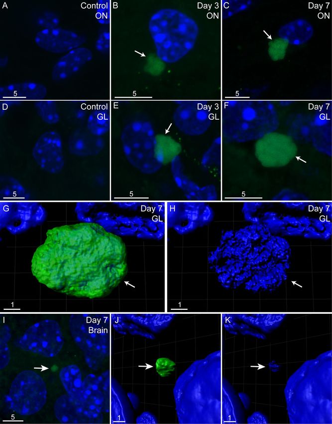
www.nature.com/scientificreports/
mice have also suggested a link between C. pneumoniae and late-onset dementia. C. pneumoniae DNA, antigen
and/or live bacteria have been detected in the brain of inoculated mice15–18, which resulted in Aβ deposition16,17
or altered appearance of Aβ deposits18. Importantly, the mice used in these studies were wild-type mice and not
mouse models of Alzheimer’s disease, suggesting that C. pneumoniae can contribute to the neuropathologies
associated with late-onset dementia.
Regardless of whether C. pneumoniae is a contributing factor to neurodegeneration, it is clear that this bacte-
rium can infect the brain and potentially contributes to chronic CNS pathologies. To date, it remains unknown
exactly how C. pneumoniae reaches the CNS. C. pneumoniae can infect lung macrophages, which migrate through
the mucosal barrier and enter the blood; the bacteria can disseminate to vasculature by surviving intracellularly
in blood monocytes, which can then cross the blood–brain barrier (BBB)19. It is also, however, possible that C.
pneumoniae may enter the CNS via alternative routes. The nerves that extend between the nasal cavity and the
brain, the olfactory and trigeminal nerves, have been shown to be a path for CNS infection by some infectious
agents reviewed i n20. These two nerves connect with the brain at the olfactory bulb and the brainstem, respec-
tively. Interestingly, the olfactory bulb, entorhinal cortex and hippocampal formation (all olfactory structures), as
well as the brainstem, are the CNS regions that exhibit the earliest signs of pathology in both late-onset dementia
and familial Alzheimer’s d isease21–23.
After intranasal inoculation in mice, C. pneumoniae antigens and/or infectious organisms have been detected
in the olfactory mucosa and the olfactory bulb 1–4 months post intranasal inoculation16–18, and C. pneumoniae
DNA in these tissues has been detected 1 week after i noculation18, strongly suggesting that the bacteria can infect
the CNS via the olfactory nerve. Certain other bacteria, however, have been shown to very rapidly (within days)
reach the CNS via the olfactory and/or trigeminal nerves reviewed i n20. We recently demonstrated that Chlamydia
muridarum can quickly (within two days) enter the CNS via these paths24, suggesting that C. pneumoniae may
also rapidly infect the brain via the nerves.
Furthermore, it is unknown how soon Aβ starts to accumulate after C. pneumoniae inoculation. Whilst previ-
ous studies have shown that this occurs after months16–18, if the bacteria reach the CNS more rapidly, alterations
in Aβ deposition may also occur sooner. In a transgenic mouse model of familial Alzheimer’s disease (5xFAD
mice, which exhibit the human amyloid precursor protein and presenilin 1 transgenes with five mutations linked
to Alzheimer’s disease), intracranial injection with Salmonella typhimurium resulted in a dramatic increase in Aβ
deposition after only 48 h13. Whilst Aβ deposition is of course much more pronounced in Alzheimer’s disease
mouse models, Aβ secretion in response to pathogens by (wild-type) neural cells can be r apid12.
Even though cranial nerves constitute a direct path by which microbes can access the brain, CNS infections
are relatively rare, and only a small number of infectious agents are thought capable of accessing the brain
via these paths. The nerves are well-protected physically and immunologically by the nasal epithelium which
exhibits powerful innate and adaptive immune system components. Together with the associated nasopharynx-
associated lymphoid tissue (NALT), the epithelium constitutes the first defence against microbes25. Injuries to the
nasal epithelium are, however, relatively c ommon26 and may expose the underlying cranial nerves to infection.
Experimental injuries to the nasal epithelium of mice has been shown to increase the risk of bacterial invasion of
the olfactory nerve and bulb by some bacteria27,28. Most microbes are, however, likely eliminated by phagocytic
glia, olfactory ensheathing cells (OECs) and trigeminal Schwann cells (TgSCs), should they penetrate the epi-
thelium and reach the nerves29–31. The glia limitans layer between the peripheral nerves and brain, populated by
astrocytes, constitutes a further immunological barrier against CNS i nfection32,33. Whilst it is largely unknown
why certain infectious agents can infect the CNS via cranial nerves, one key mechanism is thought to be the
ability of these pathogens to infect and survive in OECs, TgSCs and astrocytes, as well as in microglia (the main
phagocytes inside CNS tissue)20,27,28,34,35.
Chlamydiae are obligate intracellular bacteria with a unique biphasic life-cycle reviewed in36. Outside of
host cells, Chlamydiae exist as infectious, biologically inactive elementary bodies (EBs), which exhibit strong
resistance to environmental stress. C. pneumoniae EBs can become internalized into host cells, including many
phagocytes1,37,38. The EBs are resistant to endosomal/lysosomal degradation, and inside the host cell transform
into reticulate bodies (RBs). RBs replicate in inclusions (modified cellular vacuoles), which expand in size as
the bacteria replicate. After approximately 72 h (in cell culture), the RBs transform into EBs, which are released
by cell lysis and can infect new cells (exit via extrusion of membrane-bound compartments can also occur39).
Chlamydiae can also persistently infect cells40 which is likely relevant for the link to chronic diseases1. Persistent
Chlamydia infection can last for many years, and the persistent Chlamydia bacteria can re-activate41,42.
In the current study, we investigated whether C. pneumoniae could rapidly (3–7 days after intranasal inocu-
lation) invade the CNS via the olfactory and/or trigeminal nerves in mice and if this resulted in any alterations
in Aβ deposition in nerve/CNS tissue. Furthermore, we investigated whether C. pneumoniae could infect and
survive in cultured primary mouse OECs, TgSCs, astrocytes and microglia. We also determined whether C.
pneumoniae infection had any role in the regulation of Alzheimer’s disease gene expression over the longer term.
Material and methods
Bacterial strains. Chlamydia pneumoniae AR39 (ATCC 53592) is a human pharyngeal isolate and was
propagated in Hep-2 cells (sourced from the ATCC CCL-23). C. pneumoniae stocks were harvested in sucrose
phosphate glutamate (SPG) and aliquots were stored at – 80 °C for future experiments. The infectious yield of C.
pneumoniae was determined by counting inclusion forming units (IFU) in HEp-2 cells.
Animals. 7–8 week old female BALB/c mice were sourced from Animal Resource Centre (ARC, Murdoch,
Western Australia) and were intranasally inoculated (under anaesthesia with isofluorane 1.5–2%) with either 10
µL of PBS (phosphate buffer saline) as vehicle control (N = 5) or C. pneumoniae (1 × 10⁶ IFU [inclusion forming
Scientific Reports | (2022) 12:2759 | https://doi.org/10.1038/s41598-022-06749-9 2
Vol:.(1234567890)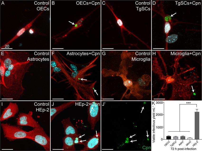
www.nature.com/scientificreports/
Figure 1. Schematics illustrating the process for quantifying the amount of viable infectious C. pneumoniae
present in various mouse tissues. (A) Mice were first intranasally inoculated with C. pneumoniae (i), some with
epithelial injury and some without. Following either 24 h, 3 days or 7 days or 28 days post inoculation, selected
tissues were collected and homogenised in tubes (ii). Homogenates were serially diluted onto HEp-2 cells and
incubated (iii). Cells were fixed and immunolabelled for C. pneumoniae inclusions, which were counted and the
number of inclusion-forming units (IFUs) per mL of homogenate was determined. Data were then compiled
into organ (tissue) load graphs (see Fig. 2). (B) Microscopic image showing a sagittal tissue section of a mouse
brain. Cell nuclei/DNA are shown in blue (DAPI stain). Key anatomical locations include the nasal cavity (NC),
olfactory epithelium (OE), olfactory bulb (OB), trigeminal nerve (Tg; not visible so approximate location is
shown by white dotted line) and the brain. Scale bar in µm.
units] per mice, N = 12), delivered as a 5 µL droplet per nostril. Mice were then sacrificed 1, 3, 7 and 28 days post
intranasal inoculation by asphyxiation with rising carbon dioxide.
Nasal epithelium injury model. In this study, we also used the methimazole injury model to investigate
whether epithelial injury can increase the risk of C. pneumoniae invasion of the olfactory/trigeminal nerves
and brain. For this purpose, 7–8 week old female BALB/c mice were injected with a single dose of methimazole
(50 mg/kg, 10 mg/mL in PBS) or vehicle (PBS only), according to our previously published p rotocol27,29. Three
days after methimazole injection, animals were intranasally inoculated with C. pneumoniae (N = 12) or vehicle
(N = 5) as outlined above.
Tissue collection. Heads and tissues including the olfactory mucosa (containing the olfactory nerve fasci-
cles), olfactory bulb, trigeminal nerve and the brain (the remainder of the brain after removal of olfactory bulbs)
were collected from euthanized mice, 1, 3, 7 days and 28 days post inoculation, for bacterial load determination
and histology.
Organ load assay. Chlamydia pneumoniae IFUs were detected by direct inoculation of tissue homogen-
ate onto HEp-2 cells which were seeded on 96-well plates with 4000 cells/well. After 72 h, the C. pneumoniae
inclusions in the entire wells were visualized by confocal microscopy and the numbers of IFUs isolated from the
homogenates (IFU/mL) were determined (see workflow in Fig. 1).
Chlamydial PCR. DNA was extracted from whole blood 2, 3 and 4 days after infection using the Qiagen
DNeasy blood and tissue kit according the manufacturer’s instructions. The quantitative Real-time PCR was car-
ried out by using Platinum SYBR Green qPCR SuperMix-UDG (ThemoFischer cat# 11733038). C. pneumoniae
was detected using 16S rRNA primers (Forward: 5′-CTCAACCCCAAGTCAGCATT-3′and Reverse: 5′-CTA
Scientific Reports | (2022) 12:2759 | https://doi.org/10.1038/s41598-022-06749-9 3
Vol.:(0123456789)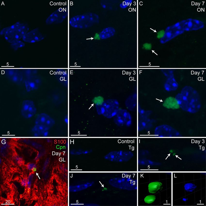
www.nature.com/scientificreports/
CGCATTTCACCGCTACA-3′. The cycling program was 10 min at 95 °C, followed by 40 cycles of 15 s at 95 °C
and 1 min at 60 °C, and a final dissociation stage. C. pneumoniae DNA was used as a positive control, and a no-
template control was also included.
Tissue preparation and sectioning. Heads were fixed in 4% paraformaldehyde (PFA) in PBS overnight
at 4 °C, followed by decalcification in 20% ethylenediaminetetraacetic acid (EDTA) for 4 weeks. The heads were
then embedded in optimal cutting temperature (OCT) medium and frozen. Sagittal sections (30 µm) were cut
using a cryostat (Leica CM1860).
Immunohistochemistry. Immunohistochemistry was performed as previously d escribed27,43,44. Speci-
mens were incubated with goat anti-C. trachomatis/C. pneumoniae (this antibody is used to detect both of these
Chlamydia species; Abcam ab20929; 1:400) and/or rabbit anti-Aβ peptide (Abcam ab201060,1:500). Secondary
antibodies were donkey anti-goat Alexa Fluor 488 (Abcam ab150129 1:400), donkey anti-rabbit 647 (Invitrogen
A31573; 1:500). Antibodies were diluted in blocking buffer (2% bovine serum albumin with 0.3% Triton X-100
in PBS). Cryostat sections were first incubated with blocking buffer for 60 min at room temperature, followed by
overnight incubation with primary antibodies at 4 °C. Sections were washed for 3 × 5 min, then incubated with
secondary antibodies for 1 h. Cell nuclei were stained with 4′6-diamidino-2-pheylindole (DAPI).
Primary glia cell culture. Olfactory ensheathing cells (OECs), trigeminal Schwann cells (TgSCs), astro-
cytes and microglia were used in this study. OECs and TgSCs were prepared from postnatal day 7 (P7) S100ß-
DsRed transgenic mice; we have previously generated and described this transgenic mouse line and the cell
isolation method45. Astrocytes and microglia were prepared from postnatal day 3 (P3) S100ß-DsRed transgenic
mice following a previous published protocol46. S100ß-DsRed transgenic reporter mice were used due to the
expression of DsRed fluorescent protein which is driven by the human S100ß promoter, such that glial cells
including OECs, Schwann cells, astrocytes and microglia express DsRed protein and facilitate easy visualisation
and identification in culture. The entire brain cell population was isolated from the brain tissue by enzymatic
digestion and mechanical dissociation using Neural Tissue Dissociation Kit with GentleMACS (Miltenyi Biotec,
130-093-231). The cell pellet consisting of a mixture of all brain cells was further subjected to magnetic cells
sorting for microglia enrichment using CD11b/c microbeads (Miltenyi Biotec, 130-093-636) or for astrocytes
using anti-GLAST (ACSA-1) microbeads kit (Miltenyi Biotec, 130–095-825) according to manufacturer’s pro-
tocol. The different glial preparations were separately plated in plastic 24-well plates and maintained in glial
medium containing Dulbecco’s Modified Eagle Medium with 10% foetal bovine serum (FBS), G5 supplement
(Gibco), gentamycin (Gibco, 50 mg/mL) and l-glutamine (200 μM) at 37 °C with 5% C O2 for 5 days. Cells were
replated into T-25 flasks and allowed to proliferate to ~ 80% confluency. Primary glial cultures with approxi-
mately 70–80% purity was used in the experiments.
In vitro infection of primary glial cells. Dilutions of C. pneumoniae bacteria were prepared in Dulbecco’s
phosphate buffered saline (DPBS). OECs, TgSCs, astrocytes and microglia were seeded at the density of 4000
cells/well in 96-well plate (Costar) in glial medium. After 24 h, bacteria (multiplicity of infection (MOI): 1:1)
were added and incubated with cells for 72 h. Following the infection, the cells were then rinsed in 1 × DPBS and
were fixed for 20 min in 4% PFA in DPBS. Subsequently, cells were washed and incubated in blocking buffer for
1 h. Cells were then incubated with the following primary antibodies overnight at 4 °C; goat anti-C. pneumoniae/
Chlamydia trachomatis (Abcam, ab20929; 1:400) and rabbit anti-glial fibrillary acidic protein (GFAP) antibody
(Thermofisher Scientific, PA5-16291; 1:200) or rabbit anti-ionized calcium-binding adaptor molecule 1 (IBA1)
microglia (Abcam, ab178847; 1:100). The following day, cells were washed with DPBS and incubated with sec-
ondary antibody donkey anti-goat Alexa Fluor 488 (Thermofisher Scientific, A11055; 1:400) and goat anti-rabbit
647 (ThermoFisher Scientific, A32733; 1:400) for 1 h. Nuclei were stained with DAPI. Hep-2 cells were visualized
by CellMask Orange Plasma membrane stain (Thermofisher Scientific, C100455; 1:10,000).
Viability assay for glial cells. Primary glial cells were infected with C. pneumoniae as described above.
Cultures were harvested after 72 h in SPG with 5 mM l-glutamine and stored at − 80 °C. Culture plates were
thawed and probe sonicated for 10 s (Sonics Vibra-Cell VCX 130, amp 1). Cell lysates (containing bacteria) were
collected and serially diluted on a monolayer of HEp-2 cells and, 72 h later, washed with PBS and fixed with 4%
PFA. Following immunocytochemistry, the infectious yield C. pneumoniae was determined by counting of the
inclusion forming units (IFU) ml−1 in Hep-2 cells.
RNA extraction and nanostring nCounter gene expression analysis. 7–8 week old female BALB/c
mice were infected as previously described. Mice were then sacrificed at 7 and 28 days post intranasal inocula-
tion by asphyxiation with rising carbon dioxide. RNA from brain lysate (the remainder of the brain after removal
of olfactory bulbs) of 7 and 28 days post C. pneumoniae inoculation and control mice (N = 3 for all groups) was
extracted using Maxwell® RSC simplyRNA tissue kits (Promega, AS1340) using the manufacturer’s protocol.
RNA was eluted in 50 µL of nuclease-free water and quality/quantity of RNA was assessed. Following RNA
elution, gene expression analysis was undertaken using the NanoString nCounter analysis system (NanoString
Technologies, Seattle, WA) using the commercially available nCounter Alzheimer’s disease panel kit (Cat num-
ber: XT-CSO-MAD1-12). The Alzheimer’s disease panel contains 23 neurodegeneration pathways, targeting 770
genes including 10 internal reference/housekeeping genes. A master mix was made following manufacturer’s
protocol with 70ul hybridisation buffer added to Reporter probes. Individual reactions for each sample were
Scientific Reports | (2022) 12:2759 | https://doi.org/10.1038/s41598-022-06749-9 4
Vol:.(1234567890)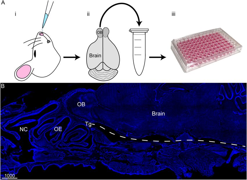
www.nature.com/scientificreports/
made with 8 μL master mix, 5 μL extracted RNA diluted to 125 ng and 2 μL Capture probe. Each reaction was
hybridised in a thermal cycler (Eppendorf) at 65 °C for 20 h. Samples were processed on the NanoString Prep
Station and the target-probe complex was immobilised onto the analysis cartridge. Cartridges were scanned by
the nCounter Digital Analyser for digital counting of molecular barcodes corresponding to each target at 555
fields of view.
Image capture. Images were captured using Nikon confocal microscope and Olympus FV3000 laser scan-
ning confocal microscope. Three-dimensional reconstructions were made using Imaris × 64 (Version 7.4.2). For
comparison between groups, the same image capture settings, laser intensity and focal depths were used. Images
were colour balanced uniformly across the field of view using Adobe Photoshop Creative Cloud 2019 (20.0.4)
and compiled into panels using Adobe Illustrator Creative Cloud 2019 (23.0.3).
Statistical analysis. Data are shown as means ± SEM. Statistical significance was analyzed using either a
two-way ANOVA with Bonferroni’s post hoc test or a one-way ANOVA with Tukey’s post hoc test. Statistical
analysis was performed using GraphPad Prism 9.0 software, and statistical significance was set at p < 0.05.
Gene expression data was processed using the Advanced Analysis Module in the nSolver Analysis Software
version 4.0 from NanoString Technologies (NanoString Technologies, WA, USA). Quality control was assessed,
and the data was analysed using Rosalind software (partner open-source software). Normalised data were gen-
erated by the software followed by fold change and p value. p value was adjusted using Benjamini–Hochberg
method of estimating false discovery rate. Venn diagram was generated using an open-source software (http://
bioinformatics.psb.ugent.be/webtools/Venn) normalised to day 7 and day 28 non-infected control. Principal
component analysis (PCA) and volcano plot was generated using Graphpad prism 9.0. Hierarchical clustering
was generated using Morpheus software with Pearson correlation and average linkage method across the samples
for the most significant genes (adjusted p value < 0.05). Molecular process was generated from REACTOME
database linked to Rosalind software with cut-off set at p value < 0.05. Pathway profile score was generated from
nSolver Analysis Software using the 23 neurodegenerative pathways mentioned previously.
Ethics and biosafety. The experimental procedures used in the study were conducted with the approval
of the Griffith University Biosafety Committee (NLRD/09/15_var7) and the Griffith University Animal Eth-
ics Committee (MSC/08/18/AEC) in accordance with guidelines of the Australian Commonwealth Office of
Gene Technology Regulator and the National Health and Medical Research Council of Australia. All the animal
experiments in this study are reported in accordance with ARRIVE guidelines (https://arriveguidelines.org).
Results
Chlamydia pneumoniae infects the olfactory mucosa, olfactory bulb and cerebral cortex within
3 days after intranasal inoculation. The key aim of the current study was to determine whether C.
pneumoniae could invade the brain via the olfactory/trigeminal nerve routes in the shorter term (≤ 1 week)
after intranasal exposure, as has been shown for C. muridarum24. We intranasally inoculated adult mice with C.
pneumoniae, then 3 days, 7 days and 28 days later mice were sacrificed. The 3 day time-point was chosen as C.
pneumoniae has a life-cycle of approximately 72 h1,37. We then determined whether infectious (viable, inclusion-
forming) C. pneumoniae could be isolated from the following homogenized mouse tissues: (1) olfactory mucosa
(consisting of the neuroepithelium, underlying lamina propria and the many nerve fascicles that constitute the
olfactory nerve), (2) olfactory bulb, (3) trigeminal nerve and (4) brain (beyond the olfactory bulb), as outlined in
Fig. 1. Tissue homogenates were diluted onto HEp-2 cells and inclusion-forming units per mL of tissue homoge-
nate were determined after 72 h.
At 3 and 7 day time-points, infectious C. pneumoniae were isolated from all four tissues (Fig. 2A). No C.
pneumoniae were detected in tissue lysate from control (vehicle-inoculated) mice (n = 2). For the olfactory bulb,
the number of IFUs was significantly higher at 3 days than 7 days post inoculation (p.i.) (Fig. 2A,B), whereas the
reverse occurred for the trigeminal nerve (Fig. 2A,B non-injury). At 28 days, low levels of infectious C. pneu-
moniae were isolated only from trigeminal nerve p.i. (not shown). We also determined whether C. pneumoniae
was present in the blood 2, 3 and 4 days post intranasal inoculation using PCR. Based on the curves, all samples
showed an absence of C. pneumoniae in blood (Fig. 2C). Note: the injury results presented in Fig. 2A,B are
reported in the section “Injury to the nasal epithelium increases peripheral infection”.
We also analysed tissue sections from the olfactory nerve, olfactory bulb, trigeminal nerve and brain (beyond
the olfactory bulb) for the presence of C. pneumoniae using immunohistochemistry. In addition to the 3 days,
7 days and 28 days mice, we also examined mice that had been sacrificed only 24 h after intranasal inoculation.
24 h after inoculation C. pneumoniae was detected within the olfactory mucosa and olfactory bulb and infec-
tious C. pneumoniae were isolated from both the olfactory mucosa and olfactory bulb (Supplementary Fig. 1).
However, as the bacteria at 24 h was likely to be from the inoculum we did not analyse this tissue further. At
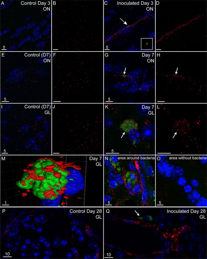
later time-points which are sufficient for at least one life cycle, C. pneumoniae inclusions were detected in the
olfactory nerve (Fig. 3A–C), glomerular layer of olfactory bulb (Fig. 3D–F) and trigeminal nerve (Fig. 3H–L) at
both 3 and 7 days p.i. Within the olfactory bulb, C. pneumoniae was only detected within the nerve fibre layer
and glomerular layer, with C. pneumoniae inclusion bodies being present inside OECs (Fig. 3G). C. pneumoniae
was also detected in the glomerular layer of the olfactory bulb at 28 days (Fig. 5Q).
Despite being able to isolate viable C. pneumoniae from the brain (beyond the bulb), we did not find defini-
tive C. pneumoniae inclusions in brain tissue sections from these mice (not shown), suggesting that inclusions
in brain tissue were too small or sparse to be confirmed by histology when screening tissue sections.
Scientific Reports | (2022) 12:2759 | https://doi.org/10.1038/s41598-022-06749-9 5
Vol.:(0123456789)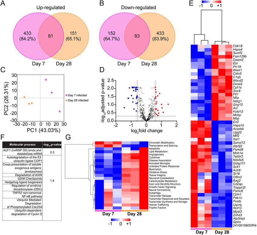
www.nature.com/scientificreports/
Figure 2. C. pneumoniae infects the nasal peripheral nerves and brain in mice with or without pre-injured nasal
epithelium after intranasal inoculation. (A,B) Graph showing the amounts of C. pneumoniae IFUs isolated from
various tissues of mice with or without pre-injured olfactory epithelium at 3 and 7 days post C. pneumoniae
inoculation. Infectious C. pneumoniae organisms were isolated from the olfactory mucosa, olfactory bulb,
trigeminal nerve and brain (n = 9/group). No C. pneumoniae was isolated from various tissues of control mice
(vehicle only, n = 2). Data are shown as the mean number of inclusions ± SEM, n = 9/group. *p ≤ 0.05, **p ≤ 0.01
***p ≤ 0.001, two-way ANOVA with Bonferroni’s post hoc test. For comparisons between non-injury Day 3
versus Day 7: a-a′ p ≤ 0.01, b-b′ p ≤ 0.01; for comparisons between injury Day 3 versus Day 7: c–c′ p ≤ 0.01,
d-d′ p ≤ 0.001, two-way ANOVA with Bonferroni’s post hoc test. (C) PCR amplification curves of blood for C.
pneumoniae (Cpn) at 2, 3 and 4 days post intranasal inoculation. (+) shows the positive control in each graph.
Samples tested include the control (vehicle only; N = 2), methimazole only (N = 2), methimazole + Cpn (N = 9)
and Cpn only (N = 9).
Injury to the nasal epithelium increases peripheral infection. It has previously been shown that
experimental injury to the olfactory neuroepithelium facilitates invasion of the olfactory nerve and bulb by
certain bacteria27,28. To investigate whether epithelial injury could also affect C. pneumoniae infection of nerves
and brain, we used our well-established methimazole-mediated model of nasal epithelial injury. Methimazole
causes degeneration of the nasal epithelium in rodents47. We have shown that this mode of injury leads to patchy,
dispersed injuries to the epithelium, separated by normal epithelium; this constitutes a model better resembling
“natural” nasal injuries than other models, such as chemical irrigation m odels27,29.
Mice were treated with methimazole, and 3 days later, when epithelial degeneration p eaks48, the mice were
inoculated intranasally with C. pneumoniae. The 3-day time-point was also chosen to limit any potential unrelated
effects of methimazole, as methimazole at this stage has been largely cleared49. We examined mice that had been
sacrificed only 24 h after intranasal inoculation. The methimazole treatment clearly damaged the epithelial layer
(Fig. 4A–C) and C. pneumoniae was found within the lamina propria underlying the epithelial layer and in the
nerve fibre layer of the olfactory bulb (Supplementary Fig. 2); as the bacteria is likely to be from the inoculum
we did not analyse this tissue further.
Mice were then sacrificed 3 and 7 days after inoculation, followed by determination of the amounts of viable
C. pneumoniae (IFUs) in tissues (Fig. 2A,B), as well as immunohistochemistry of tissue sections (Fig. 4). Whilst
described separately here for better clarity, these experiments were conducted simultaneously to the experiment
groups described for Fig. 2 (so that methimazole-induced epithelial injury followed by C. pneumoniae inocula-
tion could be compared to C. pneumoniae inoculation alone).
We compared the C. pneumoniae load (IFUs) between (1) mice inoculated with C. pneumoniae alone and
(2) mice with pre-injured nasal epithelium. Epithelial injury resulted in an increased C. pneumoniae load in the
olfactory mucosa (which includes the olfactory nerve; both at day 3 and day 7 post inoculation), olfactory bulb
and trigeminal nerve (day 7 only) in comparison to mice without injury (Fig. 2A,B). However, epithelial injury
did not result in a significant difference in C. pneumoniae load in the brain.
For both the pre-injured mouse group and the group that was not pre-injured, the bacterial load in the
trigeminal nerve was higher on day 7 than day 3 post inoculation (Fig. 2A,B). In the pre-injured group, olfactory
Scientific Reports | (2022) 12:2759 | https://doi.org/10.1038/s41598-022-06749-9 6
Vol:.(1234567890)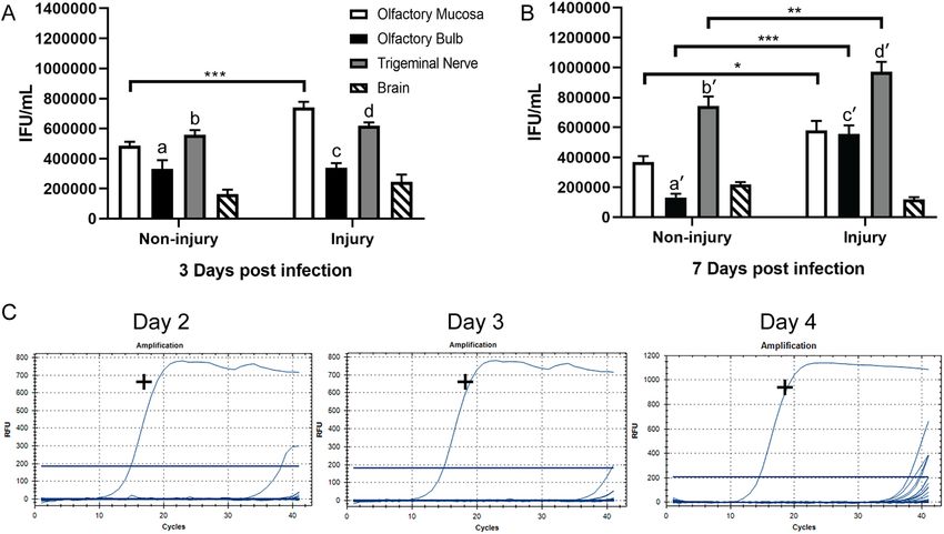
www.nature.com/scientificreports/

                                  Figure 3.  C. pneumoniae infects the nasal peripheral nerves and brain in mice after intranasal inoculation.
                                  Panels show images of tissue sections from control (vehicle only) and inoculated mice (C. pneumoniae) for
                                  both 3 days and 7 days post inoculation (p.i.). Images are representative ones from the olfactory nerve (ON)
                                  and olfactory bulb (OB) of 3 control mice, 3 C. pneumoniae-inoculated mice at 3 days p.i. and 3 inoculated
                                  mice at 7 days p.i. C. pneumoniae inclusions are shown in green (C. pneumoniae immunolabelling) and
                                  indicated by arrows. Nuclei/DNA is shown in blue (DAPI stain). Panels show maximum projection of confocal
                                  microscopy z-stacks. (A–C) Olfactory nerve (ON). (A) Control (vehicle only). (B) 3 days p.i. (C) 7 days p.i.
                                  (D–F) The glomerular layer (GL) of the OB. (D) control. (E) 3 days p.i. (F) 7 days p.i. (G) The GL at 7 days p.i.,
                                  immunostained with anti-S100 antibodies. (H–L) Trigeminal nerve (Tg). (H) control. I: 3 days p.i. (J) 7 days
                                  p.i. (K,L) 3D reconstruction of panel J. C. pneumoniae inclusions. (K) Inclusions (green) are within the Tg
                                  and contain DNA (L; blue, DAPI stain). Scale bars in µm. For a low-power image showing the approximate
                                  localization of the images, see Fig. 1B.

                                  bulb infection also increased with time, whereas the opposite occurred in mice that were not pre-injured
                                  (Fig. 2A,B). Immunolabelling confirmed the presence of C. pneumoniae in the olfactory nerve (Fig. 4A–C) and
                                  bulb (Fig. 4D–H). Three-dimensional reconstructions provided clear visualisation of the inclusions within the
                                  glomerular layer, with the DNA of the bacteria being clearly distinct from the host cell DNA (Fig. 4G,H). In one
                                  mouse, in the pre-injured group, C. pneumoniae IB/s were also found in the olfactory piriform cortex, with three-
                                  dimensional reconstruction again showing the bacterial DNA being distinct from the host cell DNA (Fig. 4I–K).

                                  Aβ is associated with regions of C. pneumoniae infection in the olfactory bulb. To determine
                                  whether C. pneumoniae inclusions were associated with Aβ deposits, we also immunolabeled the sections for
                                  Aβ. We found diffuse/patchy Aβ immunolabelling in olfactory nerve and bulb tissues from all animals, including
                                  control mice (Fig. 5A,B,E,F,I,J,P). However, we found that in tissues from inoculated mice, distinct Aβ deposits

Scientific Reports |   (2022) 12:2759 |                https://doi.org/10.1038/s41598-022-06749-9                                                      7

                                                                                                                                               Vol.:(0123456789)
www.nature.com/scientificreports/
Figure 4. C. pneumoniae infection of the olfactory nerve, olfactory bulb and brain after injury to the olfactory
epithelium. Panels show confocal images (maximum projection of z-stacks, A–I) of tissue sections from vehicle
control and C. pneumoniae-inoculated mice, all with pre-injured olfactory epithelium before inoculation/
vehicle treatment. Images are representative from n = 3 animals per group. C. pneumoniae inclusions are shown
in green (immunolabelling) with nuclei/DNA in blue (DAPI stain). (A–C) The olfactory nerve (ON) in (A)
control mice and mice inoculated with C. pneumoniae, (B) 3 days and (C) 7 days post inoculation. (D–F) The
glomerular layer (GL) of the olfactory bulb in (D) control mice and inoculated mice (E) 3 days and (F) 7 days
after inoculation. (G) A 3D reconstruction of the C. pneumoniae inclusion in panel (F) (green; arrow); (H) the
same 3D reconstruction as panel (G), showing only the DAPI staining (blue), where bacterial DNA within C.
pneumoniae inclusion is distinct from host cell DNA (arrow pointing to bacterial DNA). (I) Image showing a
C. pneumoniae inclusion in the olfactory piriform cortex seven days after inoculation (green; arrow); (J,K) a 3D
reconstruction and render of the C. pneumoniae inclusion shown in (I), with C. pneumoniae in green (J) and
bacterial DNA shown in K, arrow). Scale bars in µm.
Scientific Reports | (2022) 12:2759 | https://doi.org/10.1038/s41598-022-06749-9 8
Vol:.(1234567890)www.nature.com/scientificreports/
Figure 5. C. pneumoniae is associated with Aβ peptide accumulation in both the olfactory nerve (ON) and the
glomerular layer (GL) of the olfactory bulb (OB). Images show maximum projections of confocal microscopy z-stacks
from vehicle control mice and C. pneumoniae-inoculated mice, 3, 7 and 28 days p.i. Images are representative ones
from the ON or OB of 3 control and 3 inoculated mice. Immunolabelling shows Aβ peptide (anti-Aβ1-42) (red),
C. pneumoniae (green) and DNA (DAPI, blue). (A–D) At 3 days, the ON of (A,B) a control mouse and (C,D) a C.
pneumoniae-inoculated mouse. Arrows show the location of C. pneumoniae inclusions (green in C). Panels (B) and (D)
show only Aβ immunolabelling (red). (E–H) Similarly, at 7 days the immunolabelling of the ON in (E,F) control and
(E,F) inoculated mice. (I–L) At 7 days, the GL in (I,J) a control mouse and (K,L) a C. pneumoniae-inoculated mouse.
Panels (J and L): Aβ immunolabelling only (red). Arrows indicate C. pneumoniae inclusions. (M) A 3D reconstruction
of panel (K) showing Aβ (red) surrounding a C. pneumoniae inclusion (green). (N–O) Images of GL regions within the
same tissue section of a C. pneumoniae-inoculated mouse, showing areas where C. pneumoniae inclusions are, or are
not, localized. (N) A GL area were C. pneumoniae inclusions were detected. Inclusions (green) were associated with Aβ
accumulation (red). (O) An adjacent GL region where inclusions were not detected. Only diffuse Aβ immunolabelling
is seen (red). (P,Q) At 28 days, the GL of (P) a control mouse and (Q) a C. pneumoniae-inoculated mouse. Arrows
indicate C. pneumoniae inclusions; Aβ (red). Scale bars in µm.
Scientific Reports | (2022) 12:2759 | https://doi.org/10.1038/s41598-022-06749-9 9
Vol.:(0123456789)www.nature.com/scientificreports/
accumulated near C. pneumoniae inclusions. At 3 and 7 days after inoculation, we detected Aβ deposits near C.
pneumoniae inclusions in the olfactory nerve (Fig. 5C,D,G,H). At 7 days, Aβ deposits near C. pneumoniae inclu-
sions were detected in the glomerular layer of the olfactory bulb (Fig. 5K–N). The Aβ deposits were not detected
in adjacent tissue regions where inclusions were not present (Fig. 5O). At 28 days, Aβ deposits continued to be
detected near C. pneumoniae inclusions in the glomerular layer of the olfactory bulb (Fig. 5Q), while control
uninfected mice exhibited diffuse Aβ deposits (Fig. 5P) which is similar to previous reports16,18,50. C. pneumoniae
inclusions, as well as associated Aβ deposits, were detected sporadically within tissues. For this reason, we were
not able to quantify the difference in Aβ levels between tissues from the different time-points after inoculation,
as well as between inoculated and control animals. Correlating with the fact that we could not detect C. pneumo-
niae inclusions in the brain beyond the olfactory bulb, we also did not detect any evidence of distinct Aβ deposits
in these areas (not shown).
C. pneumoniae can infect primary glial cells. The capacity to infect and survive inside glial cells is
thought to be a key mechanism for the ability of bacteria to invade the CNS via cranial nerves34,35. Therefore,
we next examined whether glia from the olfactory and trigeminal nerves, olfactory bulb and brain could con-
stitute host cells for C. pneumoniae infection. OECs, TgSCs, astrocytes and microglia were inoculated with C.
pneumoniae (MOI: 1:1, i.e., 1 IFU/cell) for 72 h. For comparison and as a positive control, HEp-2 cells, which
are highly susceptible to C. pneumoniae infection and in which the bacteria have strong capacity for intracellular
survival51, were also included. Cells were either (1) fixed and immunolabelled for C. pneumoniae, or (2) lysed for
determination of C. pneumoniae IFUs (viable, infectious organisms). Immunolabelling showed C. pneumoniae
inclusions in all cell types (Fig. 6A–J). The HE-p2 cells had distinctly more pronounced inclusions compared
to the other cells, with the DNA of the bacteria being strongly visible (Fig. 6J-J’). The different glia had similar
levels of viable C. pneumoniae, but significantly lower amounts of viable C. pneumoniae were recovered from all
the glia than from HEp-2 cells (Fig. 6K).
C. pneumoniae infection modulates Alzheimer’s disease related gene expression. To inves-
tigate if C. pneumoniae infection had any role in the regulation of Alzheimer’s disease gene expression at the
transcriptional level, we profiled 7 and day 28 day infected and non-infected mice brains using NanoString
nCounter Alzheimer’s disease Panel. We first mapped the total number of genes which were up-regulated and
down-regulated in day 7 and day 28 infected samples out of 760 genes which were all normalised to their respec-
tive non-infected samples. We found that a total of 514 genes were up-regulated in day 7 samples compared to
232 genes in day 28, out of which 81 genes were common to both and 433 (84.2%) were exclusively up-regu-
lated in day 7 while 151 (65.1%) were exclusively up-regulated by day 28 (Fig. 7A). Interestingly, for genes that
were down-regulated, 152 (64.7%) in day 7 and 433 (83.9%) genes in day 28 were exclusively down-regulated
(Fig. 7B). We also investigated whether duration of infection had a role in differential gene expression (DGE) of
Alzheimer’s disease genes. The normalised expression of all the 760 genes from day 7 and day 28 infected sam-
ples were used to construct correlation mapping using dimensional reduction technique (Principal component
analysis—PCA). The maximum variance across the expressions was 43.03% (PC1 value) between the day 7 and
day 28 infected samples (Fig. 7C). This analysis showed that the duration of infection had a definite role as the
individual biological replicates from respective time points clustered together but away from each other as a
group.
Next, we determined the significantly different genes with large changes in fold expression in infected sam-
ples of day 28 compared to day 7 in the form of a volcano plot. A volcano plot of the genes from day 28 infected
samples using day 7 infected samples as a baseline and cut-offs at ± 1.5 on log twofold change and 0.5 on − log 10
adjusted p value was performed (Fig. 7D). A total of 53 genes were differentially regulated out of which 22 genes
were down-regulated (blue) and 31 genes were up-regulated (red). We further explored these genes to investigate
the relationship between the infected samples at the respective time points. A heatmap was constructed based
on the z-score of the normalised gene expressions followed by hierarchical clustering using Pearson correla-
tion method (Fig. 7E). We observed separate gene clustering of day 7 and day 28 infected mice. A total of 107
molecular processes were regulated (from REACTOME database using the Rosalind software) based on the DGE
of day 28 infection compared to day 7. Out of these, 10 pathways were sorted based on their − log 10 p value
with a cut-off at p < 0.05 (Fig. 7F). Interestingly, all these processes were down-regulated as the genes related to
these processes were significantly down-regulated in day 28 infected samples.
We also used the pathway profile scores from nCounter to construct a heat map and compared the pathway
modulation between day 7 and day 28 infected samples. A total of 23 pathways were compared which were
already pre-selected in the Alzheimer’s disease Nanostring Panel (Fig. 7G). Most of the pathways were over-
expressed in day 28 infected mice in comparison to day 7 infected mice. These trends also corroborated with
the findings of the REACTOME database where most of the processes, especially ubiquitin-mediated protein
degradation, were down-regulated reflecting the over-activation of “unfolded protein response” in day 28 infected
mice pathway profile score.
In summary, these findings suggest that C. pneumoniae infection leads to a differential regulation of Alzhei-
mer’s disease genes with long term infection (day 28) down-regulating most of the genes related to folding of
proteins and aiding in misaggregation.
Discussion
In the current study, we showed that (1) C. pneumoniae rapidly infected both the olfactory and trigeminal nerves
in mice, (2) C. pneumoniae entered the CNS via nerves within 24–72 h after intranasal inoculation and without
concurrent blood infection, (3) injury to the nasal epithelium exacerbated peripheral nerve infection, but reduced
Scientific Reports | (2022) 12:2759 | https://doi.org/10.1038/s41598-022-06749-9 10
Vol:.(1234567890)www.nature.com/scientificreports/
Figure 6. C. pneumoniae can infect glial cells from the olfactory and trigeminal nerves, olfactory bulb and
brain. Cultured glia (OECs, TgSCs, astrocytes and microglia) from S100β-DsRed mice, in which glia express
DsRed, were inoculated with C. pneumoniae (Cpn) for 72 h, along with HEp-2 cells. (A–J′) Maximum
projection images from confocal microscopy z-stacks of control cells and C. pneumoniae-inoculated cells. Nuclei
are stained with Hoechst (cyan). (A,C,E,G,I) Cells treated with cell culture medium alone (control; glia in red).
(B,D,F,H,J) Cells inoculated with C. pneumoniae. Immunolabelling showed C. pneumoniae inclusions in the
cells (green; arrows). (J-J′) Merged image shown in (J), C. pneumoniae inclusions alone shown in (J′). Scale bars
in µm. (K) Graph showing the amounts of C. pneumoniae IFUs isolated from the cell cultures. The infectious
yield of C. pneumoniae was significantly different between glia and HEp-2 cells (***p ≤ 0.001, one-way ANOVA
with Tukey’s post hoc test). Data shown as the mean number of inclusions ± SEM.
brain infection, (4) C. pneumoniae inclusions in the olfactory nerve and bulb were associated with accumulations
of Aβ, (5) the glial cells populating the olfactory/trigeminal nerves and brain supported C. pneumoniae replica-
tion, and (6) C. pneumoniae infection leads to differential regulation of Alzheimer’s disease related genes. Thus,
C. pneumoniae can very rapidly spread from the periphery to the CNS via the nerves extending between the
nasal cavity and the brain, without blood infection. To our knowledge, this study is the first report of Aβ deposi-
tion in response to C. pneumoniae infection of the primary olfactory nervous system, and the first time such
rapid (72 h) deposition of Aβ in response to any bacterium in wild-type animals in vivo has been demonstrated.
The time-frame for infection of the CNS by C. pneumoniae was considerably faster than what has previously
been shown (1 week–3 months16–18), which may be due to differences in the inoculation dose since we used a
higher inoculation dose than two previous s tudies16,50 but lower than a nother18. Nevertheless, the time-frame is
comparable to CNS invasion via cranial nerves by Burkholderia pseudomallei32,43,44,52, Streptococcus pneumoniae53,
Neisseria meningitidis54, Listeria monocytogenes55 and now recently another Chlamydia species, C. muridarum24.
The amoeba Naegleria fowleri56, as well as herpes simplex type virus type 1 (HSV-1)57, severe acute respiratory
syndrome coronavirus 2 (SARS-CoV-2)58–60 and other coronaviridae61 can also invade the CNS via these two
paths (shown in humans and/or animals).
Within the olfactory bulb, C. pneumoniae inclusions were detected in OECs within the nerve fibre layer/
glomerular layer. Another bacteria, Burkholderia pseudomallei also accumulated within the nerve fibre layer/
glomerular layer after intranasal inoculation, suggesting that the glia limitans acts to restrict further progression
of bacteria into the deeper regions. However, with C. pneumoniae while we could easily detect the inclusion bod-
ies, the much smaller infectious elementary bodies would likely be missed in our analyses of the tissue sections;
Scientific Reports | (2022) 12:2759 | https://doi.org/10.1038/s41598-022-06749-9 11
Vol.:(0123456789)www.nature.com/scientificreports/
Figure 7. Differential gene expression (DGE) involved in Alzheimer’s disease for short (day 7) and long term
(day 28) infection (n = 3/group). (A) Numbers and percentages of genes up-regulated for day 7 (pink) and day
28 (orange) infections normalised to their respective non-infected controls. (B) Numbers and percentages of
downregulated genes for day 7 (pink) and day 28 (orange) infections normalised to their respective non-infected
controls. (C) Principal component analysis of all the biological replicates of day 7 (pink) and day 28 infections
(orange) for all gene expressions normalised to their respective controls. Axes show the percentage variation in
each PC plots with maximum at 43.03% for PC1 followed by 26.31% for PC2. (D) Volcano plot of log twofold
change of DGE of day 28 infection (normalised to day 7 infections) against –log 10 adjusted p value. Cut-offs for
log twofold and –log 10 adjusted p value, set at ± 1.5 (± 0.585) and 0.5 (0.301), respectively. Blue dots represent
down-regulated and red dots represent up-regulated genes. (E) Heat map of significant DGE plotted from (D)
with hierarchical clustering on day 7 and day 28 infected replicates. Colour of the map is based on z-score from
log twofold change for each gene (across rows). Blue represents down-regulated and red represents up-regulated
genes. (F) Significant molecular processes (p < 0.05) in day 28 infection highlighted from REACTOME database
with corresponding –log 10 p value. (G) Heat map of biological pathway profile scores compared between day 7
and day 28 infected samples from nSolver data for 23 pathways enlisted in Nanostring Alzheimer’s disease panel.
thus it is possible that elementary bodies were present deeper in the olfactory bulb. As inclusion bodies were
detected in the olfactory piriform cortex, it suggests that C. pneumoniae did progress deeper into the olfactory
bulb as previously r eported15,16,18,50.
Injury to the nasal epithelium has been shown to increase infection of the olfactory nerve and bulb by B.
pseudomallei27 and to allow the entry of S. aureus, which does not normally invade cranial nerves, to enter the
olfactory bulb28. We therefore hypothesized that epithelial injury may lead to increased C. pneumoniae invasion
Scientific Reports | (2022) 12:2759 | https://doi.org/10.1038/s41598-022-06749-9 12
Vol:.(1234567890)www.nature.com/scientificreports/
of the olfactory/trigeminal nerves, olfactory bulb and remaining parts of the brain. We found that epithelial
injury resulted in increased C. pneumoniae load in the olfactory mucosa (which contains the fascicles of the
olfactory nerve), olfactory bulb and trigeminal nerve. In contrast, injury did not alter C. pneumoniae invasion of
the brain after 7 days. We have previously observed a similar result for B. pseudomallei in some mice, in which
the nasal infection in itself caused massive peripheral infection and destruction of the nasal epithelium (more
pronounced than in our epithelial injury model used in the current study). In these mice, B. pseudomallei inva-
sion of the CNS was n egligible20. We then hypothesized that this may be because glia in the olfactory nerve and
outer layers of the bulb responded to both the injury and bacteria, secreting large amounts pro-inflammatory
factors which limited CNS infection; this may also be the case for C. pneumoniae infection in the current study.
The ability to infect glia is considered key for CNS invasion via the cranial nerve p aths20,27,28,34,35. We here
showed that C. pneumoniae could infect, survive in and replicate (form inclusions) within glia from the PNS
(OECs and TgSCs) and the CNS (astrocytes and microglia). This is the first-time infection of OECs and TgSCs (or
other Schwann cells) by C. pneumoniae has been reported, however, we have recently shown that C. muridarum
can infect OECs and TgSCs24. Whilst C. pneumoniae infection of cultured primary astrocytes and microglia has
not been described, infection of astrocyte and microglial cell lines has been d emonstrated62–66. Most relevantly,
however, C. pneumoniae antigens have been detected inside both astrocytes and microglia in post-mortem human
brains9,11,67,68. OECs, Schwann cells and astrocytes are all innate immune cells which can respond to and phagocy-
tose bacteria, and microglia (the macrophages of the CNS) are well characterized professional phagocytes31,69,70.
The fact that C. pneumoniae can form inclusions in these cells suggest that the bacteria, at least to some extent,
can overcome phagocytic destruction; this may be one important mechanism by which this bacterium can invade
and establish long-term infection of the CNS.
We also detected localized deposition of Aβ adjacent to C. pneumoniae IBs and in the olfactory bulb after
7 days and 28 days post inoculation. Diffuse/scattered Aβ immunoreactivity was also present in these tissues of
control mice, however, the co-localisation of Aβ deposits and C. pneumoniae inclusions in inoculated mice was
clear and distinct. Previous studies have demonstrated Aβ deposits near C. pneumoniae-infected areas of the
cerebral cortex 1–4 months post intranasal i noculation16. One study reported that whilst there were not neces-
sarily more Aβ deposits in the cortex of C. pneumoniae-infected animals, Aβ deposits in infected animals were
morphologically different from those in control animals18. A previous long-term study showed that C. pneumo-
niae infection of the cerebral cortex preceded the peak of Aβ d eposition17. In combination with the findings of
the current study, it appears that Aβ secretion occurs in response to the infection. One reason may be that Aβ
is secreted as an antimicrobial a gent12 but alternatively it may be secreted in response to infection because of
pathway activation for the processing of the APP protein into Aβ which is then secreted; future work can clarify
the secretion and role of Aβ in this context.
The secretion of Aβ may thus be a normal immune response to any microbe that may invade the nervous
system, and if infection clears, the deposited Aβ can be cleared by phagocytic g lia71. It is, however, possible that
if bacteria are not cleared and instead become persistent or latent in neural cells, continued Aβ deposition may
occur, contributing to late-onset dementia and/or accelerating Aβ deposition in familial Alzheimer’s disease7.
In the case of C. pneumoniae, one study in wild-type mice demonstrated that Aβ deposits resulting from infec-
tion were subsequently cleared17, whilst another study showed that the deposits did not disappear over several
months16.
It is interesting that we observed Aβ deposits in the olfactory nerve earlier than in the bulb, as one study in
an Alzheimer’s disease mouse model (APP/PS1 mice) showed that the terminal end of the olfactory nerve within
the nasal olfactory epithelium is the first nervous system area to exhibit Aβ deposition, which then progresses
to the olfactory bulb and other CNS a reas72. As the mice in that study were kept in a standard animal holding
facility (not specific pathogen free), perhaps exposure to infectious agents may have contributed to this early,
peripheral deposition of Aβ (which likely would be much more pronounced in an Alzheimer’s disease model
than in wild-type mice).
Chlamydia pneumoniae infection also resulted in up-regulation of key pathways involved in Alzheimer’s
disease pathogenesis. The pathologic features of Alzheimer’s disease like activated microglia, production of
inflammatory mediators and reactive oxygen species (ROS) were highly regulated in infected brain tissue at
28 days post inoculation as compared to 7 days post inoculation. Theses neuroinflammatory responses are
considered a major driving factor in patients with neurodegeneration and Alzheimer’s disease pathology, which
starts early in the course of the disease, prior to the formation of Aβ plaques in the brain73. Previous studies
have shown that microglia and astrocytes act as host cells of C. pneumoniae in Alzheimer’s disease b rain9. It has
been shown that following infection, activated microglia and astrocytes secrete pro-inflammatory cytokines,
including IL-1β, TNFα and IL-6 which are neurotoxic and may directly increase Aβ production via activa-
tion of β-secretase (BACE)66,74. BACE cleaves amyloid precursor protein and initiates the amyloid cascade.
Microglia activation reduces the accumulation of Aβ in the brain by increasing its phagocytosis, clearance, and
degradation75. However, the neuroinflammation associated with Alzheimer’s disease could be a double-edged
sword because persistent microglia activation stimulated by the binding of microglia to Aβ can increase the
production of inflammatory mediators and reactive oxygen species (ROS), which further amplifies the neuro-
inflammatory response causing chronic inflammation and n eurodegeneration76.
Disturbance of endoplasmic reticulum (ER) function is emerging as a relevant factor driving neurodegenera-
tion in Alzheimer’s disease77. Several reports have described manifestations of ER stress in post-mortem brain
samples from Alzheimer’s disease p atients78. Protein folding in the endoplasmic reticulum (ER) is an essential
cell function and to safeguard protein production and ensure quality control, ER-stress triggers the activation
of several biochemical pathways collectively referred to as the unfolded protein response (UPR). Chlamydia
infection can induce cellular stress that impacts protein folding, thus inducing UPR activation however it is also
proposed to modulate the UPR to promote their survival and replication79. Interestingly, we found UPR pathway
Scientific Reports | (2022) 12:2759 | https://doi.org/10.1038/s41598-022-06749-9 13
Vol.:(0123456789)You can also read