Micro-fusion inhibition tests: quantifying antibody neutralization of virus-mediated cell-cell fusion
←
→
Page content transcription
If your browser does not render page correctly, please read the page content below
RESEARCH ARTICLE
Thakur et al., Journal of General Virology 2021;102:001506
DOI 10.1099/jgv.0.001506 OPEN
ACCESS
Micro-fusion inhibition tests: quantifying antibody neutralization
of virus-mediated cell–cell fusion
Nazia Thakur1, Carina Conceicao1, Ariel Isaacs2, Stacey Human1, Naphak Modhiran2, Rebecca K. McLean1,
Miriam Pedrera1, Tiong Kit Tan3, Pramila Rijal3, Alain Townsend3, Geraldine Taylor1, Paul R. Young2, Daniel Watterson2,
Keith J. Chappell2, Simon P. Graham1 and Dalan Bailey1,*
Abstract
Although enveloped viruses canonically mediate particle entry through virus–cell fusion, certain viruses can spread by cell–
cell fusion, brought about by receptor engagement and triggering of membrane-bound, viral-encoded fusion proteins on the
surface of cells. The formation of pathogenic syncytia or multinucleated cells is seen in vivo, but their contribution to viral
pathogenesis is poorly understood. For the negative-strand paramyxoviruses respiratory syncytial virus (RSV) and Nipah virus
(NiV), cell–cell spread is highly efficient because their oligomeric fusion protein complexes are active at neutral pH. The recently
emerged severe acute respiratory syndrome coronavirus 2 (SARS-CoV-2) has also been reported to induce syncytia formation
in infected cells, with the spike protein initiating cell–cell fusion. Whilst it is well established that fusion protein-specific anti-
bodies can block particle attachment and/or entry into the cell (canonical virus neutralization), their capacity to inhibit cell–cell
fusion and the consequences of this neutralization for the control of infection are not well characterized, in part because of the
lack of specific tools to assay and quantify this activity. Using an adapted bimolecular fluorescence complementation assay,
based on a split GFP–Renilla luciferase reporter, we have established a micro-fusion inhibition test (mFIT) that allows the iden-
tification and quantification of these neutralizing antibodies. This assay has been optimized for high-throughput use and its
applicability has been demonstrated by screening monoclonal antibody (mAb)-mediated inhibition of RSV and NiV fusion and,
separately, the development of fusion-inhibitory antibodies following NiV vaccine immunization in pigs. In light of the recent
emergence of coronavirus disease 2019 (COVID-19), a similar assay was developed for SARS-CoV-2 and used to screen mAbs
and convalescent patient plasma for fusion-inhibitory antibodies. Using mFITs to assess antibody responses following natural
infection or vaccination is favourable, as this assay can be performed entirely at low biocontainment, without the need for live
virus. In addition, the repertoire of antibodies that inhibit cell–cell fusion may be different to those that inhibit particle entry,
shedding light on the mechanisms underpinning antibody-mediated neutralization of viral spread.
INTRODUCTION immunity against virus infection. nAbs are principally
involved in virus particle neutralization, the process by which
The development of neutralizing antibodies (nAbs) following virion entry is blocked by antibodies that inhibit receptor
infection or immunization is a central pillar of long-lasting engagement, block uptake outright, inhibit endocytosis, cause
Received 22 July 2020; Accepted 16 September 2020; Published 15 October 2020
Author affiliations: 1The Pirbright Institute, Ash Road, Pirbright, Woking, GU24 0NF, UK; 2University of Queensland, Brisbane, Queensland 4071, Australia;
3
MRC Human Immunology Unit, MRC Weatherall Institute of Molecular Medicine, University of Oxford, Oxford OX3 9DS, UK.
*Correspondence: Dalan Bailey, dalan.bailey@pirbright.ac.uk
Keywords: cell–cell fusion; enveloped virus; mFIT; neutralizing antibodies; Nipah virus; RSV; SARS-CoV; SARS-CoV-2; vaccines.
Abbreviations: BHK, baby hamster kidney; bRSV, bovine respiratory syncytial virus; COVID-19, coronavirus disease 2019; DMEM, Dulbecco’s modified
Eagle’s medium; FBS, foetal bovine serum; GCU, green count units; HEK, human embryonic kidney; HeV, Hendra virus; hRSV, human respiratory
syncytial virus; IC50, 50% inhibition of luciferase; IC90, 90% inhibition of luciferase; LoD, limit of detection; mAb, monoclonal antibody; MeV, measles
virus; mFIT, micro-fusion inhibition test; MOTA, Motavizumab; mVNT, micro virus neutralization test; nAb, neutralizing antibody; NiV, Nipah virus; NiV
mcsF, molecular clamp stabilized Nipah virus fusion protein; NiVpp, Nipah virus pseudoparticles; NiVsG, secreted Nipah virus attachement protein;
PBS, phosphatebuffered saline; PRF-DMEM, phenol red-free Dulbecco’s modified Eagle’s medium; PVM, pneumonia virus of mice; RBD, receptor-
binding domain; RLU, relative light units; rLuc-GFP, green fluorescent protein–Renilla luciferase reporter; RSV, respiratory syncytial virus; SARS-CoV,
severe acute respiratory syndrome coronavirus; SARS-CoV-2, severe acute respiratory syndrome coronavirus 2; SARS-CoV-2-pp, severe acute
respiratory syndrome coronavirus 2 pseudoparticles; SARS-CoV-pp, severe acute respiratory syndrome coronavirus pseudoparticles; vGP, viral
glycoprotein; VNT, virus neutralization test.
Five supplementary figures and two supplementary tables are available with the online version of this aricle.
001506 © 2021 The Authors
This is an open-access article distributed under the terms of the Creative Commons Attribution License. The Microbiology Society waived the open access fees for this article.
1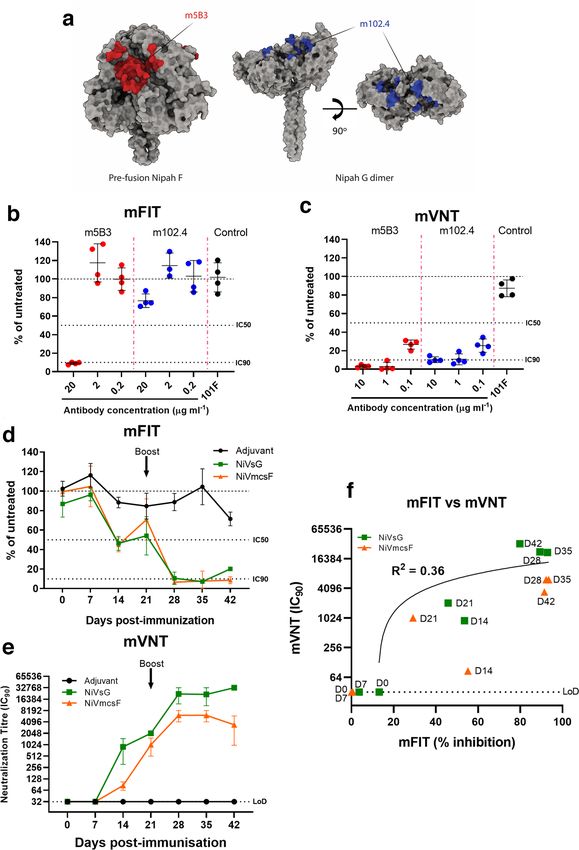
Thakur et al., Journal of General Virology 2021;102:001506
aggregation and/or trigger complement activation [1, 2]. with robust repeatability [13, 15] and also rapidly adaptable,
Accordingly, the majority of assays to identify nAbs focus on for example to SARS-CoV-2 [19]. This system is based on
mimicking this process, involving derivations of the ubiqui- the use of a split green fluorescent protein–Renilla luciferase
tous virus neutralization test (VNT) with live virus particles reporter (rLuc-GFP) to monitor cytoplasmic mixing after
or pseudotyped surrogates [2–4]. Certain viruses, however, cell–cell fusion [20]. Briefly, ‘effector’ cells expressing rLuc-
can also spread by directly inducing cell–cell fusion, resulting GFP 1–7 (beta strands 1–7 of GFP) and individual viral glyco-
in multinucleated cells or syncytia [5], including severe acute proteins (vGPs) are co-cultured with ‘target’ cells expressing
respiratory syndrome coronavirus 2 (SARS-CoV-2), the the rLuc-GFP 8–11 (beta strands 8–11 of GFP) component
recently emerged coronavirus responsible for the ongoing and the viral receptor. When receptor engagement triggers
coronavirus disease 2019 (COVID-19) pandemic [6, 7]. cell–cell fusion, the cytoplasm of target and effector cells mix
Importantly, not all particle-neutralizing nAbs are capable and the corresponding elements of the rLuc-GFP reporter
of inhibiting cell–cell fusion and vice versa, and there are reconstitute, becoming biologically active and quantifiable.
currently few robust methodologies available to specifically In the micro-fusion inhibition test (mFIT) described herein,
dissect the development and properties of fusion-inhibitory we have modified this assay to allow the incubation of effector
nAbs, which may be less abundant than standard nAbs, but cells with sera or monoclonal antibodies (mAbs), to quantify
necessary as an additional barrier to stop viral spread. their fusion-inhibition phenotype (Fig. 1). Whilst related
assays have been described in the past [21], we believe that
For enveloped viruses the viral glycoproteins found on the
the mFIT provides a simple, high-throughput and tractable
surface of virions represent the major target of nAbs. Whilst
assay that is easily adapted to different viruses.
the exact mechanism of particle attachment and membrane
varies, some essential principles are maintained [8]. Briefly, Our initial focus in this study was the development of mFITs
viral glycoproteins, often oligomeric, are embedded in the for three medically and agriculturally important paramyxovi-
virion surface by transmembrane domains, with the majority ruses; human and bovine respiratory syncytial virus (h/b RSV;
of the protein being exterior to the virion (the ectodomain). closely related orthopneumoviruses) and NiV (a henipavirus).
These complexes are assembled in a pre-fusion state, main- hRSV is a significant cause of respiratory disease, morbidity
tained by intra or inter-molecular constraints, with a hydro- and mortality in children and the elderly, whilst bRSV causes
phobic fusion loop or peptide buried within the interior respiratory disease resulting in large losses to the global dairy
of the oligomer. The capacity to initiate membrane fusion and cattle industry. NiV is an emerging zoonotic pathogen,
is normally primed by proteolytic cleavage of the fusogen currently restricted to South and South-East Asia, which
polypeptide and triggered by ligand binding. The latter step can be transmitted from its natural reservoir – fruit bats – to
represents the area with the greatest mechanistic heteroge- humans, causing a highly fatal respiratory and neurological
neity. Viruses such as respiratory syncytial virus (RSV) and disease. In the first outbreak of NiV in Malaysia in 1999 the
SARS-CoV-2 have a single trimeric fusogen (their fusion (F) virus also infected pigs, which served as an amplification
and spike (S) proteins, respectively) that mediate both attach- reservoir allowing spread to farm workers [22]. For both NiV
ment and fusion, while for others, such as Nipah virus (NiV), and hRSV there are no licensed vaccines for use in humans;
a separate protein (glycoprotein, G) maintains the fusion (F) with the only licensed therapeutic being a monoclonal
protein in its pre-fusion state, with triggering of F controlled antibody against the hRSV F protein [23, 24]. Following the
by receptor binding by G. Nevertheless, the final steps are emergence of SARS-CoV-2 in late 2019, and its pandemic
orthologous, with the fusogens undergoing large structural spread from early 2020, we also developed an equivalent
rearrangements to embed the fusion peptide in the host assay for this coronavirus, alongside SARS-CoV, to facilitate
membrane, leading to the so-called post-fusion state. This the examination and characterization of fusion-inhibitory
process ultimately leads to the juxtaposition of viral and host antibodies against this virus. Importantly, for all the viruses
membranes, the formation of a fusion pore and viral genome described here there is established evidence (or emerging for
entry. From a nAb perspective, the development of antibodies SARS-CoV-2 [25–27]) that viral syncytia form in the tissues
against the pre-fusion state of the viral fusogen is considered of infected individuals or animal models of disease [3, 28–33],
to be favourable, as this is more likely to inhibit particle entry. highlighting the importance of this route of spread. We hope
that the development of mFIT assays for these viruses will
In previous studies, we and others have developed assays to
enable a broader understanding of the mechanisms of virus
reliably quantify cell–cell fusion induced by viruses [9–14].
neutralization and the immune response to fusogenic viruses,
Indeed, these systems have been successfully applied to
and in doing so facilitate the development of novel vaccines
examine virus host range [15], protein–protein interactions
and therapeutics.
within the attachment complex [16], the mechanism of
attachment and entry [11, 17], and other biologically relevant
questions. Seeking to adapt these systems to examine fusion-
inhibitory antibodies has proven technically challenging, as
RESULTS
many of these assays are not easily adaptable to protocols that Optimization of cell–cell fusion assays
require titration of sera or purified antibodies. However, the Previous iterations of the cell–cell fusion assay have relied on
cell–cell fusion assay we have developed for paramyxoviruses transient expression (via transfection) of both the vGPs and
[16, 18] has proven to be applicable to a 96-well plate format the rLuc-GFP reporter [9, 13, 34]. To simplify our approach,
2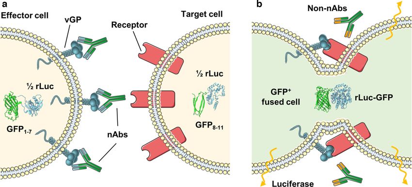
Thakur et al., Journal of General Virology 2021;102:001506
Fig. 1. The micro-fusion inhibition test (mFIT). Samples containing antibodies are incubated with effector cells (HEK293T Lenti rLuc-GFP
1–7) expressing the viral glycoprotein (vGP) of interest. The antibody–effector cell mix is then co-cultured with target cells (HEK293T
Lenti rLuc-GFP 8–11) expressing the corresponding vGP’s cellular receptor and incubated for 18–24 h. In (a) the presence of fusion-
inhibitory neutralizing antibodies (nAbs) prevents the reconstitution of the rLuc-GFP reporter in fused cells, while in (b) the absence of
specific neutralizing antibodies (non-nAbs), allows vGP-mediated cell–cell fusion to occur. Subsequent mixing of the target and effector
cell cytoplasm leads to reconstitution of the split reporter and increased GFP and luciferase signals. This figure was generated using
modified images from SMART Servier Medical Art By Servier, used under CC BY 3.0, https://smart.servier.com/, accessed June 2020.
we clonally selected stable cell lines that express the rLuc-GFP and 18 did not (Fig. 2b). Unsurprisingly, the site I-binding
reporter elements (Fig. S1, available in the online version of antibody 4D7 showed no capacity for inhibiting hRSV-F-
this article) and compared their activity to standard transient mediated fusion in our mFIT, correlating with this mAb
transfection (Fig. S2), confirming their suitability for cell–cell binding only the post-fusion variant of this epitope. In
fusion experiments. Within our laboratory we have developed contrast, antibody binding sites II and IV, which are able to
cell–cell fusion assays for a number of vGPs [13, 15, 18, 19]; access their epitopes on both the pre- and post-fusion forms
however, the conditions required for fusion are rarely main- of RSV-F, showed much greater capacity for inhibition in our
tained between these proteins. For this study, the mass and mFIT. 101F (site IV) showed robust inhibition of hRSV–F
ratio of DNA transfected, the length of co-culture, as well
fusion even at 1.25 µg ml−1, while inhibition by MOTA (site
as the duration and temperature of nAb incubation, were all
II) gradually titred out between 5 1 and 1.25 µg ml−1. AM14
conditions that required optimization for hRSV, bRSV, NiV,
severe acute respiratory syndrome coronavirus (SARS-CoV) (site V) and MPE8 (site III), which bind to the pre-fusion form
and SARS-CoV-2 mFITs (Figs S3 and S4). of RSV-F, were also able to potently inhibit RSV fusion even
at the lowest concentration assessed, 1.25 µg ml−1 (Fig. 2c). As
RSV mFITs to examine mAb neutralization of fusion an additional control we also included monoclonal antibodies
Using the established hRSV mFIT we began by character- to measles virus (MeV) H, which had little appreciable effect
izing the fusion-inhibition properties of a number of recog- on RSV-mediated fusion. Of note, MOTA is a derivative of
nized human F-specific mAbs, including 101F, AM14, 4D7, the licensed RSV mAb Palivizumab and MPE8 is known to
Motavizumab (MOTA) and MPE8 (Fig. 2; additional mAb cross-compete with Palivizumab [41]. The binding epitope of
details provided in Table S1) [35–39]. We also included four MPE8 is well conserved between all orthopneumoviruses –
RSV F-specific murine antibodies, which we have previously hRSV, bRSV and pneumonia virus of mice (PVM) [41] – so
shown to either inhibit (mAb 19 and 20) or not inhibit (mAb we carried out a side-by-side comparison of these antibodies
16 and 18) RSV fusion, albeit in semi-quantitative assays in a bRSV-F mFIT, which demonstrated a similar trend of
[40]. The RSV-F protein, a functional trimer, has a number inhibition with both the murine and human mAbs (Fig. S5).
of well characterized surface epitopes, which are differentially Our findings highlight the antigenic similarity of these viruses
exposed on the pre- and post-fusion variants of this complex and the likely conservation of epitopes between related F
[41] (Fig. 2a), with our panel of mAbs covering a diversity of proteins, which are roughly 80 % identical at the amino acid
these epitopes (Fig. 2a). level [42]. More broadly, we demonstrate that the mFIT can
Consistent with previous findings [40], murine RSV-specific be rapidly used to screen the fusion-inhibitory properties of
mAbs 19 and 20 inhibited hRSV–F fusion, while mAbs 16 a panel of vGP-specific mAbs.
3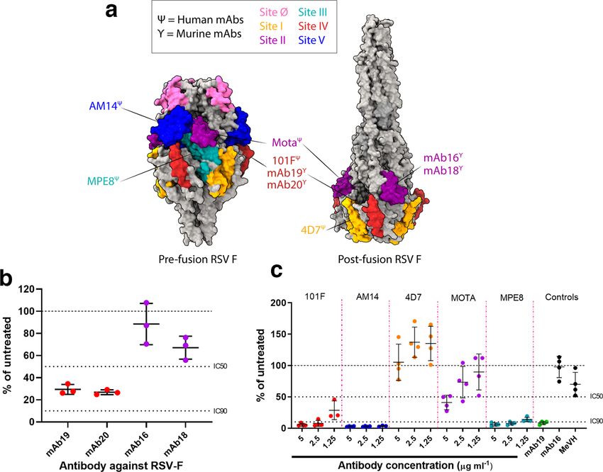
Thakur et al., Journal of General Virology 2021;102:001506
Fig. 2. Examining the neutralization of cell–cell fusion by monoclonal antibodies in human RSV mFITs. (a) Molecular surface representation
of RSV F trimer in the pre-fusion (left; PDB 4MMV) and post-fusion (right; PDB 3RRR) forms with antigenic sites coloured as follows:
site ø, pink; site I, orange; site II, purple; site III, turquoise; site IV, red; site V, blue. RSV F-specific mAbs are annotated and coloured
according to the corresponding antigenic binding site. Ψ represents human mAbs, while γ represents murine mAbs. Molecular graphics
and analyses were performed in UCSF’s ChimeraX program. (b) Murine mAbs (1 : 160 working dilution) and (c) human mAbs (5, 2.5 and
1.25 µg ml−1) were tested in hRSV-F mFITs. mAb 19, positive control; mAb16, specific negative control; MeVH, non-specific negative
control (Table S1). Data are expressed as a percentage of the average luciferase readings seen in no-sera/negative controls with 50 or
90 % inhibition (IC50 and IC90) lines indicated. Error bars represent mean±sd.
NiV mFITs to examine mAb neutralization of fusion (Fig. 3b). In contrast to our mFIT results, both m102.4 and
and immune responses during vaccination m5B3 were able to inhibit NiV particle entry at 10 µg ml−1 in
To broaden our study to examine neutralization of other fuso- micro virus neutralization tests (mVNTs) using pseudotyped
genic viruses, we performed NiV mFITs with two henipavirus- NiV, with this inhibition beginning to titre out at 0.1 µg ml−1
specific mAbs. Firstly, the human monoclonal m102.4, which (Fig. 3c). Of note, we included the RSV F mAb 101F as a
specifically interacts with the receptor- binding domain negative control, with this mAb showing no significant inhibi-
(RBD) of the NiV attachment protein (G) and has strong tion of NiV-mediated fusion in either assay. These differences
cross-reactivity between NiV and the closely related Hendra highlight how mFITs and VNTs can be used in tandem to
virus (HeV) [43] (Fig. 3a). m102.4 was shown to be protective probe the neutralizing properties of antibodies against vGPs.
in several animal studies and has also been administered to
individuals at high risk of exposure to HeV [3, 44–46]. The Beyond mAb characterization, we also assessed the applica-
second monoclonal (humanized m5B3) recognizes the pre- bility of the mFIT in vaccine immunogenicity studies. As part
fusion forms of NiV and HeV-F proteins and has been shown of an ongoing study, we are assessing the immunogenicity
to inhibit membrane fusion by holding F in its pre-fusion and efficacy of candidate NiV vaccines in pigs. Two of the
form [47] (Fig. 3a). We found that the F-binding m5B3 was vaccine candidates undergoing analysis are recombinant,
able to robustly inhibit fusion at 20 µg ml−1, which titred out secreted variants of the NiV glycoproteins; NiV F (mcsF)
at 2 µg ml−1. Interestingly, the G-specific m102.4 was also able (Young P.R. et al., in press) and NiV G (sG) [48]. These
to inhibit NiV fusion at 20 µg ml−1, albeit at reduced levels two proteins are the major targets for particle-neutralizing
4Thakur et al., Journal of General Virology 2021;102:001506
Fig. 3. Using NiV mFITs to characterize neutralization of cell–cell fusion by monoclonal antibodies and sera from NiV vaccinated pigs.
(a) Molecular surface representation of NiV-F pre-fusion trimer (left, PDB 5EVM) and NiV G dimer (right, PDB 2VWD). The stalk domain
of G (residues 62–117) is modelled after parainfluenza virus 5 stalk (PDB 4JF7). The 5B3 epitope on Nipah F is coloured red, and the
m102.4 epitope on Nipah G is coloured blue. Molecular graphics and analyses performed with UCSF ChimeraX. Antibodies against
NiV-F- and NiV-G-specific mAbs were tested in a NiV-FG (b) mFIT (20, 2 and 0.2 µg ml−1) and (c) mVNT using NiV viral pseudotypes (10, 1
and 0.1 µg ml−1). A negative control, RSV F mAb (101F), was also included. (d) Sera from individual representative NiV mcsF- or NiV sG-
vaccinated pigs were tested longitudinally in a NiV-FG mFIT (1 : 5 working dilution) and by (e) mVNT using NiV viral pseudotypes. (f) An xy
scatter plot illustrating the correlation between mFIT results (% reduction) and mVNTs (IC90) from the immunogenicity study performed
in pigs (Table S2). A linear line of regression is shown together with the calculated R2 value. Data are expressed as a percentage of the
average luciferase readings seen in no-sera/negative controls. Error bars represent mean±sd with 50 or 90 % inhibition (IC50 and IC90)
and limit of detection (LoD) lines are indicated.
5Thakur et al., Journal of General Virology 2021;102:001506
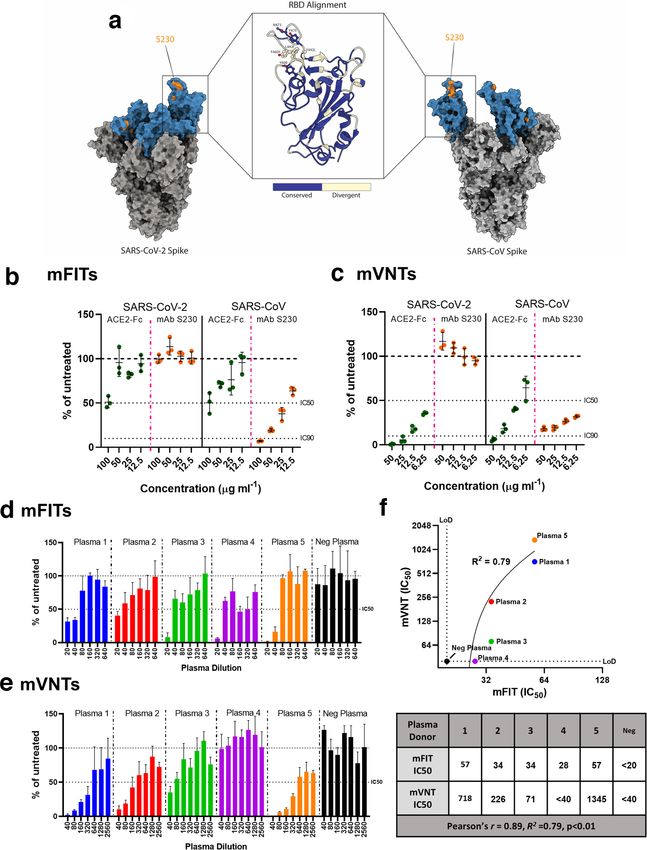
antibodies, which correlates well with protection from disease There is also an urgent need to develop a better understanding
in in vivo models [3]. To date, however, little is known about of antibody responses to SARS-CoV-2. For example, very little
the cell–cell fusion-inhibitory phenotype of these nAbs. For is known about whether antibodies can block S-mediated
NiV vaccines the development of fusion-inhibitory responses cell–cell fusion. With this in mind, we performed mFITs
may correlate significantly with immunity, since syncytia on plasma from convalescent donors who have recovered
cell formation has consistently been observed in the infected from COVID-19, alongside a negative plasma pool (from
tissues of experimentally infected animals [29, 49–52]. Using healthy donors collected prior to 2019). We found that all five
a homologous prime (day 0) and boost (day 21) regime, our donors were able to inhibit SARS-CoV-2 cell–cell fusion by
NiV-F and NiV-G vaccine candidates were inoculated into >50 % when their plasma was used at a 1 : 20 dilution, although
pigs and blood samples were taken every week until 42 days this dropped to 2/5 at the next dilution (1 : 40). As expected,
post-vaccination. Sera from some of these vaccinated animals, the negative plasma was unable to inhibit fusion (Fig. 4d).
as well as from an adjuvant-only control group, were exam- We also demonstrated a good positive correlation (Pearson
ined for cell–cell fusion-inhibitory Abs. mFIT results from R=0.89, R2=0.79) between mFIT IC50 values calculated for
single representative animals from each group demonstrate each of these donors and the corresponding IC50 values from
that a fusion-inhibitory response is generated by both NiV-F mVNTs (Fig. 4e, f), allowing us to conclude that SARS-CoV-2
and NiV-G vaccines pre-boost and that this response is infection can lead to the development of cell–cell fusion-
significantly boosted following the second inoculation of the inhibitory nAbs.
recombinant protein on day 21 (Fig. 3d). The development
of fusion-inhibitory Abs is specific to the vaccine groups, DISCUSSION
as the adjuvant-only immunized animal did not develop
Herein we have demonstrated the broad applicability of the
equivalent responses (Fig. 3d). We were also able to show
viral mFIT, examining neutralization of cell–cell fusion by
a positive correlation (Pearson R=0.66, R2=0.43) between
monoclonal antibodies and recombinant proteins as well
fusion inhibition and particle neutralization titres using NiV
as sera from vaccinated animals and plasma from naturally
pseudotypes in mVNTs (Fig. 3e, f, Table S2). In summary, the infected individuals. This assay allows the de facto calculation
mFITs carried out using NiV-specific mAbs and sera from of fusion-inhibitory IC50 and IC90, which we demonstrated
vaccinated animals highlight the broad applicability of this correlated well with more classical approaches such as
assay and the presence of nAbs that inhibit particle fusion, mVNTs. While virus particle neutralization and antibody-
cell–cell fusion, or both. mediated inhibition of viral-induced cell–cell fusion are
likely to functionally overlap, based on the mutual recogni-
SARS-CoV-2 and SARS-CoV mFITs to examine mAb tion of epitopes on the surface of viral glycoproteins and the
neutralization of fusion subsequent blocking of their related mechanisms of action,
Both SARS-CoV and SARS-CoV-2 S proteins, functional there may be distinct nAb clones that neutralize particles
trimers (Fig. 4a), were rapidly adaptable to our cell–cell but cannot block fusion or vice versa. The combined use of
fusion system. We first examined whether a soluble version mFITs and VNTs could prove useful in delineating these
of the ACE2 receptor fused to the human IgG1 Fc (hinge- differences. This assay builds on existing techniques for the
CH2-CH3) (ACE2-Fc) was able to inhibit S-mediated fusion. identification of antibody-mediated inhibition of cell–cell
ACE2-Fc was able to inhibit approximately 50 % of SARS- fusion, including classical observation of syncytia formation
by microscopy, computational analysis of images to quantify
CoV and SARS-CoV-2 fusion when used at 100 µg ml−1 with
syncytia formation, HIV-based variations of the dual-reporter
corresponding SARS-CoV and SARS-CoV-2 pseudopar-
assay [12] and assays based on the quantification of cellular
ticles being neutralized at lower concentrations in mVNTs
electrical impedance [55]. Clearly, from our data, the mFIT
(Fig. 4b, c). As proof of principle, we also examined whether
can be applied to different viruses, dependent only on the
the SARS-CoV-specific mAb S230 could inhibit SARS-CoV
availability of a cloned viral glycoprotein capable of inducing
fusion in an mFIT. S230 binds the SARS-CoV S receptor- fusion. On this, we have also demonstrated that this assay is
binding domain RBD; however, this epitope is not completely rapidly adaptable, in our case for SARS-CoV-2. The detec-
conserved in SARS-CoV-2 S (Fig. 4a) [53, 54]. While S230 tion of fusion-inhibitory responses in human plasma from
was able to potently inhibit SARS-CoV cell–cell fusion and COVID-19 convalescent individuals highlights the clinical
pseudoparticle entry, this was not the case for SARS-CoV-2 relevance and potential uses of this assay, especially important
(Fig. 4b, c). This finding is congruent with previous studies when considering the emerging evidence that SARS-CoV-2
investigating S230 escape mutants, demonstrating that residue causes syncytia formation in the lungs of infected indi-
L443 in SARS-CoV S is central and key for S230 binding [53]. viduals [25, 27]. We propose that the mFIT could be used
An alignment of SARS-CoV and SARS-CoV 2 RBDs revealed in the assessment of COVID-19 vaccine immunogenicity
this residue to be divergent between the two coronaviruses, and efficacy trials as well as in the assessment of candidate
resulting in reduced S230 binding and functionality (Fig. 4a). therapeutic mAbs, and hypothesize that the development of
Nevertheless, these results highlight the potential importance a highly fusion-inhibitory antibody response may correlate
of the mFIT for screening SARS-CoV-2 therapeutic candi- well with protection from infection. Separately, the mFITs for
dates mAbs. bRSV, hRSV and NiV have allowed us to functionally probe
6Thakur et al., Journal of General Virology 2021;102:001506
Fig. 4. Examining neutralization of fusion by monoclonal antibodies and convalescent patient plasma in SARS-CoV-2 mFITs. (a) Molecular
surface representation of SARS-CoV-2 and SARS-CoV spike trimers, with the S230 binding epitope highlighted in orange, and an RBD
alignment between the two spikes shown. Molecular graphics and analyses performed with UCSF ChimeraX. A soluble ACE2-Fc and a
mAb targeting SARS-CoV, S230, were tested in (b) SARS-CoV-2 and SARS-CoV mFITs (100, 50, 25 and 12.5 µg ml−1) and (c) mVNT using
SARS-CoV-2 and SARS-CoV viral pseudotypes (50, 25, 12.5 and 6.25 µg ml−1). Convalescent human plasma from COVID-19 recovered
patients and a negative plasma pool from healthy donors were tested in a (d) SARS-CoV-2 mFIT (1 : 20 final dilution) and by (e) mVNT
using SARS-CoV-2 viral pseudotypes. (f) An xy scatter plot illustrating the correlation between IC50 results from (d) and (e). A linear line
of regression is shown together with the calculated R2 value and the Pearson’s correlation factor, R, all calculated from the tabulated
data (under). Data are expressed as a percentage of the average luciferase readings seen in no-sera/negative controls with 50 or 90 %
inhibition (IC50 and IC90) and limit of detection (LoD) lines are indicated. Error bars represent mean±sd.
7Thakur et al., Journal of General Virology 2021;102:001506
the fusion-inhibitory activity of well-characterized RSV F and than mVNT, the identification and characterization of such
NiV mAbs, including those approved for use or in clinical a protein, antibody or antiviral in the future would be of broad
trial. The sensitivity of RSV fusion to the mAbs tested supports interest. There are other technical aspects to this assay that
their therapeutic use, since this virus is known to spread via also require consideration. One of the key conclusions to be
fusion in the lungs of infected individuals [56]. This inhibi- drawn from our optimization is that too much transfected
tion also correlated well with our understanding of the RSV DNA is inhibitory, perhaps due to vGP overexpression and
F protein and which epitopes are available for Ab-binding on cytotoxicity. In addition, the kinetics of fusion can vary. For
the pre- and post- fusion variants of the protein. In related example, NiV effector cells are markedly more fusogenic
immunogenicity studies we were also able to monitor the 2 days post-transfection, while for RSV 1 day leads to supe-
emergence of a fusion-inhibitory nAb response following NiV rior fusion (Fig. S3). In addition, the varying fusogenicity of
vaccination in pigs. When interpreting the inhibitory capacity vGPs may impact on antibody-mediated inhibition of cell–
of antibodies used in this assay, it is important to consider the cell fusion. To inhibit NiV fusion, for example, 20 µg ml−1 of
potential mechanisms of action of these antibodies. Inhibi- inhibitory antibody is required, whereas for RSV 5 µg ml−1
tion may not necessarily correlate to direct inhibition of the or less was needed. However, NiV is demonstrably more
glycoprotein fusion machinery, but instead inhibition of the fusogenic than RSV in our assay [higher luciferase signal
steps leading up to fusion. We therefore propose three mecha- and greater number and size of GFP-positive syncytia (Fig.
nisms of action: (1) antibodies block attachment, preventing S3)]; as such, side-by-side comparison of different viruses and
receptor engagement and downstream activation of fusion, the antibody concentrations required for inhibition of fusion
e.g. m102.4, which interacts with the RBD of NiV-G, not might not be appropriate.
NiV-F; (2) antibodies bind sites distal to the RBD, blocking To summarize, we propose the mFIT as a complementary assay
the activation of F by preventing conformational changes to classical and pseudotype-based VNTs, allowing additional
from a pre- to post- fusion state; (3) antibodies bind on or aspects of the nAb response to infection and vaccination to be
near the fusion peptide, directly inhibiting this domain being probed. This assay may prove broadly applicable for charac-
embedded in the host cell membrane. To date, our system has terizing nAb responses to vaccines or therapeutics, allowing
been optimized for viral glycoproteins that are physiologically its development as a diagnostic tool for related viruses. This
active at neutral pH; however, we believe adaptation for glyco- depth of understanding promises to greatly inform imme-
proteins that require a lower pH to trigger fusion is feasible. diate requirements for efficacious SARS-CoV-2 vaccines and
Indeed, we have already demonstrated that lowering the pH mAb therapeutics, and to improve our understanding of the
in VSV-G-transfected cells leads to cell–cell fusion (data not dominant epitopes for nAbs on SARS-CoV-2 S.
shown) and we are currently performing similar experiments
with influenza HA, using a transient pH activation of effector
cells to activate the fusogen prior to co-culture. METHODS
In general, the amount of mAb or sera required to elicit Cell lines
neutralization of fusion is roughly twice that required for Human embryonic kidney (HEK) 293T and baby hamster
mVNTs. This reflects both the prevalence of fusion-inhibitory kidney (BHK)-21 cells (Central Services Unit, The Pirbright
nAbs in sera and the level of viral glycoproteins available to Institute, UK) were used for pseudoparticle generation and
induce fusion on the surface of transfected cells. There is mVNTs and were maintained using DMEM-10%: Dulbecco’s
likely to be more vGP present on the surface of an effector modified Eagle’s medium (DMEM; Sigma-Aldrich) supple-
cell in our fusion assay compared with that on the surface of mented with 10 % foetal bovine serum (FBS; Life Science
a pseudoparticle in a mVNT. Indeed, the constructs used to Production), 1 % sodium pyruvate, NaP solution (Sigma-
drive vGP expression are codon-optimized for human cells, Aldrich) and 1 % penicillin/streptomycin (Pen- Strep;
which probably increases the amount of antibody required to 10 000 U ml−1; Life Technologies Ltd). HEK293T cells (Cell
inhibit fusion. This aspect of our assay in particular requires Servicing Unit, The Pirbright Institute, UK) stably expressing
careful optimization as a balance needs to be struck between Lenti-rLuc-GFP 1–7, or separately, Lenti-rLuc-GFP 8–11
fusion and establishing a window for antibody-mediated were used for all fusion assays and mFITs and were main-
inhibition of this process. Of note, RSV codon optimization tained using PRF-DMEM-10%: phenol red-free DMEM
is fundamental to the success of the mFIT, as expression of (Sigma-Aldrich) supplemented with 10 % FBS (Life Science
non-codon-optimized, RSV wild-type F ORF does not result Production), 1 % NaP (Sigma- Aldrich), 1 % Pen- Strep
in detectable levels of fusion (data not shown). There are some (10 000 U ml−1; Life Technologies Ltd) and 1 % l-glutamine
notable exceptions to this general trend, however, specifically 200 mM (Sigma-Aldrich).
those antibodies or proteins that target receptor interactions
(ACE2 and m102.4). In this case neutralization in the mVNTs Stable cell line generation
was far more efficient than in the mFITs, likely because cells HEK293T cells were transduced with lentiviruses expressing
are already in close proximity in the cell–cell fusion assay, halves of a split Renilla–GFP luciferase (rLuc-GFP) reporter,
with vGP abundance and Ab accessibility presumably rLuc-
GFP 1–7 or rLuc- GFP 8–11 [20]. Lentiviral plas-
constraining neutralization. Although we did not identify mids expressing these constructs were generated using
any instance where mFIT neutralization was more potent plasmids provided by Zene Matsuda, University of Tokyo
8Thakur et al., Journal of General Virology 2021;102:001506
as a template along with primers rLuc-GFP 1–7 (forward: by intramuscular injection at 0 and 21 days). Convalescent
AATTACTAGTGCCACCATGgcttccaaggtgtacgacccc, plasma from donors that recovered from COVID-19 (cat no.
reverse: AATTACGCGTTTATcacttgtcggcggtgatgta) or 20/118, plasma 1–4; cat no 20/130, plasma 5) and a negative
rLuc-GFP 8–11 (forward: AATTACTAGTGCCACCAT- plasma pool from healthy donors collected before 2019 (cat
Gcagaagaacggcatcaaggcc, reverse: AATTACGCGTTTAt- no. 20/118, neg plasma), were obtained from NIBSC, South
tactgctcgttcttcagcac), generating amplicons via PCR using Mimms, UK. Sera and plasma were used for subsequent
KOD DNA polymerase (Novagen) as follows: 95 °C for 2 min, virus-specific mFIT and mVNT assays. Previously published
followed by 30 cycles of 95 °C for 20 s, 58 °C for 10 s and 70 °C purified recombinant henipavirus-specific mAbs (m102.4
for 15 s, and yielding PCR products of ~951 and ~711 bp, [43], m5B3 [47]), RSV-F specific (mAb16, mAb18, mAb19,
respectively (Fig. S1a). These products were then cloned sepa- mAb20 [40], 101F [38], AM14 [36], 4D7 [37], motavizumab
rately into a lentiviral vector encoding a puromycin selection [39], MPE8 [35]; Table S1) mAbs, the SARS-CoV-specific
marker (pdlNotIMC S’R’Pk – Lenti wild-type empty) using mAb S230 [54] (Absolute Antibody) and a full-length recom-
SpeI and MluI restriction enzymes (New England Biolabs). binant ACE2-Fc were also used in virus-specific mFITs (all)
Lentiviral vectors encoding the protein of interest (1 µg) were and mVNTs (NiV, SARS-CoV and SARS-CoV-2).
transfected into HEK293T cells along with two helper plas-
mids encoding the VSV-G (0.5 µg) and Gag/Pro/Pol, Tat and Cell–cell fusion assay
Rev proteins of HIV-1 (0.5 µg) in the pcDNA3.1 backbone
HEK293T Lenti rLuc-GFP 1–7 (effector cells) and HEK293T
using the TransIt-X2 Dynamic Delivery System (Geneflow)
Lenti rLuc-GFP 8–11 (target cells) were seeded separately at
as per the manufacturer’s recommended protocol. After 48 h,
7.5×10⁵ per well in a six-well dish in 3 ml of PRF-DMEM-10%
cell supernatants containing the lentivirus were collected,
and incubated overnight at 37 °C, 5 % CO2. Transfection mixes
centrifuged at 1800 g for 10 min and filtered through a 0.45 µm
were set up in 200 µl Opti-MEM (Gibco) with the TransIT-X2
syringe filter. One millilitre of the lentivirus-containing
Dynamic Delivery System as per the manufacturer’s recom-
medium was then transduced onto HEK293T cells with
mendations (Mirus). Viral glycoproteins (vGP; hRSV- F,
5 µg ml−1 polybrene (Sigma-Aldrich) for 2 h, after which cells
bRSV-F, NiV-F+NiV G, SARS-CoV S or SARS-CoV-2 S)
were supplemented with 2 ml DMEM-10 % and incubated for
were transfected into effector cells and the corresponding
a further 48 h. Cells were then expanded and clonally selected
receptor (hACE2 for SARS-CoV S and SARS-CoV-2 S) into
under 1 µg ml−1 puromycin (Gibco) selection.
target cells. For NiV and RSV, target cells were not transfected
with receptor as HEK293T cells are already fusion competent.
Plasmids A mock-transfected (pcDNA3.1 empty plasmid, - vGP) and
rLuc-GFP 1–7 and rLuc-GFP 8–11 plasmids (available upon positive transfection control (250 ng rLuc-GFP 8–11 plasmid)
request under MTA from Zene Matsuda, University of Tokyo was also set up. Transfections were incubated at 37 °C, 5 %
[20]) were used to test the stable cell lines that were generated. CO2 for 24–48 h. Cells were then carefully washed with 2 ml
RSV-specific fusion assay optimization and mFITs in combi- fresh PRF-DMEM-10 % and harvested, and similarly trans-
nation with mAbs were carried out using bRSV-F (Snook fected wells were pooled and diluted to 2×10⁵ ml−1. Diluted
strain, Y17970.1) or a codon-optimized hRSV-F (A2 strain, effector cells were added in 100 µl volume (2×10⁴ cell/well) to
EF566942.1). pGEN2.1 plasmids expressing codon-optimized each well of a white-bottomed, sterile 96-well plate (Corning).
NiV-F and NiV-G ORFs (Malaysia strain, AY816748.1 and Effector cells were then co-cultured with 100 µl of diluted
AY816745.1, respectively), tagged at their C termini with target cells (2×10⁴ cell/well) for 18–24 h at 37 °C, 5 % CO2,
haemagglutinin (HA) and myc, respectively, were used after which GFP-positive syncytia and Renilla luciferase were
for initial optimization of fusion assays and subsequently quantified (see Methods: Luciferase assays and IncuCyte).
used in an equivalent ratio for mFIT and mVNT assays in Negative controls (effector cells only, target cells only) and
combination with animal sera or mAbs. pcDNA3.1 plasmids positive transfection controls (HEK293T Lenti rLuc-GFP
expressing codon-optimized SARS-CoV-2 spike, S (Wuhan 1–7 cells transfected with rLuc-GFP 8–11 plasmid) were
strain QHR63290.2) or SARS-CoV S (ShanghaiQXC2 strain, always included.
AAR86775.1), tagged at their C termini with FLAG, and
human angiotensin-converting enzyme 2 (hACE2, Addgene), Micro-fusion inhibition test (mFIT)
were used to optimize fusion assays and subsequently used in
Effector and target cells were set up as described in the
mFITs and mVNTs with mAbs or human plasma.
cell–cell fusion assay above. For the mFIT, sera or antibodies
were diluted to optimized dilutions in sterile 1.5 ml tubes
Sera and monoclonal antibodies using serum-free PRF-DMEM and plated at 25 µl/well in a
Weekly sera samples were obtained from 10-week-old, female, white-bottomed, sterile 96-well plate (Corning), including
Large White–Landrace–Hampshire cross-bred pigs immu- no sera/antibody controls. The sera/antibody were incubated
nized with recombinant vaccine candidates against the NiV with 2×10⁴ effector cells in 50 µl at 37 °C, 5 % CO2 for 1 h,
glycoproteins (a molecular clamp stabilized NiV-F; NiV mcsF, after which target cells were co-cultured to corresponding
or a secreted NiV-G; NiV sG), or an adjuvant-only control, as wells and incubated for 18–24 h (Fig. 1, appendix 1: Quick
part of a yet unpublished study that followed a homologous mFIT protocol). Of note, for mAbs or polyclonal sera with
prime–boost regime (immunization with 100 µg protein unknown neutralization properties preliminary assays with a
9Thakur et al., Journal of General Virology 2021;102:001506
broad range of dilutions were performed to establish an initial incubated with sera or mAb for 1 h at 37 °C, 5 % CO2. BHK-21
working dilution, prior to the addition of effector cells for use target cells were then added at a density of 2×10⁴ in 100 µl and
in our standard mFITs. For hRSV-F and bRSV-F, this dilution incubated at 37 °C, 5 % CO2 for 72 h. For SARS-CoV-2 and
was determined to be 1 : 160, and for NiV-FG, a 1 : 5 dilution of SARS-CoV mVNTs, convalescent plasma was diluted (final
sera was shown to be the most inhibitory (Fig. S4c). dilution 1 : 40) in serum-free DMEM and 50 µl was added to a
96-well plate in triplicate and titrated twofold. The S230 mAb
IncuCyte and recombinant ACE2-Fc were added in triplicate at 50, 25,
To quantify GFP expression, cells were plated in clear flat- 12.5 and 6.25 µg ml−1. SARS-CoV-2-pp or SARS-CoV-pp was
bottomed 96-well plates (Nunc) and imaged every hour using added at a dilution equivalent to ~106 signal luciferase units
the IncuCyte S3 live cell imaging system (Essen BioScience). in 50 µl and incubated with sera or Ab for 1 h at 37 °C. Target
Five fields of view were taken per well at 10× magnification, HEK293T cells previously transfected with a hACE2 expres-
and GFP expression was determined using the total integrated sion plasmid were then added at a density of 2×10⁴ in 100 µl
intensity metric included in the IncuCyte S3 software (Essen and incubated at 37 °C, 5 % CO2 for 72 h. Firefly luciferase
BioScience). To analyse images generated on the IncyCyte activity was then measured (see Methods: Luciferase assays).
S3, a collection of representative images is first taken to set CSV files were exported onto a USB flash drive for subsequent
fluorescence and cellular thresholds, which allows for the analysis.
removal of background fluorescence, and selection of cell
boundaries (‘objects’) by creating ‘masks’. Following this, the Luciferase assays
total integrated intensity metric can be accurately calculated To quantify Renilla luciferase expression in fusion assays
by the software, which takes the total sum of objects’ fluores- and mFITs, media were replaced with 100 µl of phosphate-
cent intensity in the image, expressed as green count units
buffered saline (PBS) followed by 60 µl of diluted substrate,
(GCU) µm−2.
Coelenterazine-H, 1 µM (Promega) 1 : 400 with PBS. The plate
was incubated in the dark for 2 min then read on the GloMax
Generating lentiviral-based pseudoparticles Multi+ Detection System (Promega) To quantify firefly lucif-
Lentiviral-based NiV pseudoparticles (NiVpp) were gener- erase in mVNTs, media were replaced with 100 µl 1 × reporter
ated as described previously [48]. Briefly, HEK293T cells lysis buffer and incubated at room temperature for 2 h on an
were transfected with NiV-G and NiV-F vGPs along with orbital shaker. Then 45 µl of lysate was transferred to a white-
p8.91 (encoding for HIV-1 gag-pol) and CSFLW (lentivirus bottomed 96-well plate (Corning) and 45 µl luciferase assay
backbone expressing a firefly luciferase reporter gene). A substrate. The plate was incubated in the dark for 2 min and
‘no-GP’ control was also set up using an empty plasmid. then read on a GloMax Multi+ Detection System (Promega)
NiV-pp were titrated 10-fold on BHK-21 target cells and as above. CSV files were exported onto a USB flash drive for
firefly luciferase activity was measured. To generate SARS- analysis.
CoV pseudoparticles (SARS-CoV-pp) and SARS-CoV-2
pseudoparticles (SARS- CoV-2-pp), HEK293T cells were
plated at 7.5×10⁵/well in six-well dishes and transfected the
Data analysis and statistics
following day with 600 ng p8.91, 600 ng CSFLW and either For each sera/antibody, luciferase values are expressed
500 ng SARS-CoV S or 500 ng SARS-CoV-2 S with 10 µl as a percentage of the mean luciferase activity seen in the
PEI, 1 µg ml−1 (Sigma). No-GP controls were included using no-sera/antibody control. Data were plotted and analysed
pcDNA3.1. The following day, media was replaced with 3 ml using GraphPad Prism v8.2.1. A percentageThakur et al., Journal of General Virology 2021;102:001506
Acknowledgements 16. Brindley MA, Takeda M, Plattet P, Plemper RK. Triggering the
We would like to thank Zene Matsuda (University of Tokyo) for providing measles virus membrane fusion machinery. Proc Natl Acad Sci U S
the rLuc-GFP constructs and Giada Mattiuzzo and Emma Bentley at A 2012;109:E3018–E3027.
National Institute for Biological Standards and Control for their help 17. Kondo N, Miyauchi K, Meng F, Iwamoto A, Matsuda Z. Conforma-
sourcing convalescent plasma reagents. We wish to thank the Animal tional changes of the HIV-1 envelope protein during membrane
Sciences and Pathology Department, Animal and Plant Health Agency, fusion are inhibited by the replacement of its membrane-spanning
UK, for the care of animals and provision of samples. domain. J Biol Chem 2010;285:14681–14688.
Author contributions 18. Gonçalves-Carneiro D, McKeating JA, Bailey D. The measles
Conceptualization: D. B., N. T. Methodology: D. B., N. T., K. J. C., P. R. Y., D. virus receptor Slamf1 can mediate particle endocytosis. J Virol
W., S. P. G., A.T. Investigation: N. T., C. C., A. I., S. H., N. M., R. K. M., M. P., T. 2017;91:e02255–02216.
K. T., P. R. Resources: D. B., S. P. G., G. T., K. J. C., P. R. Y., D. W., A. T. Writing 19. Conceicao C, Thakur N, Human S, Kelly JT, Logan L et al. The
– original draft preparation: D. B., N. T. Writing – review and editing: all SARS-CoV-2 spike protein has a broad tropism for mammalian
authors. Project Administration: D. B., N. T. Funding acquisition: D. B., ACE2 proteins. bioRxiv 2020.
S. P. G.
20. Ishikawa H, Meng F, Kondo N, Iwamoto A, Matsuda Z. Genera-
Conflicts of interest tion of a dual-
functional split-
reporter protein for monitoring
The authors declare that there are no conflicts of interest. membrane fusion using self-associating split GFP. Protein Eng Des
Sel 2012;25:813–820.
Ethical statement 21. Bossart KN, Wang LF, Flora MN, Chua KB, Lam SK et al.
All studies were conducted in accordance with the UK Animals (Scien- Membrane fusion tropism and heterotypic functional activities of
tific Procedures) Act 1986 and with approval from the Animal and Plant the Nipah virus and Hendra virus envelope glycoproteins. J Virol
Health Agency Animal Welfare and Ethical Review Body (project license 2002;76:11186–11198.
P9C86DC55).
22. Paton NI, Leo YS, Zaki SR, Auchus AP, Lee KE et al. Outbreak of
Nipah-virus infection among abattoir workers in Singapore. Lancet
References
1999;354:1253–1256.
1. Corti D, Lanzavecchia A. Broadly neutralizing antiviral antibodies.
Annu Rev Immunol 2013;31:705–742. 23. Resch B. Product review on the monoclonal antibody palivizumab
for prevention of respiratory syncytial virus infection. Hum Vaccin
2. Klasse PJ. Neutralization of virus infectivity by antibodies: old
Immunother 2017;13:2138–2149.
problems in new perspectives. Adv Biol 2014;2014:1–24.
24. Young J. Development of a potent respiratory syncytial virus-
3. Thakur N, Bailey D. Advances in diagnostics, vaccines and thera-
specific monoclonal antibody for the prevention of serious lower
peutics for Nipah virus. Microbes Infect 2019;21:278–286.
respiratory tract disease in infants. Respir Med 2002;96 Suppl
4. Ferrara F, Temperton N. Pseudotype neutralization assays: from B:S31–S35.
laboratory bench to data analysis. Methods Protoc 2018;1:E8:8. 25. Stadlmann S, Hein-Kuhnt R, Singer G. Viropathic multinuclear
5. Sattentau Q. Avoiding the void: cell- to-cell spread of human syncytial giant cells in bronchial fluid from a patient with COVID-
viruses. Nat Rev Microbiol 2008;6:815–826. 19. J Clin Pathol 2020;73:607–608.
6. Ou X, Liu Y, Lei X, Li P, Mi D et al. Characterization of spike glycopro- 26. Rockx B, Kuiken T, Herfst S, Bestebroer T, Lamers MM et al.
tein of SARS-CoV-2 on virus entry and its immune cross-reactivity Comparative pathogenesis of COVID-19, MERS, and SARS in a
with SARS-CoV. Nat Commun 2020;11:1620. nonhuman primate model. Science 2020;368:eabb7314–1015.
7. Xia S, Liu M, Wang C, Xu W, Lan Q et al. Inhibition of SARS- 27. Xu Z, Shi L, Wang Y, Zhang J, Huang L et al. Pathological findings
CoV-2 (previously 2019-nCoV) infection by a highly potent pan- of COVID-19 associated with acute respiratory distress syndrome.
coronavirus fusion inhibitor targeting its spike protein that Lancet Respir Med 2020;8:420–422.
harbors a high capacity to mediate membrane fusion. Cell Res 28. Wong KT, Grosjean I, Brisson C, Blanquier B, Fevre-Montange M
2020;30:343–355. et al. A golden hamster model for human acute Nipah virus infec-
8. Harrison SC. Viral membrane fusion. Virology 2015;479-480:498–507. tion. Am J Pathol 2003;163:2127–2137.
9. Yamamoto M, Du Q, Song J, Wang H, Watanabe A et al. Cell-cell 29. Geisbert TW, Daddario-DiCaprio KM, Hickey AC, Smith MA,
and virus-cell fusion assay-based analyses of alanine insertion Chan Y-P et al. Development of an acute and highly pathogenic
mutants in the distal α9 portion of the JRFL gp41 subunit from nonhuman primate model of Nipah virus infection. PLoS One
HIV-1. J Biol Chem 2019;294:5677–5687. 2010;5:e10690.
10. Ou W, Xiong Y, Silver J. Quantification of virus-envelope-mediated 30. Nicholls JM, Poon LLM, Lee KC, Ng WF, Lai ST et al. Lung
cell fusion using a tetracycline transcriptional transactivator: pathology of fatal severe acute respiratory syndrome. Lancet
fusion does not correlate with syncytium formation. Virology 2003;361:1773–1778.
2004;324:263–. 31. Pickles RJ, DeVincenzo JP. Respiratory syncytial virus (RSV) and
11. York J, Nunberg JH. A cell-cell fusion assay to assess arenavirus its propensity for causing bronchiolitis. J Pathol 2015;235:266–276.
envelope glycoprotein membrane-fusion activity. Methods Mol Biol 32. Bryson DG, McConnell S, McAliskey M, McNulty MS. Ultrastructural
2018;1604:157–167. features of alveolar lesions in induced respiratory syncytial virus
12. Herschhorn A, Finzi A, Jones DM, Courter JR, Sugawara A et al. An pneumonia of calves. Vet Pathol 1991;28:286–292.
inducible cell-cell fusion system with integrated ability to measure 33. Meyerholz DK, Grubor B, Fach SJ, Sacco RE, Lehmkuhl HD et al.
the efficiency and specificity of HIV-1 entry inhibitors. PLoS One Reduced clearance of respiratory syncytial virus infection in a
2011;6:e26731. preterm lamb model. Microbes Infect 2004;6:1312–1319.
13. Kelly JT, Human S, Alderman J, Jobe F, Logan L et al. BST2/Teth- 34. Muñoz-Alía MA, Russell SJ. Probing morbillivirus antisera
erin overexpression modulates morbillivirus glycoprotein produc- neutralization using functional chimerism between measles
tion to inhibit cell-cell fusion. Viruses 2019;11:692. virus and canine distemper virus envelope glycoproteins. Viruses
14. Branigan PJ, Liu C, Day ND, Gutshall LL, Sarisky RT et al. Use of a 2019;11:688.
novel cell-based fusion reporter assay to explore the host range of 35. Corti D, Bianchi S, Vanzetta F, Minola A, Perez L et al. Cross-
human respiratory syncytial virus F protein. Virol J 2005;2:54. Neutralization of four paramyxoviruses by a human monoclonal
15. Abdullah N, Kelly JT, Graham SC, Birch J, Gonçalves-Carneiro D antibody. Nature 2013;501:439–443.
et al. Structure-Guided identification of a nonhuman morbillivirus 36. Gilman MSA, Moin SM, Mas V, Chen M, Patel NK et al. Characteriza-
with zoonotic potential. J Virol 2018;92:e01248–01218. tion of a Prefusion-Specific antibody that recognizes a quaternary,
11Thakur et al., Journal of General Virology 2021;102:001506
cleavage-dependent epitope on the RSV fusion glycoprotein. PLoS Malaysia strains in primates: implications for antibody therapy. Sci
Pathog 2015;11:e1005035. Rep 2016;6:30916.
37. Flynn JA, Durr E, Swoyer R, Cejas PJ, Horton MS et al. Stability 47. Dang HV, Chan Y-P, Park Y-J, Snijder J, Da Silva SC et al. An anti-
characterization of a vaccine antigen based on the respiratory body against the F glycoprotein inhibits Nipah and Hendra virus
syncytial virus fusion glycoprotein. PLoS One 2016;11:e0164789. infections. Nat Struct Mol Biol 2019;26:980–987.
38. Wu S-J, Schmidt A, Beil EJ, Day ND, Branigan PJ et al. Charac- 48. Pedrera M, Macchi F, McLean RK, Franceschi V, Thakur N et al. Bovine
terization of the epitope for anti-human respiratory syncytial virus Herpesvirus-4- Vectored delivery of Nipah virus glycoproteins
F protein monoclonal antibody 101F using synthetic peptides and enhances T cell immunogenicity in pigs. Vaccines 2020;8:E115:115.
genetic approaches. J Gen Virol 2007;88:2719–2723. 49. Middleton DJ, Westbury HA, Morrissy CJ, van der Heide BM,
39. Wu H, Pfarr DS, Johnson S, Brewah YA, Woods RM et al. Devel- Russell GM et al. Experimental Nipah virus infection in pigs and
opment of motavizumab, an ultra-potent antibody for the preven- cats. J Comp Pathol 2002;126:124–.
tion of respiratory syncytial virus infection in the upper and lower 50. Mungall BA, Middleton D, Crameri G, Bingham J, Halpin K et al.
respiratory tract. J Mol Biol 2007;368:652–665. Feline model of acute Nipah virus infection and protection
40. Taylor G, Stott EJ, Furze J, Ford J, Sopp P. Protective epitopes on the with a soluble glycoprotein- based subunit vaccine. J Virol
fusion protein of respiratory syncytial virus recognized by murine 2006;80:12293–12302.
and bovine monoclonal antibodies. J Gen Virol 1992;73:2217–2223. 51. Johnston SC, Briese T, Bell TM, Pratt WD, Shamblin JD et al.
41. McLellan JS. Neutralizing epitopes on the respiratory syncytial Detailed analysis of the African green monkey model of Nipah
virus fusion glycoprotein. Curr Opin Virol 2015;11:70–75. virus disease. PLoS One 2015;10:e0117817.
42. Taylor G. Bovine model of respiratory syncytial virus infection. In: 52. Lo MK, Miller D, Aljofan M, Mungall BA, Rollin PE et al. Char-
Anderson LJ, Graham BS (editors). Challenges and Opportunities for acterization of the antiviral and inflammatory responses
Respiratory Syncytial Virus Vaccines. Berlin, Heidelberg: Springer against Nipah virus in endothelial cells and neurons. Virology
Berlin Heidelberg; 2013. pp. 327–345. 2010;404:78–88.
43. Zhu Z, Bossart KN, Bishop KA, Crameri G, Dimitrov AS et al. 53. Walls AC, Xiong X, Park Y-J, Tortorici MA, Snijder J et al. Unex-
Exceptionally potent cross-reactive neutralization of Nipah and pected receptor functional mimicry elucidates activation of coro-
Hendra viruses by a human monoclonal antibody. J Infect Dis navirus fusion. Cell 2019;176:1026–1039.
2008;197:846–853. 54. Rockx B, Corti D, Donaldson E, Sheahan T, Stadler K et al. Struc-
44. Geisbert TW, Mire CE, Geisbert JB, Chan Y-P, Agans KN et al. Ther- tural basis for potent cross- neutralizing human monoclonal
apeutic treatment of Nipah virus infection in nonhuman primates antibody protection against lethal human and zoonotic severe
with a neutralizing human monoclonal antibody. Sci Transl Med acute respiratory syndrome coronavirus challenge. J Virol
2014;6:242ra82–242ra282. 2008;82:3220–3235.
45. Bossart KN, Zhu Z, Middleton D, Klippel J, Crameri G et al. A 55. Watterson D, Robinson J, Chappell KJ, Butler MS, Edwards DJ et al.
neutralizing human monoclonal antibody protects against lethal A generic screening platform for inhibitors of virus induced cell
disease in a new ferret model of acute Nipah virus infection. PLoS fusion using cellular electrical impedance. Sci Rep 2016;6:22791.
Pathog 2009;5:e1000642. 56. Johnson JE, Gonzales RA, Olson SJ, Wright PF, Graham BS. The
46. Mire CE, Satterfield BA, Geisbert JB, Agans KN, Borisevich V et al. histopathology of fatal untreated human respiratory syncytial
Pathogenic differences between Nipah virus Bangladesh and virus infection. Mod Pathol 2007;20:108–119.
Five reasons to publish your next article with a Microbiology Society journal
1. The Microbiology Society is a not-for-profit organization.
2. We offer fast and rigorous peer review – average time to first decision is 4–6 weeks.
3. Our journals have a global readership with subscriptions held in research institutions around
the world.
4. 80% of our authors rate our submission process as ‘excellent’ or ‘very good’.
5. Your article will be published on an interactive journal platform with advanced metrics.
Find out more and submit your article at microbiologyresearch.org.
12You can also read