POLE2 facilitates the malignant phenotypes of glioblastoma through promoting AURKA-mediated stabilization of FOXM1
←
→
Page content transcription
If your browser does not render page correctly, please read the page content below
www.nature.com/cddis

                  ARTICLE                  OPEN

                  POLE2 facilitates the malignant phenotypes of glioblastoma
                  through promoting AURKA-mediated stabilization of FOXM1
                                                   2✉
                  Peng Zhang1, Xu Chen                  , LingYun Zhang3, Dan Cao2, Yong Chen2, ZhengQian Guo2 and Jian Chen2

                  © The Author(s) 2022

                      Glioblastoma (GBM) is a type of brain cancer with high morbidity and mortality worldwide. The clinical significance, biological roles,
                      and underlying molecular mechanisms of DNA poly ε-B subunit (POLE2) in GBM were investigated in the study. Firstly, the Cancer
                      Genome Atlas (TCGA) database found that POLE2 was highly expressed in GBM. Immunohistochemistry (IHC) results further
                      confirmed that POLE2 was abnormally elevated in GBM. In addition, loss-of-function assays revealed that POLE2 knockdown could
                      inhibit the malignant behaviors of GBM, especially reduce cell viability, weaken cell clone formation, enhance the sensitivity of
                      apoptosis, restrain migration and inhibit epithelial-mesenchymal transition (EMT) in vitro. In vivo experiments further clarified the
                      suppressive effects of reduced POLE2 expression on tumors. Mechanically, POLE2 knockdown promoted the ubiquitination as well
                      as reduced the stability of Forkhead transcription factor (FOXM1), which is a known tumor promotor in GBM, through Aurora kinase
                      A (AURKA). Moreover, the knockdown of FOXM1 could weaken the promoting effects of POLE2 on malignant behaviors of GBM. In
1234567890();,:

                      conclusion, our study revealed crucial roles and a novel mechanism of POLE2 involved in GBM through AURKA-mediated stability of
                      FOXM1 and may provide the theoretical basis of molecular therapy for GBM.

                      Cell Death and Disease (2022)13:61 ; https://doi.org/10.1038/s41419-021-04498-7

                  INTRODUCTION                                                                             from Saccharomyces cerevisiae in 1970 [10]. DNA poly ε consists
                  Glioma is one of the most common primary central nervous                                 of four subunits POLE, POLE2, POLE3, and POLE4, of which POLE (A
                  system tumors in adults, accounting for more than 70% of                                 subunit) is the largest subunit and POLE2 (B subunit) is the second
                  malignant brain tumors [1]. In general, gliomas can be divided into                      largest with a molecular weight of 59 kDa [11]. These subunits are
                  oligodendrogliomas, meningiomas, and astrocytomas [2]. Accord-                           involved in synthesis regulation and co-factor binding [12].
                  ing to histopathological characteristics, the World Health Organi-                       Somatic exonuclease domain-mutations in POLE have been
                  zation (WHO) further divides astrocytomas into grade I                                   identified in colorectal cancer and endometrial cancer patients,
                  (astrocytoma) and grade II (diffuse astrocytoma), grade III                              and showed association with hypermutability and microsatellite-
                  (anaplastic astrocytoma), and grade IV (pleomorphic glioblastoma)                        stability [13, 14]. In recent years, numerous evidences indicated
                  (GBM) [3]. At present, the traditional treatment of glioma includes                      that POLE2 is abnormally overexpressed in lymphoma [15],
                  surgical resection, radiotherapy, and temozolomide (TMZ) adju-                           cervical cancer [16], bladder cancer [17], lung adenocarcinoma
                  vant chemotherapy [4]. Among them, grade I and II have slow                              [18], breast cancer [19], and colorectal cancer [20]. Moreover, Wu
                  growth, poor invasiveness, good prognosis, and sensitivity to                            et al., suggested that the high expression of POLE2 is a biomarker
                  treatment [5]. Unfortunately, GBM are highly invasive and lethal,                        associated with poor survival and prognosis of squamous cell lung
                  which are easy to relapse and have poor therapeutic effects due to                       cancer, and negatively correlated with immune infiltration [21].
                  resistance to chemotherapy and radiotherapy [6]. In the world, the                       The latest report demonstrated that knockdown of POLE2 can
                  incidence and mortality of GBM are in the forefront, showing an                          inhibit the tumor progression of esophageal squamous cells [22].
                  upward trend year by year [7]. In addition, GBM has a poor                               So far, the function and regulation of POLE2 in GBM have not
                  prognosis, with an overall survival of less than 15 months after                         been explored. In this context, the clinical significance, biological
                  diagnosis [8]. The development of more effective and accurate                            roles, and downstream regulatory mechanism of POLE2 in GBM
                  therapies relies on the exploration of the molecular mechanisms                          were investigated.
                  of GBM. Therefore, it is of great significance to identify potential
                  molecular targets related to the behaviors and mechanism of
                  GBM.                                                                                     MATERIALS AND METHODS
                     The human genome contains at least 15 DNA polymerase for                              Immunohistochemical (IHC) analysis
                  genome replication, DNA repair, and cell cycle control [9].                              The tissue microarray of human survival glioma (Outdo Biotech Company,
                  Eukaryotic DNA polymerase epsilon (DNA poly ε) was first isolated                         Shanghai, China) has a total of 180 points, including detailed pathological

                  1
                   Department of Neurosurgery of the First Affiliated Hospital of Zhengzhou University, Zhengzhou, No.1 Jianshe East Road, Zhengzhou City, Henan Province, China. 2Department
                  of Neurosurgery, Tongji Hospital, Tongji Medical College, Huazhong University of Science and Technology, No. 1095 Jiefang Ave, Wuhan City, Hubei Province, China. 3Department
                  of Thyroid and Parathyroid Surgery, West China Hospital, Sichuan University, No. 37 Guoxue Alley, Chengdu City, Sichuan Province, China. ✉email: drchenxu@hotmail.com
                  Edited by Professor Bertrand Joseph

                  Received: 24 August 2021 Revised: 6 December 2021 Accepted: 20 December 2021

                  Official journal of CDDpress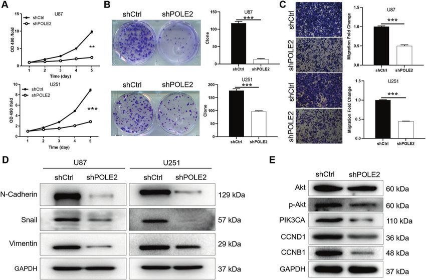
P. Zhang et al.
2
    data such as gender, age, pathological grade, and so on. All patients signed      Scientific, California, USA). The protein–protein interactions were analyzed
    informed consent to use clinical data for research purposes. Briefly,              as described in the literature [25].
    deparaffinize and rehydrate by immersing the tissue microarray through
    xylene and ethanol. After the tissue sections were washed twice with 1%
    animal serum (Thermo Fisher Scientific, California, USA, Cat. No.
                                                                                      MTT cell viability assay
    1921005PJ) in PBS with 0.4% Triton X-100 (PBS-T), the primary antibody            U87 and U251 cells were cultured in 96-well plates at 2000 cell/well density
    POLE2 (Table S1) diluted in 1% animal serum in PBS-T and incubate at              for 24 h. The 20 μL of 5 mg/mL MTT (Genview, Cat. No. JT343, Shanghai,
    room temperature for 2 h. Continue the incubation with secondary                  China) was added 4 h before the termination of the culture. After 4 h, the
    antibody overnight at 4 °C in a humidified chamber. The tissue sections            culture medium was completely absorbed and 100 μL of DMSO was added
    were stained with DAB solution and hematoxylin in turn. All tissue chips          to dissolve formazan granules. After 2–5 min of oscillation, OD value at
                                                                                      490/570 nm was detected by microplate reader (Cat. No. M2009PR, Tecan
    were photographed with microscopic, viewed with ImageScope and
                                                                                      infinite M200, Switzerland) for 5 days.
    CaseViewer. Notably, high or low expression of POLE2 was determined by
    the median of IHC scores of all tissues.
       The mice tumor tissues were fixed with 10% formalin, immersed in                Colony formation assay
    xylene and ethanol in turn. After tumor tissues were blocked with 3% PBS-         U87 and U251 cells were cultured in 96-well plates at 2000 cell/well density
    H2O2, incubated with anti-Ki67 and HRP goat anti-rabbit IgG (The detail           for 14 days. Subsequently, the cells were fixed with 1 mL 4%
    was listed in Table S1), respectively. Finally, slides were stained by            paraformaldehyde in each well for 60 min and washed with PBS. Next,
    Hematoxylin (Cat. No. 517-28-2, Sigma-Aldrich®, St. Louis, Missouri, USA)         cells were stained with GIEMSA 500 μL for 20 min, washed several times
    and Eosin (Cat. No. 548-24-3, Sigma-Aldrich®, St. Louis, Missouri, USA) as        with ddH2O and dried. Cell clones were photographed and counted under
    well as examined at ×200 objective lens microscopic.                              a fluorescence microscope.

    Cell culture                                                                      Transwell assay
    The U87 [23] and U251 [24] cell lines (Cell Bank of Chinese Academy of            After 18 h of U87 and U251 cells culture in 24-well plates at a density of
    Sciences, Shanghai, China) are commonly used as experimental models of            1 × 10⁵ cell/well, 100 μL of cell suspension was placed in each chamber
    GBM, which were tested for mycoplasma contamination. HEK 293T cells, is           (3422 corning). The 600 μL medium containing 30% FBS was added to the
    an adhesion-dependent epithelialize-like cell, often referred to as “tool         lower chamber, and 100 μL serum-free medium was added to the up
    cells”, used for lentivirus packaging production and titer determination, cell    chamber. After incubation for 2 h, the culture medium in the chamber was
    transfection. All the cells were cultivated in DMEM medium (Gibco, life           removed and transferred to the lower chamber. After 24 h of culture, the
    technologies, California, USA) supplemented with 10% fetal bovine serum           medium was removed by inverting the chamber on the absorbent paper,
    and 100 mg/mL streptomycin plus 100 UI/mL of penicillin (Gibco, life              and the non-metastatic cells were gently removed with a cotton swab. The
    technologies, California, USA) in the atmosphere of 5% CO2 at 37 °C.              400 μL staining solution was added to the 24-well plate for staining the
                                                                                      transferred cells for 20 min. Subsequently, the chamber was washed with
                                                                                      water for several times to dry. The 10% acetic acid was added to detect
    Lentiviral shRNA vector construction and cell infection
    Interfering sequences containing the target gene were synthesized using           OD570 and the film was photographed under a microscope.
    the POLE2, FOXM1, and AURKA sequence as template, which were directly
    connected to the lentiviral vector BR-V-108 (Bio Sci Res, Shanghai, China).       Wound-healing assay
    Meanwhile, the amplified sequence of POLE2 was linked to the lentiviral            U87 and U251 cells were cultured into 6-well plates (100 μL/well) at a
    vector. The lentiviral plasmid containing the target sequence was                 density of 4000 cells per well. The cells were eluted with PBS, fixed with
    transfected using Lipofectamine® 2000 (Invitrogen; Thermo Fisher Scien-           3.7% paraformaldehyde (Corning) for 15 min, stained with 1% crystal violet
    tific, California, USA) at a MOI (multiplicity of infection) of 10 to infect U87   for 10 min. Wound healing was observed at 0, 8, and 72 h under a
    and U251 cells, respectively. After 72 h, the expression of green fluorescent      microscope for image acquisition and Image J software (National Institutes
    protein (GFP) was observed under the fluorescence microscope (Cat. No.             of Health) was used to quantify the distance (μm) between the scratches.
    IX71, OLYMPUS, JPN).
                                                                                      Cell apoptosis analysis by Flow cytometry
    RNA isolation and qPCR                                                            U87 and U251 cells were cultured in 6-well plates at 2 mL/well for 5 days
    After the U87 and U251 cells were collected, they were cleaved by Trizol          and centrifuged. Then, the cell precipitation was washed successively with
    for total RNA extraction. The concentration and quality of the extracted          the 4°C pre-cooled D-Hanks (pH = 7.2–7.4) and 1×binding buffer. Annexin
    RNA were analyzed and determined by Nanodrop 2000/2000 C spectro-                 V-APC (cat. no. 88-8007-74, eBioscience, Thermo Fisher Scientific, California,
    photometer. The cDNA was obtained by reverse transcription using                  USA) 10 μL was added to stain cells at 37 °C in the dark for 10–15 min. The
    Promega M-MLV kit. The qPCR was accomplished with the SYBR Green PCR              number of apoptotic cells was detected using the FACSCanto II flow
    kit (Thermo Fisher Scientific, California, USA) and quantification was              cytometer (cat. no. Guava easyCyte HT, Millipore, Massachusetts, USA) after
    analyzed by the method of 2-ΔΔCq. Notably, the primers were listed in Table       adding 400-800 μL of 1×binding buffer.
    S2, where GAPDH as an internal control.
                                                                                      Human apoptosis antibody array
    Western blotting (WB) analysis                                                    After total proteins of U251 cells were extracted, quantified by BCA protein
    After total proteins of U87 and U251 cells were extracted, quantified by           assay kit (Cat. No. A53227, Thermo Fisher Scientific, California, USA). The
    BCA protein assay kit (Cat. No. A53227, Thermo Fisher Scientific, California,      protocol was operated according to the instruction of the human
    USA). CHX (0.2 mg/mL) refers to Cycloheximide blocking protein biosynth-          apoptosis antibody array membrane (Cat. No. ab134001, Abcam, Cam-
    esis to study the half-life of FOXM1 and AURKA proteins over time (0–8 h).        bridge, UK). Finally, the array membranes were exposed in the
    Same for the use of the proteasome inhibitor MG-132 (20 μM) to inquire            chemiluminescence imaging system.
    about the protein degradation. Equivalent amount of protein was
    separated through 10% SDS-polyacrylamide gel electrophoresis (SDS-
    PAGE) and transferred to polyvinylidene fluoride (PVDF) film at 4 °C. The           Xenograft mouse tumor model
    protein was incubated with primary antibody and secondary antibody                The animal experiments were in accordance with the Guide for Care and
    (antibody information was listed in Table S1) in turn at 4 °C for 3 h. The        Use of Laboratory animals (NIH publication number 85-23, revised at 1996)
    immune response was visualized with the Amersham ECL + plusTM                     and approved by the Ethics Committee of the First Affiliated Hospital of
    Western Blot system, and the blots were imaged by luminescent image               Zhengzhou University. Twenty 4-week-old female BALB/ C nude mice
    analyzer.                                                                         (Lingchang Biotechnology, Shanghai, China) were randomly divided into
                                                                                      two groups of shCtrl (negative control, n = 10) and shPOLE2 (POLE2
                                                                                      knockdown, n = 10). The U87 cells were prepared into cell suspension and
    Co-Immunoprecipitation (Co-IP) assay                                              subcutaneously injected with 200 μL at 4 × 10⁶ cells/mL to the right
    After the U251 cells were lysed, the protein was obtained and the                 forearm of each mouse. About a week later, the mice were anesthetized by
    concentration was measured by BCA protein detection kit (Thermo Fisher            intraperitoneal injection of 0.7% pentobarbital sodium at 10 μL/g and

                                                                                                             Cell Death and Disease (2022)13:61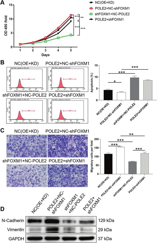
P. Zhang et al.
                                                                                                                                                      3
placed in the living imaging system (Cat. No. LB983, Berthold Technologies,
Germany) for imaging and fluorescence observation. Tumor size and mice
weight were measured every other day until 26 days after subcutaneous
injection. The mice were physically killed and the tumors were removed,
weighed and photographed.

Human Gene Chip
The U251 cells RNA with complete fragments and high purity was used for
the analysis of molecular mechanism through Affymetrix human Gene
Chip Prime View combined with Affymetrix Scanner 3000 scan (Affymetrix,
Santa Clara, CA, USA). The Human Gene Chip technology has been used to
detect differentially expressed genes (DEGs) in different groups of shPOLE2
and shCtrl in U251 cells. The volcano plot and hierarchical clustering of the
shPOLE2 and shCtrl in U251 cells were presented by the DEGs with
criterion of |Fold Change|≥1.3 and false discovery rate (FDR) ≤ 0.05.
Furthermore, the significant enrichment of DEGs in canonical pathways
as well as disease and function were explored based on Ingenuity Pathway
Analysis (IPA) (Qiagen, Hilden, Germany).

Statistical analysis
Statistical analyses were conducted by SPSS 19.0 with GraphPad Prism
8.0 software and data were expressed as the mean ± standard deviation.
The statistical significance between different groups was accomplished by
independent Student’s t test and P < 0.05 was considered statistically
significant. The correlation between POLE2 expression and clinic
characteristics of GBM was evaluated by Mann–Whitney U analysis.
Survival curves were obtained by the Kaplan–Meier method, and
differences in survival rates were assessed by the log-rank test. All
statistical tests were two-tailed and values of P < 0.05 were considered
statistically significant.

RESULTS
POLE2 is highly expressed in human GBM
Firstly, we found that the mRNA expression of POLE2 in tumor
samples (169 cases) was significantly higher compared with the
normal samples (5 cases) from the Cancer Genome Atlas
(TCGA) database (Fig. 1A). Based on the database, we further
analyzed the correlation analysis between POLE2 expression                      Fig. 1 POLE2 is highly expressed in human GBM. A The mRNA
                                                                                expression of POLE2 in GBM samples (169 cases) and normal
and the survival probability of patients with GBM. Perhaps due
                                                                                samples (5 cases) was compared from TCGA-GBM database. B The
to the sample size of the database, there is no significant                      representative picture of the expression level of POLE2 in normal
correlation between them (Fig. S1A). In order to further clarify                tissues and tumor tissues with different grades was detected by
the expression level of POLE2 in GBM, we performed IHC                          immunohistochemistry (IHC). The magnification is 200.
staining analysis in normal brain tissue [15] and tumor tissue
(165) in clinical GBM patients. According to the scores of IHC
staining, we defined POLE2 as a high expression if it is greater
than the median, otherwise as low expression. Consistently, the                  Table 1. Correlation analysis between POLE2 expression level and
results of IHC staining showed that the signal intensity of                      clinic characteristics of GBM.
POLE2 in GBM tissues was stronger than that in adjacent
normal tissues (Fig. 1B). The representative image of IHC                       Features          No. of patients       POLE2               P value
staining indicated that the higher the pathological grade, the                                                          expression
higher the expression of POLE2 in the tumor tissue. The
                                                                                                                        Low       High
relationships between POLE2 expression and clinic character-
istics of GBM were summarized in Table 1. A total of 75 (45.4%)                 All patients      165                   90        75
cases showed high levels of POLE2 expression (score 5–12). We                   Age                                                           0.057
further suggested that the POLE2 expression was positively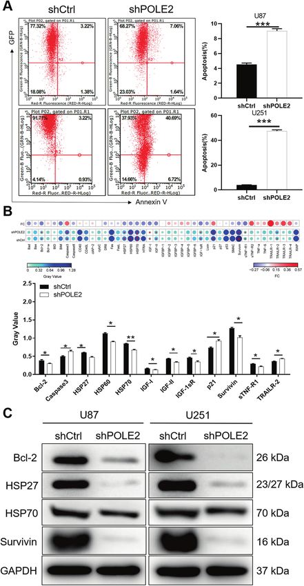
P. Zhang et al.
4

    Fig. 2 POLE2 knockdown inhibits the viability and migration of GBM cells in vitro. A MTT cell viability assay was employed to show the
    effects of POLE2 on U87 and U251 cells. B Colony-forming ability of U87 and U251 cells was detected in shPOLE2 and shCtrl groups. C The U87
    and U251 cell migration ability was accessed by Transwell assay. D The protein expression of N-cadherin, Vimentin, and snail of U87 and
    U251 cells was measured by WB. E The downstream protein expression of Akt, p-Akt, PIK3CA, CCND1, and CCNB1 of U251 cells was measured
    by WB. The representative images were selected from at least three independent experiments. Data were shown as mean ± SD. **P < 0.01,
    ***P < 0.001.

    than that in shCtrl group (Fig. S1C). Moreover, the WB results of      POLE2 knockdown enhances the apoptosis of GBM cells
    U87 and U251 cells indicated that POLE2 bands in shPOLE2 was           in vitro
    weaker than that in the control group (Fig. S1D). Therefore, POLE2     In addition, the cell apoptosis of U87 and U251 cells following
    was knocked down in U87 and U251 cells for the detection of cell       POLE2 knockdown was measured by flow cytometry. The results
    function. The results of MTT assay showed that the OD490 value         presented that the percentage of apoptosis was increased in
    of shPOLE2 group in U87 and U251 cells were lower than that of         shPOLE2 group compared with shCtrl group (P < 0.001), suggesting
    the control group, indicating that the downregulation of POLE2         that knockdown of POLE2 enhanced apoptosis of U87 and
    resulted in a significant decrease in cell viability (Fig. 2A).         U251 cells (Fig. 3A). Collectively, the results provided evidence
    Consistently, knockdown of POLE2 contributed to an obvious             that downregulation of POLE2 significantly induced the apoptosis
    decrease in the number of colonies in GBM cells (P < 0.001) (Fig.      of GBM cells, and played important roles in the regulation of cell
    2B). Transwell results further indicated that the cell migration       viability. To further explore the molecular mechanism underlying
    ability of shPOLE2 group was inhibited compared with shCtrl            cell apoptosis accelerated by POLE2 knockdown, we used the
    group (P < 0.001) (Fig. 2C). In view the fact that epithelial-         human apoptosis antibody array membrane to simultaneously
    mesenchymal transition (EMT) is a developmental procession that        detect the differential expression of 43 apoptosis-related proteins
    induces invasion and metastasis in various types of tumors [26].       in shCtrl and shPOLE2 groups. As illustrated in Fig. 3B, knockdown
    During EMT, N-cadherin, Snail, and Vimentin are the most               of POLE2 in U251 cells resulted in the abnormal expression of
    frequent detected epithelial and mesenchymal markers, respec-          apoptosis-related proteins in human apoptosis signal pathway.
    tively [27]. Thus, our results indicated that knockdown of POLE2       Specifically, the expression of Caspase3, p21, Tumor necrosis factor
    led to the downregulation of N-cadherin, Snail, and Vimentin of        (TNF)-related apoptosis-inducing ligand (TRAIL) receptors (TRAILR)-
    U87 and U251 cells (Fig. 2D). Furthermore, EMT process involves        2 was upregulated, whereas the expression of Bcl-2, heat shock
    multiple regulatory mechanisms, including phosphorylated Akt           protein 27 (HSP27), heat shock protein 60 (HSP60), heat shock
    serine/threonine kinase (p-Akt) activation and cyclin alteration       protein 70 (HSP70), insulin-like growth factor-I (IGF-I), insulin-like
    [28, 29]. The present study showed that knockdown of POLE2             growth factor-II (IGF-II), IGF system components (IGF-1sR), soluble
    resulted in the downregulation of p-Akt, PIK3CA, G1 cyclin D1          tumor necrosis factor receptor R1 (sTNF-R1) and Survivin was
    (CCND1), and cyclin B1 (CCNB1) (Fig. 2E). Collectively, knockdown      downregulated. Consistently, results of WB analysis further showed
    of POLE2 through EMT inhibited the malignant behaviors of GBM          that the protein levels of Bcl-2, HSP27/70, and Survivin were
    cells.                                                                 downregulated in GBM cells with POLE2 knockdown (Fig. 3C).

                                                                                                Cell Death and Disease (2022)13:61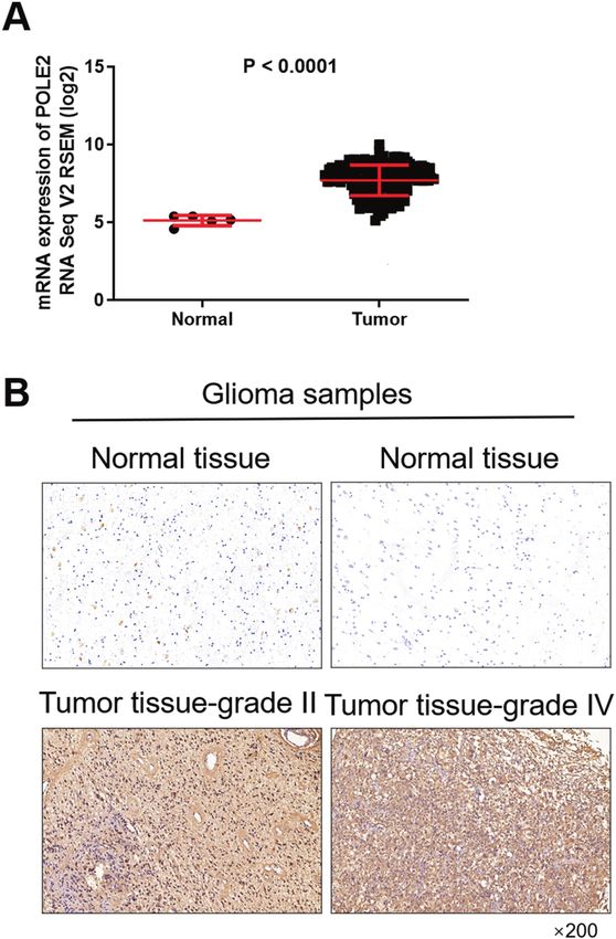
P. Zhang et al.
                                                                                                                                          5

Fig. 3 POLE2 knockdown enhances apoptosis of GBM cells in vitro. A Flow cytometry was performed to detect cell apoptosis of U87 and
U251 cells with or without POLE2 knockdown. The representative images were selected from at least three independent experiments.
B Human apoptosis antibody array was utilized to analyze the regulatory ability of POLE2 on expression of apoptosis-related proteins in
U251 cells. C The protein levels of Bcl-2, HSP27/70, and Survivin in U87 and U251 cells with or without POLE2 knockdown were further
analyzed by WB. Data were shown as mean ± SD. *P < 0.05, ***P < 0.001.

Cell Death and Disease (2022)13:61
P. Zhang et al.
6
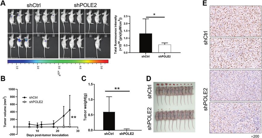
Fig. 4 POLE2 knockdown suppresses GBM growth in vivo. A In vivo imaging was performed to evaluate the tumor burden in mice of
shPOLE2 and shCtrl groups post tumor-inoculation B–C U87 cells with or without POLE2 knockdown, the volume (B) and weight (C) of tumors
formed in mice was measured and calculated at indicated time intervals. D photo of the removed tumors was taken post tumor-inoculation.
E The Ki67 level in tumors removed from mice was detected by IHC as a representation of tumor growth. Data were shown as mean ± SD.
*P < 0.05, **P < 0.01.
POLE2 knockdown suppresses GBM growth in vivo analysis based on the TCGA database shows that the mRNA
The xenograft mice tumor models were established to further expression level of AURKA and FOXM1 is abundantly expressed in
verify the role of POLE2 in regulating GBM cells in vivo. As GBM (Fig. S3A–B). Although there is no significant correlation
illustrated in Fig. 4A, the fluorescence intensity of tumors in between the expression level of AURKA and the survival of GBM
shPOLE2 group was obviously weaker than that of shCtrl, which patients, GBM patients with high AURKA expression have a high
preliminarily indicated that the downregulation of POLE2 could probability of short survival (Fig. S3C). Moreover, the relationship
reduce the ability of tumor formation. Furthermore, the tumor between the expression level of FOXM1 and the survival time of
volume of mice in the shPOLE2 group was remarkably smaller than GBM patients shows the same situation (Fig. S3D). Accordingly, we
that of mice in the shPOLE2 group after 26 days of observation hypothesized that POLE2 regulated GBM through AURKA-
(Fig. 4B). The comparison of tumor weight between shPOLE2 group mediated FOXM1 ubiquitination. In order to verify our hypothesis,
and shCtrl group showed that the reduction of POLE2 weakened we carried out the following experiment. After treatment of
tumor growth more intuitively (Fig. 4C), which can be observed protein synthesis inhibitor CHX (0.2 mg/mL), we examined the
from Fig. 4D. Besides, Ki67 is a well-known proliferation marker for protein stability of FOXM1 in U87 and U251 cells after POLE2
the evaluation of cell proliferation [30, 31]. IHC staining analysis of knockdown or AURKA knockdown, respectively (Fig. 5A, B). The
mice tumor tissues showed that the signal intensity of Ki67 in results showed that decreased expression of POLE2 led to the
shPOLE2 group was significantly weaker than that in control group weakening of FOXM1 protein stability in GBM cells (Fig. 5A).
(Fig. 4E), which further confirmed that POLE2 knockdown could Similarly, AURKA could affect the protein stability of FOXM1 (Fig.
inhibit the tumor formation. In view of the above results, 5B). Interestingly, the addition of proteasome inhibitor MG-132
downregulation of POLE2 might suppress tumor growth in vivo. (20 μM) partially eliminated the effect of POLE2 or AURKA
knockdown on FOXM1 protein stability in GBM cells (Fig. 5C, D),
POLE2 promotes AURKA-mediated FOXM1 de-ubiquitination indicating the involvement of proteasome in POLE2-induced
The potential molecular mechanism of POLE2 involved in GBM regulation of FOXM1. Subsequently, we evaluated the regulation
cells was preliminarily explored in this study. The downstream of POLE2 on FOXM1 ubiquitination, and the results showed that
molecular mechanism of POLE2 on the regulation of GBM cells knockdown of POLE2 significantly promoted FOXM1 ubiquitina-
was analyzed through human Gene Chip. The results showed that tion (Fig. 5E), thus decreasing FOXM1 stability. Considering that
knockdown of POLE2 resulted in upregulation of 1983 DEGs and previous study demonstrated that AURKA attenuated ubiquitina-
downregulation of 1383 DEGs (Fig. S2A). IPA-based analysis of the tion to stabilize FOXM1 [32], we further explored the interaction
disease and function (Fig. S2B) as well as canonical pathway (Fig. between POLE2 and AURKA (Fig. 5F). As expected, there was an
S2C) showed that these DEGs were enriched in signaling pathways interaction between AURKA and POLE2. Taken together, POLE2
associated with cell proliferation and death. In addition, the most may promote GBM through AURKA-mediated de-ubiquitination of
significant DEGs were selected by PCR (Fig. S2D) and verified by FOXM1.
WB (Fig. 2E). Aurora kinase A (AURKA) was preliminarily considered
as a downstream target of POLE2 in GBM cells. Downregulation of FOXM1 reverses the promotion of POLE2
In view the fact that AURKA could directly bind and attenuate on the malignant phenotype of GBM cells
ubiquitination of Forkhead Box M1 (FOXM1) [32], which has been To fully verify the effects of POLE2, AURKA, and FOXM1 in GBM, the
shown to promote the progression of GBM [33]. In addition, functional recovery assays was conducted. We used lentivirus
Cell Death and Disease (2022)13:61P. Zhang et al.
7
Fig. 5 POLE2 knockdown regulates AURKA-mediated FOXM1 ubiquitination. A–B The protein stability of FOXM1 in U87 and U251 cells
after POLE2 knockdown (A) or AURKA knockdown (B) was examined. C–D After treatment with MG-132, levels of FOXM1 proteins in U87 and
U251 cells with POLE2 (C) or AURKA (D) knockdown was examined. E The lysates of U87 and U251 cells were immunoprecipitated and WB was
performed to examine the ubiquitination of FOXM1. F Co-IP analysis of interaction of POLE2 and FOXM1 in U87 and U251 cells. The
representative images were selected from at least three independent experiments.
shFOXM1 or shAURKA to interfere with GBM cells, respectively, and On the other hand, FOXM1 was regarded as a downstream
constructed U251 cells with FOXM1 (Fig. S4A–D) and AURKA low target of POLE2 in the regulation of GBM. FOXM1 is a transcription
expression (Fig. S4E–H). The loss-of-function assays demonstrated that factor of the Forkhead box (Fox) protein superfamily [34]. FOXM1
knockdown of AURKA in U251 cells showed a significant inhibitory is an important component of a wide range of biological activities,
effect on the biological malignancies, which was exacerbated by the including maintenance of mitotic spindle integrity, regulation of
simultaneous downregulation of AURKA and POLE2 (Fig. S5A–E). cell cycle, angiogenesis, metastasis, apoptosis, DNA damage
Analogously, we established U251 cells overexpressing POLE2 repair, and tissue regeneration [35–37]. In addition, FOXM1 has
(POLE2 + NC-shFOXM1), simultaneously upregulating POLE2 and been identified as one of the most DEGs in most solid tumors [38].
downregulating FOXM1 (POLE2 + shFOXM1), respectively. Notably, Numerous evidences indicate that FOXM1 expression is increased
NC(OE + KD) was the cells transfected with empty plasmid, as in a variety of human cancers. Lee et al. clarified that dual
negative control; shFOXM1+NC-POLE2 was the cells transfected with inhibition of FOXM1 and its compensatory signaling pathway
lentivirus shFOXM1 and NC-POLE2 for downregulating FOXM1. decreased the survival of ovarian cancer cells [39]. In addition,
Knockdown of FOXM1 could inhibit biological behaviors of FOXM1 facilitates breast cancer cell stemness and migration [40].
U251 cells on slowing down of proliferation (P < 0.001) (Fig. 6A), Moreover, FOXM1 has been shown to promote the progression of
enhancement of apoptosis (P < 0.001) (Fig. 6B), and weakening of GBM [33]. Interestingly, FOXM1 can recruit AURKA as a cofactor to
migration (P < 0.001) (Fig. 6C) and EMT (Fig. 6D). As expected, activate FOXM1 target genes in a kinase-independent manner.
overexpression of POLE2 significantly promoted the malignant Besides, AURKA and FOXM1 inhibition by either genetic knock-
behavior of U251 cells, including increased proliferation, decreased down or pharmacologic inhibitors impair melanoma growth and
apoptosis rate, enhanced migration, and EMT (Fig. 6A–D). Further- survival [41]. AURKA and FOXM1 participate in a tightly coupled
more, we found that downregulation of FOXM1 could partially positive feedback loop to enhance the BCSC phenotype [42].
recover the promoting effects of POLE2 on GBM cells (Fig. 6A–D). However, the underlying mechanism of AURKA and FOXM1 in
GBM remains inclusive.
Previous study demonstrated that AURKA could directly bind
DISCUSSION and attenuate the ubiquitin of FOXM1 [32]. Ubiquitination is a
In recent years, various small molecules and signaling pathways widespread post-translational modification that mediates the
involved in the regulation of GBM biological behaviors have been localization, metabolism, function, regulation, and degradation
extensively investigated. Therefore, it is necessary to thoroughly of proteins in cells [43]. Moreover, ubiquitination plays a central
explore the molecular mechanism of GBM to identify more effective role in the onset of cancers and cardiovascular diseases [44].
molecular targets for GBM. In this study, the biological function of AURKA directly binds and attenuates the ubiquitin of FOXM1,
POLE2 in GBM was explored. A significant finding of this study was which enhances paclitaxel resistance in triple‐negative breast
the discovery of a promoting role of POLE2 in GBM. We found that cancer [32]. Here, we demonstrated that POLE2 regulated AURKA-
POLE2 was highly expressed in GBM. Furthermore, knockdown of mediated FOXM1 ubiquitination in GBM. Furthermore, down-
POLE2 could inhibit the biological behaviors of GBM in vitro and regulation of FOXM1 could partially reverse the promoting effect
in vivo. Specifically, CENPO knockdown inhibited cell proliferation, of POLE2 overexpression on GBM. In conclusion, POLE2 promoted
enhanced cell sensitivity, weakened migration, and EMT of U251 cells. the biological behaviors of GBM through promoting
Cell Death and Disease (2022)13:61P. Zhang et al.
8
Fig. 6 Downregulation of FOXM1 weakens the promoting effects of POLE2 on GBM. U251 cells were subjected to the detection of viability
(A), apoptosis (B), migration (C), EMT marker expression (D). Notably, NC(OE + KD) was the cells transfected with the empty plasmid, as
negative control; POLE2 + NC-shFOXM1 was the cells transfected with lentivirus POLE2 and NC-shFOXM1 for upregulating POLE2; shFOXM1
+NC-POLE2 was the cells transfected with lentivirus shFOXM1 and NC-POLE2 for downregulating FOXM1; POLE2 + shFOXM1 was the cells
transfected with lentivirus POLE2 and shFOXM1 for upregulating POLE2 and downregulating FOXM1. The representative images were
selected from at least three independent experiments. Data were shown as mean ± SD. *P < 0.05, **P < 0.01, ***P < 0.001.
AURKA-mediated stabilization of FOXM1, which may provide the knockdown could inhibit the malignant behaviors of GBM in vitro
theoretical basis of molecular therapy for GBM. and in vivo. POLE2 facilitated the biological behaviors of GBM
through promoting AURKA-mediated stabilization of FOXM1.
However, there were still some shortcomings in this study. It
CONCLUSION was worth mentioning that the clinical sample size was small and
A significant finding of this study was the discovery of a only had reference value. Secondly, the regulatory role of POLE2
promoting role of POLE2 in human GBM. We identified that and AURKA had not been clearly elucidated, which required
POLE2 was highly expressed in GBM. Knockdown of POLE2 further exploration.
Cell Death and Disease (2022)13:61P. Zhang et al.
9
DATA AVAILABILITY 28. Kishore C, Sundaram S, Karunagaran D. Vitamin K3 (menadione) suppresses
The data used and analyzed during the current study are available from the epithelial-mesenchymal-transition and Wnt signaling pathway in human color-
corresponding author on reasonable request. ectal cancer cells. Chem Biol Interact. 2019;309:108725.
29. Li B, Cheng J, Wang H, Zhao S, Zhu H, Li C, et al. CCNB1 affects cavernous sinus
invasion in pituitary adenomas through the epithelial-mesenchymal transition. J
REFERENCES Transl Med. 2019;17:336.
1. McFaline-Figueroa JR, Lee EQ. Brain tumors. Am J Med. 2018;131:874–82. 30. Yang C, Zhang J, Ding M, Xu K, Li L, Mao L, et al. Ki67 targeted strategies for
2. Chen R, Smith-Cohn M, Cohen AL, Colman H. Glioma subclassifications and their cancer therapy. Clin Transl Oncol. 2018;20:570–5.
clinical significance. Neurotherapeutics. 2017;14:284–97. 31. Menon SS, Guruvayoorappan C, Sakthivel KM, Rasmi RR. Ki-67 protein as a
3. Gusyatiner O, Hegi ME. Glioma epigenetics: from subclassification to novel tumour proliferation marker. Clin Chim Acta. 2019;491:39–45.
treatment options. Semin Cancer Biol. 2018;51:50–8. 32. Yang N, Wang C, Wang J, Wang Z, Huang D, Yan M, et al. Aurora kinase A
4. Bush NA, Chang SM, Berger MS. Current and future strategies for treatment of stabilizes FOXM1 to enhance paclitaxel resistance in triple-negative breast can-
glioma. Neurosurg Rev. 2017;40:1–14. cer. J Cell Mol Med. 2019;23:6442–53.
5. McKhann GM, Duffau H. Low-grade glioma: epidemiology, pathophysiology, 33. Zhang C, Han X, Xu X, Zhou Z, Chen X, Tang Y, et al. FoxM1 drives ADAM17/EGFR
clinical features, and treatment. Neurosurg Clin N. Am. 2019;30:xiii–xiv. activation loop to promote mesenchymal transition in glioblastoma. Cell Death
6. Xiong L, Wang F, Qi Xie X. Advanced treatment in high-grade gliomas. J BUON. Dis. 2018;9:469.
2019;24:424–30. 34. Clark KL, Halay ED, Lai E, Burley SK. Co-crystal structure of the HNF-3/fork head
7. Siegel RL, Miller KD, Jemal A. Cancer statistics, 2020. CA Cancer J Clin. 2020;70:7–30. DNA-recognition motif resembles histone H5. Nature. 1993;364:412–20.
8. Rajaratnam V, Islam MM, Yang M, Slaby R, Ramirez HM, Mirza SP. Glioblastoma: 35. Costa RH. FoxM1 dances with mitosis. Nat Cell Biol. 2005;7:108–10.
pathogenesis and current status of chemotherapy and other novel treatments. 36. Laoukili J, Kooistra MR, Bras A, Kauw J, Kerkhoven RM, Morrison A, et al. FoxM1 is
Cancers. 2020;12:937. required for execution of the mitotic programme and chromosome stability. Nat
9. Zhou Q, Effati R, Talvinen K, Pospiech H, Syvaoja JE, Collan Y. Genomic changes of Cell Biol. 2005;7:126–36.
the 55 kDa subunit of DNA polymerase epsilon in human breast cancer. Cancer 37. Wonsey DR, Follettie MT. Loss of the forkhead transcription factor FoxM1
Genomics Proteom. 2008;5:287–92. causes centrosome amplification and mitotic catastrophe. Cancer Res.
10. Foiani M, Marini F, Gamba D, Lucchini G, Plevani P. The B subunit of the DNA 2005;65:5181–9.
polymerase alpha-primase complex in Saccharomyces cerevisiae executes an 38. Okabe H, Satoh S, Kato T, Kitahara O, Yanagawa R, Yamaoka Y, et al. Genome-
essential function at the initial stage of DNA replication. Mol Cell Biol. 1994;14:923–33. wide analysis of gene expression in human hepatocellular carcinomas using
11. Loeb LA, Monnat RJ Jr. DNA polymerases and human disease. Nat Rev Genet. cDNA microarray: identification of genes involved in viral carcinogenesis and
2008;9:594–604. tumor progression. Cancer Res. 2001;61:2129–37.
12. Briggs S, Tomlinson I. Germline and somatic polymerase epsilon and delta 39. Lee DW, Lee W, Kwon M, Lee HN. Dual inhibition of FOXM1 and its compensatory
mutations define a new class of hypermutated colorectal and endometrial can- signaling pathway decreased the survival of ovarian cancer cells. Oncol Rep.
cers. J Pathol. 2013;230:148–53. 2021;45:390–400.
13. Yoshida R, Miyashita K, Inoue M, Shimamoto A, Yan Z, Egashira A, et al. Con- 40. Sun HL, Men JR, Liu HY, Liu MY, Zhang HS. FOXM1 facilitates breast cancer cell
current genetic alterations in DNA polymerase proofreading and mismatch repair stemness and migration in YAP1-dependent manner. Arch Biochem Biophys.
in human colorectal cancer. Eur J Hum Genet. 2011;19:320–5. 2020;685:108349.
14. Church DN, Briggs SE, Palles C, Domingo E, Kearsey SJ, Grimes JM, et al. DNA 41. Puig-Butille JA, Vinyals A, Ferreres JR, Aguilera P, Cabre E, Tell-Marti G, et al.
polymerase epsilon and delta exonuclease domain mutations in endometrial AURKA overexpression is driven by FOXM1 and MAPK/ERK activation in mela-
cancer. Hum Mol Genet. 2013;22:2820–8. noma cells harboring BRAF or NRAS mutations: impact on melanoma prognosis
15. Hartmann E, Fernandez V, Moreno V, Valls J, Hernandez L, Bosch F, et al. Five- and therapy. J Invest Dermatol. 2017;137:1297–310.
gene model to predict survival in mantle-cell lymphoma using frozen or formalin- 42. Yang N, Wang C, Wang Z, Zona S, Lin SX, Wang X, et al. FOXM1 recruits nuclear
fixed, paraffin-embedded tissue. J Clin Oncol. 2008;26:4966–72. Aurora kinase A to participate in a positive feedback loop essential for the self-
16. Liu D, Zhang XX, Xi BX, Wan DY, Li L, Zhou J, et al. Sine oculis homeobox homolog renewal of breast cancer stem cells. Oncogene. 2017;36:3428–40.
1 promotes DNA replication and cell proliferation in cervical cancer. Int J Oncol. 43. Nakamura N. Ubiquitin System. Int J Mol Sci. 2018;19:1080.
2014;45:1232–40. 44. Popovic D, Vucic D, Dikic I. Ubiquitination in disease pathogenesis and treatment.
17. Zekri AR, Hassan ZK, Bahnassy AA, Khaled HM, El-Rouby MN, Haggag RM, et al. Nat Med. 2014;20:1242–53.
Differentially expressed genes in metastatic advanced Egyptian bladder cancer.
Asian Pac J Cancer Prev. 2015;16:3543–9.
18. Li J, Wang J, Yu J, Zhao Y, Dong Y, Fan Y, et al. Knockdown of POLE2 expression ACKNOWLEDGEMENTS
suppresses lung adenocarcinoma cell malignant phenotypes in vitro. Oncol Rep. The author(s) received no specific funding for this work.
2018;40:2477–86.
19. Pearlman A, Rahman MT, Upadhyay K, Loke J, Ostrer H. Ectopic Otoconin 90
expression in triple negative breast cancer cell lines is associated with metastasis AUTHOR CONTRIBUTIONS
functions. PLoS ONE. 2019;14:e0211737. XC designed this program. PZ, LYZ, DC, YC, and ZQG operated the cell experiments,
20. Rogers RF, Walton MI, Cherry DL, Collins I, Clarke PA, Garrett MD, et al. CHK1 PZ performed animal experiments. JC and LYZ conducted the data collection and
inhibition is synthetically lethal with loss of B-family DNA polymerase function in analysis. PZ produced the manuscript which was checked and revised by XC. All the
human lung and colorectal cancer cells. Cancer Res. 2020;80:1735–47. authors have confirmed the submission of this manuscript.
21. Wu Z, Wang YM, Dai Y, Chen LA. POLE2 serves as a prognostic biomarker and is
associated with immune infiltration in squamous cell lung cancer. Med Sci Monit.
2020;26:e921430.
22. Zhu Y, Chen G, Song Y, Chen Z, Chen X. POLE2 knockdown reduce tumorigenesis ETHICS STATEMENT
in esophageal squamous cells. Cancer Cell Int. 2020;20:388. The study was approved by the Ethics Committee of the First Affiliated Hospital of
23. Beckman G, Beckman L, Ponten J, Westermark B. G-6-PD and PGM phenotypes of Zhengzhou University.
16 continuous human tumor cell lines. Evidence against cross-contamination and
contamination by HeLa cells. Hum Hered. 1971;21:238–41.
24. Ponten J, Macintyre EH. Long term culture of normal and neoplastic human glia. COMPETING INTERESTS
Acta Pathol Microbiol Scand. 1968;74:465–86. The authors declare no competing interests.
25. Lin JS, Lai EM. Protein-protein interactions: co-immunoprecipitation. Methods
Mol Biol. 2017;1615:211–9.
26. Li L, Li W. Epithelial-mesenchymal transition in human cancer: comprehensive ADDITIONAL INFORMATION
reprogramming of metabolism, epigenetics, and differentiation. Pharm Ther. Supplementary information The online version contains supplementary material
2015;150:33–46. available at https://doi.org/10.1038/s41419-021-04498-7.
27. Yeung KT, Yang J. Epithelial-mesenchymal transition in tumor metastasis. Mol
Oncol. 2017;11:28–39. Correspondence and requests for materials should be addressed to Xu Chen.
Cell Death and Disease (2022)13:61P. Zhang et al.
10
Reprints and permission information is available at http://www.nature.com/ appropriate credit to the original author(s) and the source, provide a link to the Creative
reprints Commons license, and indicate if changes were made. The images or other third party
material in this article are included in the article’s Creative Commons license, unless
Publisher’s note Springer Nature remains neutral with regard to jurisdictional claims indicated otherwise in a credit line to the material. If material is not included in the
in published maps and institutional affiliations. article’s Creative Commons license and your intended use is not permitted by statutory
regulation or exceeds the permitted use, you will need to obtain permission directly
from the copyright holder. To view a copy of this license, visit http://creativecommons.
org/licenses/by/4.0/.
Open Access This article is licensed under a Creative Commons
Attribution 4.0 International License, which permits use, sharing,
adaptation, distribution and reproduction in any medium or format, as long as you give © The Author(s) 2022
Cell Death and Disease (2022)13:61You can also read


