The effects of the general anesthetic sevoflurane on neurotransmission: an experimental and computational study - Nature
←
→
Page content transcription
If your browser does not render page correctly, please read the page content below
www.nature.com/scientificreports
OPEN The effects of the general
anesthetic sevoflurane
on neurotransmission:
an experimental
and computational study
Jonathan Mapelli1,2,6*, Daniela Gandolfi1,6, Enrico Giuliani3,6, Stefano Casali4, Luigi Congi1,
Alberto Barbieri3, Egidio D’Angelo4,5 & Albertino Bigiani1,2
The brain functions can be reversibly modulated by the action of general anesthetics. Despite a wide
number of pharmacological studies, an extensive analysis of the cellular determinants of anesthesia at
the microcircuits level is still missing. Here, by combining patch-clamp recordings and mathematical
modeling, we examined the impact of sevoflurane, a general anesthetic widely employed in the
clinical practice, on neuronal communication. The cerebellar microcircuit was used as a benchmark to
analyze the action mechanisms of sevoflurane while a biologically realistic mathematical model was
employed to explore at fine grain the molecular targets of anesthetic analyzing its impact on neuronal
activity. The sevoflurane altered neurotransmission by strongly increasing GABAergic inhibition while
decreasing glutamatergic NMDA activity. These changes caused a notable reduction of spike discharge
in cerebellar granule cells (GrCs) following repetitive activation by excitatory mossy fibers (mfs).
Unexpectedly, sevoflurane altered GrCs intrinsic excitability promoting action potential generation.
Computational modelling revealed that this effect was triggered by an acceleration of persistent
sodium current kinetics and by an increase in voltage dependent potassium current conductance.
The overall effect was a reduced variability of GrCs responses elicited by mfs supporting the idea that
sevoflurane shapes neuronal communication without silencing neural circuits.
The selective interaction between general anesthetics and membrane proteins modulates synaptic transmission,
membrane potential and signaling in n eurons1,2. It is well established that the action of general anesthetics is
characterized by a generalized neuronal hyperpolarization subtended by an increased inhibition or by a reduced
synaptic excitation1,2. Among anesthetics, halogenated molecules are the most widely employed in medical
practice. Nevertheless, their action mechanism is not yet fully understood, and their use is primarily governed
by empirical rules. These molecules are allosteric modulators of synaptic r eceptors3. It has been shown in fact
that they increase GABA-A and Glycine receptors a ctivity3 whereas they typically downregulate the activity of
cholinergic and NMDA-type glutamate receptors4. At the cellular level, halogenated anesthetics inhibit neuronal
voltage-gated potassium5 and sodium c hannels6,7 and potentiate two-pore domain potassium c hannels8. As a side
effect of their action, these anesthetics impair synaptic long-term potentiation hampering neuronal ability to store
information9. In epileptic patients these drugs increase seizure a ctivity10 and induce delirium and agitation during
the recovery phases11. At the integrative level, the disruption of the information transfer among brain areas is
supposed to be an essential step for the action of a nesthetics12. The anesthesia could act by reducing the number
of discriminable functional states in an integrated system as well as the complexity of the overall neural s tate13.
1
Department of Biomedical, Metabolic and Neural Sciences, University of Modena and Reggio Emilia, Sezione di
Fisiologia e Neuroscienze, via G. Campi 287, 41125 Modena, Italy. 2Center for Neuroscience and Neurotechnology,
University of Modena and Reggio Emilia, 41125 Modena, Italy. 3Department of Medical and Surgical Sciences
for Children and Adults, University of Modena and Reggio Emilia, 41125 Modena, Italy. 4Department of Brain
and Behavioral Sciences, University of Pavia, 27100 Pavia, Italy. 5Brain Connectivity Center, IRCCS Mondino
Foundation, 27100 Pavia, Italy. 6These authors contributed equally: Jonathan Mapelli, Daniela Gandolfi and Enrico
Giuliani. *email: jonathan.mapelli@unimore.it
Scientific Reports | (2021) 11:4335 | https://doi.org/10.1038/s41598-021-83714-y 1
Vol.:(0123456789)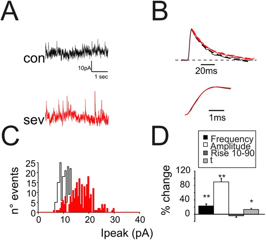
www.nature.com/scientificreports/
Although these findings represent the state of the art in the knowledge of the effects of anesthetics at integrative
level, a more detailed analysis of the changes in neuronal communication induced by anesthetics is still required.
The cerebellar cortical circuit is an ideal benchmark for the analysis of the effects of anesthetics on neuro-
transmission since GrCs show the unique characteristic among neurons of having a low number of dendrites (4.6
on average14), a very well detailed set of ionic channels and synaptic receptors, a compact electrotonic structure
allowing stable electrophysiological recordings and the development of reliable computational m odels15.
16
While the cerebellum has long been considered a marginal target for general a nesthetics , it is now known
that these cause (i) a reduction in PET and fMRI signals together with cortical and thalamic a reas17 which
participate with the cerebellum to peripheral sensory integration, (ii) a marked decrease of the frequency of
spontaneous activity of the cerebellar cortex18, (iii) the appearance of coherent oscillations in the cerebellar cortex
together with a decrease of the overall entropy of the s ystem19. Recently, the cerebellum was shown to participate
in cognitive20 and in sensory motor c ontrol21, furthermore, from consciousness to general anesthetics-induced
unconsciousness, rich-clubs of nodes in functional brain networks are switched from the high-order cognitive
function networks to sensory and cerebellum networks22. Compared with natural sleep, nodal efficiency of
cerebellum (among others) significantly decreased during propofol-induced unconsciousness and functional
connectivity between the cortex and subcortical centers (centralized in cerebellum) were significantly attenu-
ated under s edation23. Therefore, the cerebellum is now turning out to be specifically involved in the process of
consciousness regulation during anesthesia. The involvement of the cerebellum may reflect its strong connectivity
with prefrontal and frontal a reas20. The preferential reduction of low-frequency fluctuations in the anterior frontal
regions and cerebellum is consistent with frontal to sensory-motor cortical disconnection and may contribute to
the suppression of consciousness during general anesthesia24. Understanding the cellular mechanisms through
which the cerebellum is modulated by general anesthetics could thus help understanding the mechanisms of
induction and recovery from anesthesia.
The analysis of neurotransmission has been classically performed through experimental methods such as
electrophysiology25, molecular b iology26 and i maging27. More recently, the use of mathematical models to mimic
the activity of neuronal circuits is increasingly becoming an efficient tool to predict brain d ynamics28. Biologically
realistic models can faithfully reproduce the electrical behavior of single neurons and synapses embedded in
neural circuits performing computational t asks29. Despite the power of these methods, computational approaches
are mostly employed in the analysis of large-scale n etworks30 whereas simulations are rarely employed to dissect
microcircuits activity for pharmacological purposes.
Here, by using electrophysiological recordings and mathematical simulations, we investigated the cellular
mechanisms underlying the changes induced by sevoflurane on neurotransmission between mossy fibers and
granule cells at the input stage of the cerebellum.
Results
In the cerebellar cortex, information from mossy fibers (mf) activate granule cells (GrCs) and Golgi cells (GoCs)
through glutamatergic synapses. GoCs, which are also excited by feedback loops from GrCs, inhibit the same
GrCs through GABAergic synapses (Fig. 1A). A similar circuit architecture with a functional organization
composed by reciprocal excitatory and inhibitory connections can be found in various central and peripheral
neural circuits31. We have employed the cerebellar micro-circuitry as an experimental model of the information
processing in the CNS to investigate the impact of sevoflurane on neurotransmission.
Sevoflurane inhibits excitatory neurotransmission on granule cells. Excitatory Post-Synaptic
Currents (EPSCs) were recorded from GrCs voltage clamped at -− 70 mV in response to mf bundle stimulation
(Fig. 1A). The activation of inhibitory loops through Golgi cells did not produce significant inhibitory cur-
rents because the reversal potential of chloride was about − 60 mV32,33. In response to a 4-pulse, 100-Hz burst
(Fig. 1B), EPSCs showed the typical short-term depression pattern34, composed of a rapid AMPA and a slow
NMDA component. The application of sevoflurane did not significantly affect EPSCs peak amplitudes (1st peak
change + 3.5 ± 1.8%, p > 0.35; n = 5, Fig. 1B). This result is in accordance with our recent findings on desflurane, a
chemical compound of the same family of sevoflurane, showing that glutamate AMPA receptors are not targeted
by the anesthetic33. When membrane potential is lower than − 40 mV, the voltage-dependent magnesium block
is expected to dampen NMDA currents. However, a residual component of this glutamatergic current can be
effectively detected at more hyperpolarized values34. This late component is unmasked by measuring the amount
of excitatory currents 50 ms after the end of the stimulation pattern (Fig. 1B top, box) and sevoflurane indeed
reduced the residual NMDA component of NMDA current (− 27.8 ± 2.3%, p < 10–5; n = 9, Fig. 1B). In order to
further isolate NMDA currents, GrCs were voltage clamped at − 40 mV and the Mg2+ was removed. In the
presence of both AMPA (NBQX) and GABA-A receptor (Gabazine) blockers (Fig. 1B, bottom traces) currents
showed a marked temporal summation peaking in about 20 ms after the last stimulus (− 2.6 ± 0.2 pA n = 8; at
the peak of the current). This effect was completely abolished by the application of sevoflurane (− 95.5 ± 2.1%
n = 8; p < 10–5) supporting the evidence that NMDA channels are indeed targeted by the anesthetic4. In addition,
sevoflurane rapidly and transiently reduced Excitatory Post-Synaptic Potentials (EPSPs) both in peak amplitude
(− 33.9.1 ± 3.5%, p < 0.001, n = 7, Fig. 1C) and in total depolarization (EPSP area − 45.4 ± 7.2%, p < 0.01 n = 7,
Fig. 1C). The NMDA current is known to favor the temporal summation of concomitant inputs leading to a
sustained membrane depolarization. We therefore evaluated the impact of NMDA blocking induced by sevo-
flurane onto the generation of EPSPs (Fig. 1C, lower traces) in the presence of gabazine. Unexpectedly, peak
amplitude (− 6.9 ± 2.3%, p < 0.05, n = 4, Fig. 1D) and EPSP area (− 5.1 ± 0.9%, p < 0.05, n = 4, Fig. 1D) were only
slightly reduced indicating that, although the excitatory neurotransmission was affected by the block of the
NMDA current, the anesthetic mostly impacted the inhibitory component of neurotransmission. Interestingly,
Scientific Reports | (2021) 11:4335 | https://doi.org/10.1038/s41598-021-83714-y 2
Vol:.(1234567890)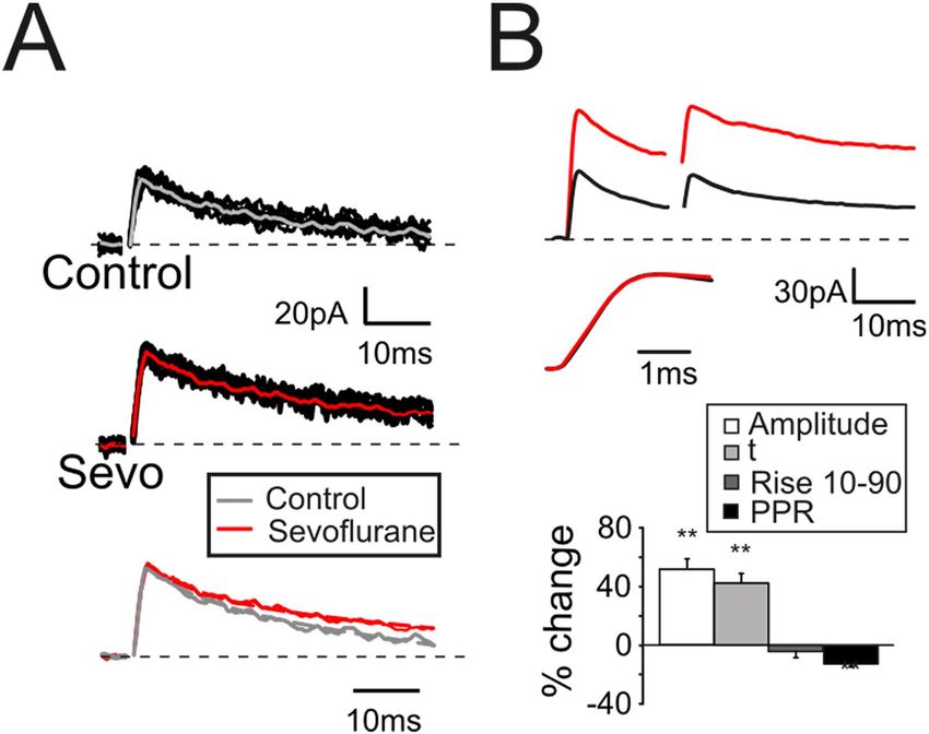
www.nature.com/scientificreports/

                                  Figure 1.  Modulation of excitatory neurotransmission by sevoflurane. (A) Scheme of the granular layer
                                  microcircuit. The stimulating electrode (stim) is positioned onto the mossy fiber bundle (mf) in order to
                                  activate excitatory synapses. GoC, Golgi cell (local interneuron); GrC, granule cell (output cell). (B) Top, EPSCs
                                  elicited in response to 4 pulses at 100 Hz and recorded from a GrC voltage clamped at − 70 mV (n = 9). Note the
                                  effects on the residual current induced by sevoflurane (gray trace, box). Bottom. In a different cell EPSCs were
                                  evoked from a GrCs voltage clamped at − 40 mV and in the presence of gabazine and NBQX (n = 8). Sevoflurane
                                  completely abolished the NMDA current. (C) EPSPs elicited by sub-threshold stimuli in GrCs at – 60 mV (10
                                  superimposed traces) in control conditions (left) and in the presence of sevoflurane (middle). The average traces
                                  (right) show the marked decrease of EPSP amplitude and area induced by sevoflurane (gray trace). Histogram
                                  summarizes the effects induced by sevoflurane on EPSP rise time, amplitude, and area (n = 7). In this and in
                                  the following figures: *p < 0.05; **p < 0.01. (D) EPSPs were recorded from GrCs at – 60 mV (10 superimposed
                                  traces) and in the presence of gabazine (top traces) to unmask the NMDA component (n = 4) in control (left)
                                  and in the presence of sevoflurane (middle). The average traces (right) show that sevoflurane decreases both
                                  peak amplitude and EPSP area. Histogram summarizes the effects induced by sevoflurane on EPSP rise time,
                                  amplitude, and area.

                                  in both cases, we observed an acceleration of the rise time of EPSP indicating changes in the overall kinetics of
                                  the synaptic machinery (Fig. 1D, histograms).

Scientific Reports |   (2021) 11:4335 |                https://doi.org/10.1038/s41598-021-83714-y                                                  3

                                                                                                                                             Vol.:(0123456789)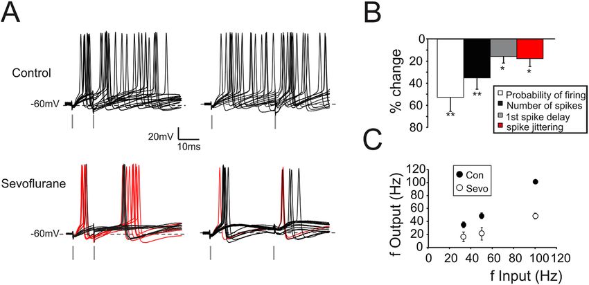
www.nature.com/scientificreports/
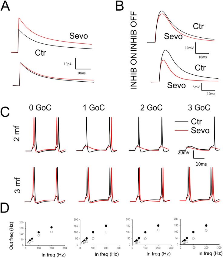
Figure 2. Modulation of GrCs firing activity by sevoflurane. (A) Top. Spikes from GrC elicited in response to
a pair of stimuli at 100 Hz (left) and 30 Hz (right) (15 superimposed traces). Bottom. Sevoflurane reduces the
total number of spikes and the probability of firing in response to the first stimulus. Note that the first spike is
anticipated and elicited with a less variable delay. Red traces show responses in which stimuli elicited two spikes.
(B) Histogram summarizes the effects induced by sevoflurane on spike related parameters (n = 7). (C) The plot
shows the relationship between input frequency and output frequency in control (Con) and during sevoflurane
perfusion (Sevo) (n = 7).
One of the mathematical relationships that better describes neurotransmission is that between input and
output variables (I/O). In response to pairs of action potentials delivered by mf at variable f requencies32, GrCs
responded with two or more spikes (Fig. 2A,B) with a quasi-linear relationship (Fig. 2C). The presence of sevo-
flurane profoundly altered the GrCs I/O by reducing the probability of eliciting spikes (− 52.8 ± 14.3%, p < 0.01;
n = 7; Fig. 2C), as well as the total number of emitted spikes (− 35.2 ± 9.1%, p < 0.01; n = 7; Fig. 2C). Furthermore,
GrCs mostly responded to low frequency inputs with EPSPs or at most single spikes (44 ± 2.9% singlet; 54.3 ± 3.4%
EPSPs over total responses at 33 Hz; Fig. 2B bottom, black traces n = 7). Only in few cases, doublets of action
potentials were generated (2.9 ± 1.8% doublets over total responses at 33 Hz; Fig. 2B bottom, red traces n = 7)
while the average firing frequency was markedly reduced (− 58.6 ± 12.2% at 100 Hz, p < 0.01; n = 7; Fig. 2A–C).
The overall result was a downward shift of the frequency dependence curve in accordance with the reduction
of NMDA currents and with the increased GABAergic inhibition. Surprisingly, the first spike delay and its vari-
ability were reduced (delay: − 15.7 ± 5.9%, p < 0.05; n = 7; Fig. 2C; variability: − 17.7 ± 8.1%, p < 0.05; n = 7; Fig. 2C).
Sevoflurane potentiates GABAergic neurotransmission on granule cells. The large majority of
anesthetics, including halogenated ones affects neurotransmission by potentiating GABAergic c urrents3,33. We
therefore evaluated the effect of sevoflurane on the GABAergic synapse whose activity was monitored by voltage
clamping GrCs at 0 mV. The GABAergic currents were identified as positive current d eflections32,33 elicited by
the direct stimulation of GoC axonal plexus (Fig. SM-1A). Inhibitory currents were pharmacologically isolated
by adding 20 μM NBQX and 50 μM D-APV to the extracellular solution to block excitatory neurotransmission
and preventing the activation of poly-synaptic pathways (see Fig. SM-1A). Spontaneous Inhibitory Post-Synap-
tic Currents (sIPSCs), which were present in almost all recordings (13/14 cells) occurred at an average frequency
of 3.1 ± 0.6 Hz (Fig. SM-1B, n = 13). Stimulation of GoC axons with two pulses at 50 Hz (see Materials and
Methods) elicited pairs of evoked Inhibitory Post-Synaptic Currents (eIPSCs; Fig. SM-1C) that were abolished
together with sIPSCs following the perfusion of 10 μM gabazine (Fig. SM-1C inset; n = 5). These results also con-
firmed the absence of slow GABA-B receptor-mediated responses in granule cell inhibitory currents. The time
courses of sIPSCs and eIPSCs shared similar kinetics both for the current rise (sIPSC r ise10–90 1.39 ± 0.26 ms,
n = 13; eIPSCs rise10–90 1.45 ± 0.46 ms, n = 13) and for the decay component (sIPSCs τ = 17.8 ± 4.7 ms; n = 13
Fig. SM-1D black trace; eIPSCs τ = 18.3 ± 4.1 ms; n = 13, Fig. SM-1D gray trace), confirming that the stimula-
tion protocol elicited currents similar to the ones spontaneously evoked by Golgi cells. A sustained slow decay
component could be also observed in eIPSCs, consistently with an indirect receptors’ activation through GABA
spillover into the cerebellar g lomerulus35 and this slow current was further unmasked by repetitive stimulation
(Fig. SM-1D, red trace).
In contrast with the effect induced by d esflurane33, sevoflurane altered spontaneous IPSCs by increasing
frequency (+ 25.5 ± 4.9%, n = 7; p < 0.01; Fig. 3A,D) and peak amplitude (+ 87.5 ± 9.6%, n = 7; p < 0.01, Fig. 3A,D).
Furthermore, the presence of anesthetic did not significantly change sIPSC rise time ( rise10–90; − 3.9 ± 2.7%, n = 7;
p > 0.35, Fig. 3B bottom traces, Fig. 3D) while slowed down the current decay (τ = 15.4 ± 4.9% n = 7; p < 0.05
Fig. 3B top traces, Fig. 3D) indicating that the anesthetic modified the GABAergic synaptic complex.
Scientific Reports | (2021) 11:4335 | https://doi.org/10.1038/s41598-021-83714-y 4
Vol:.(1234567890)www.nature.com/scientificreports/
Figure 3. Modulation by sevoflurane of spontaneous inhibitory synaptic currents. (A) sIPSCs recorded from a
granule cell before (black) and after (red) the application of sevoflurane. Note the increased frequency and peak
amplitude. (B) Normalized sIPSCs recorded from a granule cell before (black) and during (red) sevoflurane
application. The mono-exponential fitting (dashed lines) of the current relaxation reveals small changes in decay.
Bottom: rise time of the sIPCSs shown in the upper panel. (C) Distribution of sIPSCs peak amplitudes detected
during a recording of 3 min in control condition (black histogram) and in the presence of sevoflurane (red). (D)
Histogram shows the effects induced by sevoflurane on sIPCS biophysical properties (n = 13).
The analysis of eIPSCs confirmed that sevoflurane, according to the observations for sIPSCs, increased both
peak amplitude (+ 47.6 ± 7.1%, n = 11; p < 10–4, Fig. 4A,B top traces and histogram) and decay time course (τ
= + 43.8 ± 6.5%, n = 11; p < 10–3, Fig. 4A bottom traces and Fig. 4B histogram) while the rise time was unaffected
(rise10–90; − 3.9 ± 2.5%, n = 11; p > 0.4, Fig. 4B middle traces). Finally, together with the increase in sIPSC fre-
quency and peak amplitude, the reduction of the eIPSC PPR (− 12.7 ± 2.3%, n = 11; p < 10–3, Fig. 4B top traces
and histogram) suggested a modification in the presynaptic release machinery. As a whole, these results indicated
that sevoflurane induced an increase in vesicle release probability as well as a change in post-synaptic receptor
activity by increasing the total transferred charge evaluated as IPSC area (+ 109.1 ± 17.9%, n = 11; p < 10–3, data
not shown).
Sevoflurane increases intrinsic excitability of granule cells. The neuronal firing is primarily
dependent on the ratio between excitatory and inhibitory input but is also tightly bound to ionic mechanisms
bringing membrane potential to spike threshold. We have investigated the role of sevoflurane on GrC intrinsic
excitability by collecting GrCs voltage responses to current injections in current clamp configuration, in the
presence of 50 mM D-APV and 20 mM NBQX to block the contribution of glutamatergic afferences and 20 mM
gabazine to discard the contribution of GABergic inhibition. The zero-current potential, which can give an esti-
mate of resting membrane potential in patch-clamp experiments36, was monitored throughout the recordings:
no significant variations could be observed during sevoflurane perfusion (Table SM-1).
In response to depolarizing current injection, GrCs generated repetitive spike discharges (Fig. 5A, Control).
During sevoflurane perfusion, the current needed to generate action potentials was significantly reduced (from
5.9 ± 0.8 pA in control, to 4.1 ± 0.8 pA with sevoflurane, p < 0.01 n = 7; Fig. 5C) by virtue of a spike threshold
decrease (− 46.5 ± 2.9 mV in control and − 56.3 ± 3.1 mV in sevoflurane, n = 7 p < 0.01; Fig. 5C). Moreover, an
increased number of emitted spikes (+ 68.3 ± 15.9%, p < 0.01, n = 7; not shown in the histogram) together with
enhanced average (Fig. 5A; + 25.2 ± 7.1%, p < 0.05, n = 7; not shown in the histogram) and instantaneous firing
frequency (Fig. 5A; + 46.7 ± 6.8%, p < 0.01, n = 7; not shown in the histogram) supported the idea that sevoflurane
Scientific Reports | (2021) 11:4335 | https://doi.org/10.1038/s41598-021-83714-y 5
Vol.:(0123456789)www.nature.com/scientificreports/
Figure 4. Modulation by sevoflurane of evoked inhibitory synaptic currents. (A) eIPSCs elicited by a single
stimulus and recorded from a GrC in control conditions (top 20 superimposed traces) and during the
application of sevoflurane (middle 20 superimposed traces). Note the increased peak amplitude and the slower
decay. Bottom: normalized averaged eIPSCS. (B) eIPSCs elicited by a pair of stimuli at 50 Hz and recorded from
a GrC before (black) and during (red) the application of sevoflurane. Middle traces: rise time of the eIPSCs
shown in the upper panel. Histogram summarizes the effects induced by sevoflurane on the eIPSCS biophysical
properties (n = 11).
could alter the GrC intrinsic excitability, despite leaving unaffected spike waveform (Fig. 5B). These findings are
summarized by the plot representing the relationship between the injected current and the number of emitted
spikes and the average firing frequency (Fig. 5D,E).
An important aspect of the action of anesthetics is the kinetics which heavily impacts the recovery processes.
We have therefore investigated the time courses of the action of sevoflurane by eliciting GABAergic or NMDA
currents every 10 s and monitoring currents properties during sevoflurane perfusion and during the subsequent
wash-out (Fig. 6). On average, GABAergic currents started to increase around 30 s after the beginning of the
perfusion (Fig. 6A, left; n = 4) and reached the steady state level in about 100 s (Fig. 6A, left; n = 4). In response
to the anesthetic wash out, GABAergic currents started to decrease after about 50 s (Fig. 6A, right; n = 4) while
the initial conditions were restored after 150 s. Similarly, the number of spikes generated by GrCs in response to
current injection was monitored throughout perfusion and washout procedures (Fig. 6B). The plateau of varia-
tion, which started in less than 50 s, was reached in about 100 s (Fig. 6B left, n = 4), whereas the recovery phase
started 50 s after the beginning of wash out and terminated after 100 s (Fig. 6B right, n = 4).
Conversely, NMDA peak currents showed slower kinetics both for the perfusion (50 s for the initial decrease
and more than 130 s to steady state level, Fig. 6C left; n = 4) and wash out phase (100 s to start the recovery ad
250 s to restore the initial conditions, Fig. 6C right, n = 4).
Modeling the effect of sevoflurane on neurotransmission. In order to understand the mechanisms
underlying the changes in the intrinsic excitability caused by sevoflurane, we have employed a mathemati-
cal model of the GrC derived from previous versions37,38 that incorporates a detailed representation of all the
expressed ionic conductance (see Materials and Methods). According to experimental observations, the simula-
tions showed that GrCs responded to injected depolarizing current with repetitive spike discharges arising at
− 47 mV.
The altered excitability observed in the presence of sevoflurane was mimicked by analyzing the impact of the
ionic channels that are mainly involved in the regulation of spike threshold (Nav, Kv and leakage). Preliminarily,
the contribution of all other channels which are known to be expressed in cerebellar GrCs and are incorporated
in the model37,38 have been explored and none of the tested conditions could reproduce the experimental obser-
vations (data not shown).
Scientific Reports | (2021) 11:4335 | https://doi.org/10.1038/s41598-021-83714-y 6
Vol:.(1234567890)www.nature.com/scientificreports/
Figure 5. Sevoflurane increases GrCs intrinsic excitability. (A) GrC voltage responses to current injections
(bottom traces 1 pA/step) in control conditions, during sevoflurane perfusion and following wash-out of the
anesthetic. Note, during sevoflurane, less injected current is needed to generate action potentials, the number
of elicited spikes is increased, the firing threshold is lowered (arrow) and firing discharge becomes regular. (B)
Comparison of action potential waveform obtained in control (black), in the presence sevoflurane (red trace)
and following wash-out (gray trace). Note that sevoflurane did not affect the spike shape. (C) Histogram shows
the changes induced by sevoflurane on the current needed to bring GrCs to the firing zone (current inj), spike
threshold (spike thr), spike after hyperpolarization (spike AHP) and spike half-width (spike HW) (n = 7). (D)
Relationship between the injected current and the total number of emitted spikes (n = 7 cells). E Relationship
between the injected current and the average firing frequency of the emitted spikes (n = 7 cells).
The altered excitability observed in the presence of sevoflurane was therefore modelled by changing the dif-
ferent components of the voltage dependent sodium channels (see Materials and Methods), which are known
to be involved in the modulation of spike threshold. The increase of both activation and deactivation kinetics
of the persistent component of Na+ current—Nap—(Aon from 0.75 to 1.5 ms and Aoff from 0.005 to 0.05 ms,
Scientific Reports | (2021) 11:4335 | https://doi.org/10.1038/s41598-021-83714-y 7
Vol.:(0123456789)www.nature.com/scientificreports/
Figure 6. Time courses of the effect of sevoflurane. (A) Left; Time courses of the effect of sevoflurane (1st
vertical dashed line 300 s) and subsequent wash out (2nd dashed line 600 s) on eIPSCs peak amplitude changes.
Right. Inset of the panel shown on the left. Note that the return to the initial level starts 50 s after the beginning
of washout and the steady state is obtained in about 100 s. (B) Left Time courses of the effect of sevoflurane on
the number of spikes evoked in response to a 500 ms 5 pA depolarizing step current. Right. Inset of the panel
shown on the left. Note that the return to the initial level starts about 40 s after the beginning of washout and
the steady state is obtained in about 100 s. (C) Left Time courses of the effect of sevoflurane on the NMDA peak
current changes. Right. Inset of the panel shown on the left. Note that the return to the initial level starts about
100 s after the beginning of washout and the steady state is obtained in about 200 s.
Fig. 7A) allowed to lower the GrC firing threshold (from − 47 to − 54.8 mV, Fig. 7A). Furthermore, by measur-
ing the voltage response to current injection (Fig. 7C), the model reliably reproduced the responses obtained
experimentally (cfr Fig. 5E). However, the analysis of I/O curve, in terms of number of spikes (Fig. 7B) and aver-
age firing frequency (Fig. 7C), revealed that changes of Nap kinetics could not explain the overall GrC behavior
observed experimentally. In particular, while GrCs tended to show a linear increase in the difference between
the number of spikes in control and in the presence of sevoflurane at increasing current injections (Fig. 7C),
the sole modifies in Nap kinetics induced a saturation of the difference between curves at large current injec-
tion (Fig. 7C, gray circles). Conversely, by increasing the overall conductance of voltage dependent Na current
(0.03 S/cm2 to 0.04 S/cm2 in the hillock and 0.02 S/cm2 to 0.03 S/cm2 in the axon) the spike discharge behavior
approached the one observed experimentally (Fig. 7A,C). Finally, the changes induced in the spike overshoot
and after hyperpolarization were compensated by increasing the overall potassium conductance (from 0.003 to
0.005 S/cm2 both in the hillock and in the axonal compartments).
The effects of sevoflurane on cerebellar neurotransmission were further explored by incorporating changes
in conductance into the GrC model and investigating the synaptic parameters space. The GABAergic currents
Scientific Reports | (2021) 11:4335 | https://doi.org/10.1038/s41598-021-83714-y 8
Vol:.(1234567890)www.nature.com/scientificreports/
Figure 7. Simulation of GrCs intrinsic excitability. (A) Simulated GrC voltage responses to current injections (1
pA/step) in control conditions (left panel) and mimicking the presence of sevoflurane (right panel) by increasing
the persistent sodium conductance (Nap) and voltage dependent potassium conductance (Kv). Note that, as
observed experimentally, less injected current is needed to generate action potentials with sevoflurane. Also
note the increased number of elicited spikes and the reduced firing threshold in the presence of sevoflurane.
(B) The plot shows the number of spikes generated in response to current injection in control, when Nap is
increased, and when both Nap and Kv are increased. Note that simulations with increased Nap tend to saturate
more rapidly than those obtained when both Nap and Kv are increased. (C) The plot shows the relationship
between the current injected and the average firing frequency in control condition and when both Nap and Kv
are increased.
were reproduced by simulating the intracellular recordings of chloride currents in GrC at a holding potential of
− 60 mV activated by a single GoC (Fig. 8A). The changes observed experimentally (see Figs. 3, 4, 5) were reliably
reproduced by modifying the parameters accounting for both pre- and post-synaptic activity (see Table SM-2).
Given these changes on the inhibitory synapses, we have simulated the effects of sevoflurane on neurotrans-
mission (Fig. 8A,B) by removing NMDA conductance, potentiating GABAergic currents and altering the GrC
intrinsic excitability in a simplified version of the cerebellar microcircuit. According to anatomical fi ndings39,
a reduced version of the granular layer circuitry was assembled by connecting the GrC with a variable number
of excitatory mfs connections and Golgi Cells (from 1 to 4 and from 0 to 7 respectively40). Similarly, to experi-
mental observations, IPSCs evoked by single stimuli were increased in peak amplitude (+ 46.3%) and in the total
transferred charge (+ 99.6%) by GABAergic currents (Fig. 8A. Furthermore, we simulated the generation of
EPSPs in the presence and without the activity of inhibitory currents. Also, simulations reliably reproduced the
behavior observed experimentally. The EPSPs were in fact markedly decreased in peak amplitude (− 32.4%) and
total depolarization (− 57.1%) when GABAergic synapses were active while the effect of sevoflurane was barely
measurable when inhibition was disactivated (− 8.9% peak amplitude, − 10.2% total depolarization, Fig. 8B).
Unlike experimental conditions, where the number of excitatory and inhibitory afferences can only be tenta-
tively estimated, we have explored different combinations of mfs and GoCs to evaluate the parameters affecting
neurotransmission, such as first spike delay, number of spikes and average firing frequency. In response to pairs
of action potentials elicited at variable frequency (from 10 to 200 Hz), simulation showed a rather homogeneous
behavior independently from the excitatory, inhibitory (E/I) balance (black trace Fig. 8C). Of note, in case of null
inhibition the first spike delay was anticipated, and the I/O frequency curve was poorly affected by sevoflurane
(Fig. 8D). Conversely, similarly to experimental observations, the first spike delay was prolonged by sevoflurane
proportionally to the number of active GoCs (Fig. 8C), while I/O frequency curve showed a linear tendency
Scientific Reports | (2021) 11:4335 | https://doi.org/10.1038/s41598-021-83714-y 9
Vol.:(0123456789)www.nature.com/scientificreports/
Figure 8. Simulation of synaptic activity. (A) (Top) IPSCs generated by a single stimulus in control (black)
and mimicking the presence of sevoflurane (red). (Bottom) Normalized IPSCs show an increase in the current
tail resulting from changing postsynaptic parameters. (B) (Top) EPSPs generated by activating a single mossy
fiber in control condition (black) and mimicking the presence of sevoflurane (red). Note the reduction of the
late EPSP phase caused by NMDA receptor block. (Bottom) EPSPs generated by activating three mfs and a
single GoC in control condition and mimicking the presence of sevoflurane. (C) The effect of sevoflurane on
GrCs is shown in terms of spike discharging at different E/I combinations in control (black) and in the presence
of sevoflurane (red). Note that in the presence of active GoCs, sevoflurane decreases the number of spikes
and posticipates spike generation. Conversely spikes are anticipated in case of null inhibition. (D) I/O curves
referred to responses obtained with 3 mf and a variable number of GoCs (from 0 to 3) were obtained changing
the stimulation frequency from 10 to 200 Hz simulating control conditions (black circles) and mimicking the
presence of sevoflurane (white circles).
with a slope strongly decreased by sevoflurane. Finally, the number of total emitted spikes in response to pairs of
stimuli was significantly reduced in all the combination of E/I balance where there was at least one active GoC
Scientific Reports | (2021) 11:4335 | https://doi.org/10.1038/s41598-021-83714-y 10
Vol:.(1234567890)www.nature.com/scientificreports/
(data not shown). Interestingly, by taking into account the number of emitted spikes, sevoflurane reduced the
total amount of emitted spikes independently from the number of inhibitory inputs (Fig. 8C).
Discussion
In this work we have analyzed the impact of sevoflurane, a general anesthetic widely employed in the clinical
practice, on neurotransmission properties in a reduced model of brain circuit, the cerebellar cortical microcir-
cuitry. By combining experimental observations with simulations made through a biologically realistic math-
ematical model, we have dissected the cellular and molecular determinants of the effect of the anesthetic.
Several studies have shown that volatile anesthetics interfere with GABA-mediated synaptic machinery3,4 by
acting primarily on the postsynaptic side. Sevoflurane increases the total charge transfer through the reduction of
peak amplitudes and by slowing current d ecay41,42. Notably, in hippocampal and cortical preparations sevoflurane
increases the frequency and, in some degree, the peak amplitude of both sIPSCs and TTX-insensitive miniature
IPSCs (mIPSCs), raising the doubt that changes in the presynaptic release machinery could also o ccur43. Our
findings show that presynaptic changes are supported by (i) an increase of sIPSCs frequency, (ii) an accelera-
tion of current kinetics, (iii) an increase of peak currents and (iv) a decrease of the eIPSCs paired pulse ratio.
Nevertheless, we could also observe a slowing of eIPCSs decay strongly pointing to the concurrent modifies of
postsynaptic kinetics. Our hypothesis is supported by mathematical simulations revealing that the effects of
sevoflurane observed experimentally could be reproduced only by simultaneously adjusting postsynaptic kinetics
and presynaptic release. The discrepancy between our findings and published data could partially reside in the
amplification of postsynaptic integration caused by the increased neuronal excitability. Additionally, halogenated
anesthetics positively modulate two-pore domain potassium channels44, which are expected to lower resting
membrane potential45, counteracting the increased release probability.
The difference between sIPSC and eIPSC decay changes induced by sevoflurane could originate from variable
amounts of neurotransmitter released in response to the activation of a variable number of fibers, which then
accumulate in the glomerular space46. At the same time, the relative smaller changes induced by sevoflurane on
IPSCs decays compared to previously reported results3,4 could be due to the small size of the GoC-GrC synapse
and to the small amount of neurotransmitter. Halogenated anesthetics can also increase GABAergic neuro-
transmission through extrasynaptic or tonic m echanisms47. The cerebellar GrCs show both phasic and tonic
GABAergic currents regulating GrCs repetitive d ischarge48 and decreasing GrCs e xcitability49 respectively. The
increase in GrCs excitability suggests that the application of sevoflurane potentiates the phasic and transient
component rather than the tonic GABAergic inhibition which, by contrast, should decrease GrCs excitability.
The potentiation of GABAergic inhibition together with the depression of excitatory NMDA currents observed in
the presence of sevoflurane bring about a reduction of the temporal summation which prevents the generation of
GrCs repetitive firing rarely occurring with low frequency input stimuli. Additionally, the fast, transient sodium
currents have been shown to be inhibited by halogenated anesthetics6,50. However, the specific isoforms affected
by halogenated a nesthetics51 are not expressed in the G rCs52. Mathematical modeling revealed that the persistent
component of sodium current, showing small amplitudes and markedly impacting membrane excitability due
to the high GrC input r esistance53, can indeed alter GrC firing threshold through an increase of conductance or
through changes of voltage sensitivity of activation and inactivation.
The marked increase of GoC inhibition had a major role in dampening membrane potential. However, a
decreased temporal summation following the block of NMDA currents can contribute to enhance the effect of the
anesthetics in the near threshold regime. We have recently shown how desflurane, a general anesthetic belonging
to the same chemical family of sevoflurane, alters the neurotransmission in the cortical cerebellar circuit by esti-
mating the changes in Mutual Information (MI) between mfs and G rCs33. Accordingly, sevoflurane alters intrinsic
excitability and potentiates GABAergic neurotransmission, whilst inhibits glutamatergic NMDA activity which
was unaffected by desflurane33. This discrepancy in the modulation of NMDA currents could account for some
of the differences evidenced in the clinical practice between the two compounds. For instance, sevoflurane shows
a slower recovery phase, that could be attributed to the block of receptors activity persisting after the complete
removal of the anesthetic (Fig. 5C). Additionally, since the hypoactivation and a reduced expression of NMDA
receptors has been reported to correlate with schizophrenia54 and with the emergence of h allucinations55, the
observed changes in NMDA activity are suitable to explain dreamlike effects such as delirium and agitation that
are commonly reported in patients recovering from sevoflurane a nesthesia56. Volatile anesthetics are in fact a risk
factor, for postoperative delirium, an often-underdiagnosed condition, with potentially severe side effects for the
patient57. Furthermore, the different effect of sevoflurane and desflurane on NMDA receptors could explain the
different cognitive impact and potentially lower incidence of delirium between the two compounds. However, it
is still debated if it translates into a measurable difference in terms of postoperative cognitive disorders.
We have shown that sevoflurane alters the capability of transferring information between neurons at the cer-
ebellar input stage without silencing firing activity. As in the case of desflurane, the potentiation of GABAergic
inhibition and the increased intrinsic excitability, together with the block of NMDA dependent excitatory neu-
rotransmission, lead to a global reduction of action potential generation yielding a more regular firing activity.
Interestingly, the linear dependency of the I/O curve shows a lower slope markedly impacting GrCs responses
to repetitive high frequency stimulation. These mechanisms, as in the case of desflurane33, could produce a
significant reduction of the amount of information transferred between neurons.
Sevoflurane markedly reduced and regularized neuronal spiking activity (see Fig. 2). This effect modulates
the communication code rather than inducing an unspecified silencing of the neuronal activity. The output
becomes less “rich”, an indication of a reduced capability to convey information33, in turn related to the reduc-
tion of alternative active states. This effect resulted from the concomitant changes of synaptic transmission and
post-synaptic excitability. Sevoflurane potentiated GrCs capability to generate action potentials, an essential
Scientific Reports | (2021) 11:4335 | https://doi.org/10.1038/s41598-021-83714-y 11
Vol.:(0123456789)www.nature.com/scientificreports/
condition to translate the outcome of synaptic integration in fine tuning of output spikes. Concomitantly, by
increasing the inhibitory peak current in response to an increased vesicles release, sevoflurane prevented the
post-synaptic membrane depolarization to enter into the repetitive firing regime. The instantaneous increase in
synaptic inhibition counteracted the enhanced intrinsic post-synaptic excitability resulting in a reduction of the
number of elicited spikes. The input–output frequency relationship was in fact remarkably reduced.
The effect of sevoflurane on neuronal intrinsic excitability could lead, in a predisposed subject, to electrically-
induced seizures. One of the primary concerns for providing anesthesia to epileptic patients is the tendency of
general anesthetics to favor seizure activity and to interact with antiepileptic d rugs10. The clinical intervention
is normally not required in healthy patients probably due to the balance between the increased synaptic inhibi-
tion and the increased neuronal excitability. These effects may also be responsible for delirium and agitation in
the recovery from general anesthesia which are often encountered in the pediatric and adolescent s ubjects11. It
should also be noted that in young population, GABAergic currents might have depolarizing effects generating
hyperexcitatory behaviors58. In any case, the increased inhibitory synaptic transmission induced by sevoflurane
could contribute to generate the increased brain metabolism observed in anesthetized mice59.
Recent experimental evidences have shown that the cerebellum is involved in the integration of cognitive
processes20. Moreover, thalamic and sub-thalamic circuits, which are known to be deactivated during anesthesia,
are tightly bi-directionally connected with the cerebellar circuit60. The cerebellum also shows low-frequency
mechanisms favoring the communication with thalamic and cortical areas. The cerebellum thus appears a suit-
able candidate for contributing to sensory and cognitive perception changes induced by a nesthesia2 and indeed
it gains control of the rich-club networks controlling the wakefulness/ unconsciousness switch in deep sleep and
propofol-induced general a nesthesia22–24. Sevoflurane, analogously to desflurane, by reducing the information
transfer thorugh the cerebellar circuit may disrupt the communication in the cerebello-thalamo-cortical loop.
These data envisage a picture in which the cerebellar activity could be altered during anesthesia as suggested by
the reduction of the cerebral blood flow observed during anesthesia with fMRI and PET s tudies17. Moreover, the
decrease of the frequency of spontaneous cerebellar activity during anesthesia18 along with the appearance of
coherent oscillation observed in anesthetized mice and the decrease in the cerebellar entropy19, could indicate
that the integration of sensory and cognitive processes taking place in the cerebellum are severely modified dur-
ing general anesthesia. In a broader context, by exerting a millisecond control on output spikes, the cerebellum
may help in maintaining the continuity of reality perception which cannot be accounted for only considering
the frequency range of cerebrocortical cognitive processing (50–100 ms). Finally, the impact of sevoflurane on
cerebellar activity may be involved in the slow return to full mobility during recovery. Although the detrimental
effects of anesthesia on cognition are well described61, the return to normal movement and the related recovery
of cerebellar functions are less studied. The cerebellum is central to most movement-related functions and most
importantly to equilibrium and memory. Given the importance of NMDA activity in the induction and expres-
sion mechanisms of several forms of plasticity, the observed longer kinetics of NMDA recovery from sevoflurane
application could contribute to yield the learning impairment observed in patients during the post-operative
recovery from sevoflurane anesthesia. Additionally, sevoflurane has been shown to induce amnesia, hypno-
sis and immobility, which have indeed been recently correlated with cerebellar lesions62, cerebellar structural
variations63 and with an increased inhibition by Purkinje cells during the perfusion of general a nesthetics64. The
effects provoked by sevoflurane could be therefore potentiated by the altered cerebellar activity. A fine titration
of the level of anesthesia, through a better understanding of cellular mechanisms that regulate cerebellar activity
and its modulation by sevoflurane and other halogenated compounds, may lead to a reduced incidence of post-
operative cognitive dysfunction. Given these results, the involvement of cerebellar circuits during anesthesia
could be further investigated with newer perspectives.
The use of mathematical models to reproduce neuronal behavior is one of the most promising tools to
explore the pharmacology of neural circuits. We provide evidence that by combining experimental findings
and biologically realistic models the activity of full circuits can be reliably reproduced with molecular precision.
This approach is an additional tool to proficiently investigate neurotransmission and can generate predictions of
neuronal functions by exploring conditions that can be hardly tested with experimental methods. The assembly
in fact of modelled neurons and synapses in large-scale networks can provide realistic simulations of physiologi-
cal and pathological conditions of neuronal cohorts allowing the testing of new molecular compounds and the
exploration of new therapeutic strategies.
In conclusion, these results identify important changes in cerebellar granule cell synaptic activation and
excitation in the presence of the general anesthetic, sevoflurane, implying that changes will reverberate on local
computation in the granular layer15. In cascade, this will alter adaptive filtering at the input stage and reverber-
ate onto the entire cerebellar network. These results prompt for further consideration of the role of cerebellum
in regulating the wakefulness/ unconsciousness switch and in mediating the action of general anesthetics in
large-scale brain networks.
Methods
Experiments were performed using Sprague–Dawley rats at postnatal day P17–P24 [internal breeding, Charles-
Rivers (Calco, Lecco, Italy)]. All experiments were conducted in accordance with international guidelines from
the European Community Council Directive 86/609/EEC on the ethical use of animals and were approved by the
Ethical committee of the Italian Ministry of Health and by the Ethical Committee of the University of Modena
and Reggio Emilia. Furthermore, the study was carried out in compliance with the ARRIVE guidelines (http://
www.nc3rs.org.uk/page.asp?id=1357).
Animals (n = 28) were chosen independently from gender and a total number of 50 cells were employed to
perform this research.
Scientific Reports | (2021) 11:4335 | https://doi.org/10.1038/s41598-021-83714-y 12
Vol:.(1234567890)www.nature.com/scientificreports/
Cerebellar slices. Parasagittal cerebellar slices were obtained as described p reviously65. Briefly, rats were
deeply anesthetized with isoflurane (Sigma-Aldrich, Saint Louis, MO, USA) and decapitated. The cerebellum
was removed, the vermis isolated and fixed on a vibroslicer stage (VT1000S, Leica Microsystems, Nussloch, Ger-
many) with cyanoacrylic glue. Acute 200-µm thick slices were cut in cold cutting solution containing (in mM):
130 K-gluconate, 15 KCl, 0.2 EGTA, 20 HEPES and 10 glucose, pH adjusted at 7.4 with NaOH. Slices were incu-
bated at 32 °C for at least 1 h before recordings in oxygenated extracellular Krebs solution containing (in mM):
120 NaCl, 2 KCl, 1.2 MgSO4, 26 NaHCO3, 1.2 K H2PO4, 2 CaCl2, 11 glucose (pH 7.4 when equilibrated with 95%
O2 and 5% C O2). Slices were then transferred to a recording chamber on the stage of an upright microscope
(Zeiss Axioexaminer A1, Oberkochen, Germany) and perfused at 1.5 ml min − 1 with oxygenated Krebs solution
maintained at 32 °C with a thermostatic controller (Multichannel system, Gmbh, Reuntlingen, Germany). Slices
were immobilized with a nylon mesh attached to a platinum Ω-wire.
Patch‑clamp recordings. Whole-cell recordings from GrCs were obtained with the patch-clamp
technique66 by using an Axopatch 200B amplifier (Molecular Devices, Union City, CA, USA) (− 3 dB; cut-off fre-
quency = 2 kHz). Recordings were digitized at 20 kHz using pClamp 9 (Molecular Devices) and a Digidata 1322A
A/D converter (Molecular Devices). Patch pipettes were made with a vertical puller (model PP-830, Narishige,
Tokyo, Japan) from borosilicate glass capillaries and filled with the following solution (in mM): 126 K-gluconate,
8 NaCl, 15 glucose, 5 HEPES, 1 M gSO4, 0.1 BAPTA-free, 0.05 BAPTA-Ca2+, 3 ATP, 100 µM GTP; pH adjusted to
7.2 with KOH. This solution maintained resting free-[Ca2+] at 100 nM and pipettes had a resistance of 7–10 MΩ
before seal formation.
Mossy fibers (excitatory inputs to GrCs; Fig. 1A) were stimulated with a bipolar tungsten electrode (Clark
Instruments, Pangbourne, UK) via a stimulus isolation unit. Stimulation intensity (± 5–15 V; 100 μs) was raised
until the excitatory synaptic activity generated at least 1 spike in GrCs at a membrane potential between − 55 and
− 65 mV (mean − 59.2 ± 1.9 n = 14). From a comparison with previous data and mathematical m odels65, in these
conditions from 2 to 4 mossy fibers were stimulated per GrC depending on the level of synaptic inhibition. Excita-
tory Post-Synaptic Potentials (EPSPs) were analyzed in terms of rise time, amplitude and total depolarization
calculated as the integral of the membrane depolarization between the onset and 50 ms from the synaptic stimula-
tion. The total depolarization was used as an index of membrane depolarization changes in different conditions.
Golgi cell axon bundles (inhibitor inputs to GrCs; Fig. SM-1A) were stimulated via bipolar tungsten electrode
with two stimuli at 50 Hz repeated at 0.1 Hz. Paired inhibitory post-synaptic currents (IPSCs) were detected
in voltage-clamp configuration by holding neurons at 0 mV and appeared as positive deflections given that the
chloride reversal potential was set at about − 60 mV. Evoked IPSCs (eIPSCs) were isolated by adding to the bath
solution 10 µM NBQX (Tocris Bioscience, Bristol, UK) and 25 µM D-APV (Tocris Bioscience, Bristol, UK) to
block glutamate AMPA and NMDA receptors, respectively. The NMDA current was isolated by voltage clamp-
ing GrCs at − 40 mV and in the presence of 10 µM SR9519 (Gabazine; Tocris Bioscience, Bristol UK), a selective
GABA-A receptor inhibitor. Peak amplitude, time to peak, rise time from 10 to 90% of peak amplitude (rise10–90)
were computed. The decay components of synaptic currents were approximated by mono-exponential fitting
between the peak and the baseline and time constant (τ) was evaluated. Total charge transfer was calculated by
measuring IPSCs area to estimate potential differences in the synaptic release probability. At the end of some
experiments IPSCs were blocked with 10 µM Gabazine.
In patch-clamp recordings, membrane currents can be influenced by modifications of series resistance, mainly
due to pipette tip clogging (access resistance). To ensure that series resistance remained stable throughout the
experiments, we analysed current relaxation induced by a 10 mV step from the holding potential (0 mV and
− 70 mV for IPSCs and EPSCs, respectively). According to previous reports, the transients were reliably fitted
with a mono-exponential function yielding membrane capacitance of 2.8 ± 0.3 pF, input resistance of 1.9 ± 0.1
GΩ, and series resistance of 17.7 ± 0.4 MΩ (n = 28). These parameters were monitored during the perfusion of
anesthetic and none of them was significantly changed by sevoflurane. Furthermore, the “resting” membrane
potential was monitored throughout the current clamp recordings. The intrinsic excitability was evaluated by
measuring the amount of current injected to elicit action potential from a resting membrane potential of − 60 mV,
while the spike after-hyperpolarization was measured as the difference between the spike threshold and the
minimum level of membrane potential after the spike.
Perfusion with anesthetic. Aqueous anesthetic solution was prepared to obtain a final concentration
in the recording chamber compatible with previously reported data. Sevoflurane concentration has been used
typically in the range of 0.2–1.5 mM (e.g.41,51). This range could be accounted for by differences in the tissue
preparation, perfusion system and actual anesthetic concentration in the tissue which is affected by the highly
hydrophobic nature of the molecule.
The desired concentration was obtained by adding 2 ml of sevoflurane (Baxter, Deerfield, IL, USA) directly
to the extracellular solution up to a total volume of 50 ml in a closed vial (4% vol/vol). Vials were shaken and left
60 min to equilibrate before filtering (0.5 μm diameter) and adding the supernatant directly to the gravity-driven
perfusion system. In some experiments (n = 4) the anesthetic concentration was determined by means of gas
chromatography coupled with mass spectrometry (GC–MS). A GC 7890A (Agilent Technologies, Waldbronn,
Germany), coupled with a single quadrupole 5975C TAD Series GC/MSD system (Agilent Technologies). Iden-
tification of sevoflurane was achieved by using mass fragmentation data and comparison with the literature.
According to GC–MS quantification, the concentration of sevoflurane in the reservoir was 0.017 ± 0.003% vol/
vol (n = 4; p < 0.01) corresponding to 1.3 ± 0.2% mM (n = 4; p < 0.01), while the solution directly taken from the
recording chamber contained sevoflurane at a concentration of 0.014 ± 0.0003% vol/vol (n = 4; p < 0.01) corre-
sponding to 1.1 ± 0.02 mM (n = 4; p < 0.01).
Scientific Reports | (2021) 11:4335 | https://doi.org/10.1038/s41598-021-83714-y 13
Vol.:(0123456789)www.nature.com/scientificreports/
Mathematical modeling. Single cell and synaptic models. The synaptic models of single neurons were
adapted from the original scheme reported in65 and could reproduce the kinetics and size of the postsynaptic
currents during repetitive synaptic transmission at the different synapses. These models accounted for vesicu-
lar dynamics, neurotransmitter spillover and receptor gating (including multiple closed, desensitized and open
states) but not for quantal release mechanisms. The dynamics of synaptic responses were fully determined by
the kinetic constants of synaptic and neuronal models. Axonal conduction times were considered negligible and
transmission delay was set 1 ms for all the synapses.
In order to conform to in vivo conditions, all models were adapted from their original temperature T orig to
Tsim = 37 °C using the correction factor Q10 = (Tsim – Torig)/10. We have used: Q10 = 3 for ionic channel gating,
Q10 = 2.4 for receptor gating, Q 10 = 1.5 for ionic channel permeation, Q 10 = 1.3 for neurotransmitter diffusion,
Q10 = 3 for Ca2+ pumps and buffers, Q 10 = 1.3 (GrC) or 1.7 (GrC) for intracellular C a2+ diffusion. Following
adaptation at 37 °C, the models were in matching with recordings at this same temperature (data not shown).
The GrC model was adapted from65 by applying appropriate Q10 corrections. In addition, the GABA leakage
conductance was increased by two times (60 µS/cm2), the inward rectifier K + conductance was increase by 1.5
times (1350 µS/cm2) and the leakage reversal potential was adjusted to restoring resting potential to − 70 mV.
With this asset, the GrC model properly reproduced responses to current injection at 37 °C (data not shown)
and spike trains observed in vivo.
The GoC model was adapted f rom67 by applying appropriate Q 10 corrections. Without needing any further
change, the GoC model properly reproduced responses to peripheral stimulation observed in vivo.
All the results shown in the manuscript have been obtained by setting the temperature in the simulation at
30°, which accounts for a thermal dispersion of the solution in the recording chamber (from 32° at the border
where the perfusing syringe is located to about 30° in the middle of the chamber).
The glutamatergic mf-GrC synapses take part to the formation of the cerebellar glomerulus and activate
AMPA and NMDA receptors. The release, diffusion and ionic receptor mechanisms were the same reported b y65.
Using a probability of release of 0.6, the model was able to faithfully reproduce postsynaptic currents recorded at
37 °C in vitro65 and in vivo68. The time constant of the recovery from depression, τREC = 8 ms, was derived from
in vivo measurements and allowed to reproduce natural dynamics of short-term plasticity (the time constants
of presynaptic facilitation and vesicle inactivation were set to τfacil = 5 ms and τI = 1 ms, respectively).
The mf-GoC synapses are similar in several aspects compared to the mf-GrC synapses. They are also located
within the cerebellar glomerulus and are glutamatergic activating both AMPA and NMDA receptors. The mf-
GoC synapse was adapted from the mf-GrC synapse model (see above) to reproduce a peak of postsynaptic
current of − 66 pA. Release probability and vesicle cycling parameters were set at the same values as at the mf-
GrC synapse.
The GrC-GoC synapses are formed by PFs onto GoC apical dendrites in the molecular layer. These glutamater-
gic synapses activate AMPA, NMDA and kainate receptors. During repetitive stimulation, the AMPA current
shows synaptic depression while the kainate and NMDA currents show slow temporal summation. AMPA and
NMDA currents were taken from the MF-GrC synapses and the kainate receptor current was modified from the
AMPA kinetic scheme. Release probability was 0.1 and vesicle cycling parameters were set at the same values
as at the MF-GrC synapse. The AA contacts GoC basolateral dendrites in the granular layer; these synapses
activate AMPA and NMDA only; their maximal conductance was estimated to be ~ 2 times higher than AMPA
and NMDA currents of PF-GoC synapses. Also, in this case AMPA and NMDA currents were taken from the
MF-GrC synapse; release probability and vesicle cycling were set at the same values, too.
The GoC-GrC synapses are GABAergic and impinge on GrC dendrites within the glomerulus. The GABA-
A receptor schemes comprised channels with fast (α1) and slow (α6) kinetics and GABA spillover generating
the transient and sustained components of inhibition observed experimentally. In order to account for experi-
mental results, the parameters describing presynaptic dynamics were: release probability = 0.35, τREC = 36 ms,
τfacil = 58.5 ms and τI = 0.1 ms, respectively.
Statistical analysis. Data are reported as means ± standard error of the mean (SEM). All the statistical
comparisons were done using Student’s t-test.
Received: 14 November 2020; Accepted: 1 February 2021
References
1. Campagna, J. A., Miller, K. W. & Forman, S. A. Mechanisms of actions of inhaled anesthetics. N. Engl. J. Med. 348, 2110–2124
(2003).
2. Rudolph, U. & Antkowiak, B. Molecular and neuronal substrates for general anaesthetics. Nat. Rev. Neurosci. 5, 709–720 (2004).
3. Garcia, P. S., Kolesky, S. E. & Jenkins, A. General anesthetic actions on GABA(A) receptors. Curr. Neuropharmacol. 8, 2–9 (2010).
4. Petrenko, A. B., Yamakura, T., Sakimura, K. & Baba, H. Defining the role of NMDA receptors in anesthesia: Are we there yet?. Eur.
J. Pharmacol. 723, 29–37 (2014).
5. Friederich, P., Benzenberg, D., Trellakis, S. & Urban, B. W. Interaction of volatile anesthetics with human Kv channels in relation
to clinical concentrations. Anesthesiology 95, 954–958 (2001).
6. Rehberg, B., Xiao, Y. H. & Duch, D. S. Central nervous system sodium channels are significantly suppressed at clinical concentra-
tions of volatile anesthetics. Anesthesiology 84, 1223–1233; discussion 1227A (1996).
7. Ouyang, W., Herold, K. F. & Hemmings, H. C. Comparative effects of halogenated inhaled anesthetics on voltage-gated N a+ channel
function. Anesthesiology 110, 582–590 (2009).
Scientific Reports | (2021) 11:4335 | https://doi.org/10.1038/s41598-021-83714-y 14
Vol:.(1234567890)You can also read