Passive Frontal Plane Knee Joint Laxity Following Anterior Cruciate Ligament Reconstruction Within 6 Months to 5 Years
←
→
Page content transcription
If your browser does not render page correctly, please read the page content below
Passive Frontal Plane Knee Joint Laxity
 Following Anterior Cruciate Ligament
Reconstruction Within 6 Months to 5 Years
 by

 Michelle Loo

 A thesis

 presented to the University of Waterloo

 in fulfillment of the

 thesis requirement for the degree of

 Master of Science

 in

 Kinesiology

 Waterloo, Ontario, Canada, 2022

 © Michelle Loo 2022
Author’s Declaration

I hereby declare that I am the sole author of this thesis. This is a true copy of the thesis, including any

required final revisions, as accepted by my examiners. I understand that my thesis may be electronically

available to the public.

 ii
Abstract

 Following an anterior cruciate ligament rupture, surgical reconstructions aim to restore the joint

stability. Increased frontal plane laxity has been observed in the anterior cruciate ligament deficient

knee, intra-operatively immediately following reconstruction compared to contralateral knees, and in

osteoarthritic knees. This indicates that surgical intervention may not have fully mitigated the increased

frontal plane laxity associated with an anterior cruciate ligament tear.

 The primary objective of this study was to compare passive frontal plane laxity in a relatively

young study cohort (aged 19-24) across three knee statuses (anterior cruciate ligament reconstructed

knees (between 6 months to 5 years post-operation), contralateral knees, and knees from a control

group), taking into account sex. It was hypothesized that the anterior cruciate ligament reconstructed

knees would have the greatest frontal plane laxity, followed by the contralateral knees, and finally the

control knees, where females would have a greater laxity compared to males across all three knee

statuses. A secondary objective of this study was to quantify the repeatability and sensitivity of the

frontal plane measurement system following design modifications that: removed the effect of the

gravitational force from the plane of measurement, applied a consistent load between participants,

allowed rotation about the knee’s natural joint center, and monitored muscle activity that ensured

passive laxity measures. It was hypothesized that the frontal plane measurement system of this study

would have a greater repeatability and sensitivity compared to previous designs reported in the

literature.

 Twenty-four university aged participants (twelve females mean age 20.5 ± 1.8 and twelve males

mean age 21.7 ± 2.3) were recruited for this cohort study. There were two groups: twelve participants

with one ACL reconstructed knee and one contralateral knee (that had no previous ACL tear or repair)

and twelve age- and sex-matched controls. Of the ACL reconstructed participants, six received a bone-

 iii
patellar tendon-bone autograft and six received a hamstring autograft during their ACL reconstruction.

Passive bilateral lower limb kinematic data was collected using infrared marker clusters while vastus

lateralis and vastus medialis electromyographic readings were recorded. The mean laxity from three

trials was measured using a free moving sled apparatus. Frontal plane laxity was defined as the passive

varus-valgus tibiofemoral angular excursion in response to a varus-valgus moment of 10 Nm. For

controls, the knee with the greatest measured mean frontal plane laxity was used. The standard error of

measurement and minimal detectable difference was calculated using the mean of the three repeated

laxity measures for the right limb across all participants. The means of the three repeated laxity

measures for each knee status (ACL reconstructed knees, contralateral knees, and controls knees) were

used in one two-way mixed model analysis of variance between ACL reconstructed knees and

contralateral knees (status x sex) with an alpha level of 0.05 and two additional two-way ANOVA

between ACL reconstructed knees and controls knees, and contralateral knees and control knees (status

x sex) with an alpha level of 0.05. One t-test with an alpha level of 0.05 was used to determine if there

were any statistically significant differences between the type of surgical reconstruction (bone-patellar

tendon-bone graft or hamstring graft).

 The standard error of measurement and mean detectable difference was 0.7° and 1.8°

respectively. No statistically significant knee status main effect, sex main effect and knee status x sex

interaction occurred (all p>0.05). There was no significant difference in laxity between reconstruction

types (p>0.05).

 This sample population achieved normal frontal plane knee laxity at short-term follow-up. This

supports the possibility that the laxity previously measured in long-term follow-up is not residual laxity

from the anterior cruciate ligament rupture that was insufficiently addressed by the reconstruction

procedure. Increased frontal plane laxity that has been observed in anterior cruciate ligament

 iv
reconstructed and osteoarthritic knees may instead be an outcome of the disease itself or other risk

factors.

 v
Acknowledgments

 The last few years can be summarized by the words of passion, excitement, challenges, pandemic,
and growth. There are not enough words to express my gratitude to the endless amount of people who
supported me personally or contributed to this thesis to make it possible.

 I would like to thank Dr. Stacey Acker for providing me with the opportunity to be a member of the
BOHM lab and for allowing me to pursue my thirst for knowledge in areas of tissue testing, ACL
reconstructions, and knee biomechanics. I am grateful for having the opportunity to learn so much from
you as my supervisor, mentor, and course instructor. Your guidance and support have helped to form
me into the researcher that I am today, pushing me to continue to pursue my true passions.

 I would also like to thank Dr. Jack Callaghan and Dr. Naveen Chandrashekar for their support and
valuable insight to my thesis as part of my examining committee. For asking me the important and tough
questions to ensure that my thesis would have the best possible outcome, while continuing to push my
knowledge forward.

 To Tamara Maciel and Jeremy Roth, thank you for allowing me to pursue my love for anatomy
and allowing The School of Anatomy to become my second home. Both of you have mentored me in
different areas of my academic pursuits and continue to provide me with endless support academically
and personally. I am grateful for having the opportunity to witness the anatomy lab evolve over these
years and expanding the anatomy family, I look forward to hearing about how it will continue to grow. I
appreciate that to this day you still haven’t kicked me out.

 To my family and friends, specifically Fallon Rathbone and Reagan Anderson, thank you for your
endless support over these tough few years and constantly being by my side through the ups and
downs. From endless coffee, late night discussions, constant heart-stopping adventures, and everything
in-between, all of you made my Waterloo experience worth it.

 I am grateful for my experience at Waterloo, and I look forward to embarking on my next chapter
and adventure.

 vi
Contents
Author’s Declaration .................................................................................................................................... ii
Abstract ....................................................................................................................................................... iii
Acknowledgments ....................................................................................................................................... vi
List of Figures ............................................................................................................................................... ix
List of Tables ................................................................................................................................................. x
List of Abbreviations ................................................................................................................................... xi
Chapter 1: Introduction................................................................................................................................ 1
Chapter 2: Objectives and Hypotheses........................................................................................................ 4
Chapter 3: Literature Review ....................................................................................................................... 6
 3.1 ACL Anatomy and Etiology of Rupture................................................................................................ 6
 3.1.1 Anatomy ....................................................................................................................................... 6
 3.1.2 Function ....................................................................................................................................... 7
 3.1.3 Mechanism of Injury – A Perspective into Joint Degeneration ................................................... 7
 3.2 Knee Osteoarthritis Following an ACL Tear ...................................................................................... 12
 3.2.1 Knee Osteoarthritis .................................................................................................................... 12
 3.2.2 Instability and Laxity .................................................................................................................. 13
 3.3 Frontal Plane Laxity ........................................................................................................................... 14
 3.4 Frontal Plane Laxity Measurement System Designs ......................................................................... 17
Chapter 4: Methods ................................................................................................................................... 20
 4.1 Study Population ............................................................................................................................... 20
 4.2 Instrumentation and Experimental Design ....................................................................................... 21
 4.2.1 Instrumentation ......................................................................................................................... 21
 4.2.2 Experimental Design .................................................................................................................. 21
 4.3 Data Processing ................................................................................................................................. 24
 4.4 Statistical Analysis ............................................................................................................................. 25
Chapter 5: Results ...................................................................................................................................... 27
 5.1 Participant Demographics ................................................................................................................. 27
 5.2 Standard Error of Measurement (SEM) and Minimal Detectable Difference (MDD) ....................... 27
 5.3 Frontal Plane Laxity ........................................................................................................................... 28
 5.3.1 Sex and Knee Status Comparisons ............................................................................................. 28
 5.3.2 Type of Surgical Reconstruction and Knee Status Comparisons................................................ 29
 5.4 Passive Motion .................................................................................................................................. 30

 vii
Chapter 6: Discussion ................................................................................................................................. 31
 6.1 Standard Error of Measurement (SEM) and Minimal Detectable Difference (MDD) ....................... 32
 6.1.1 Knee Flexion Angle ..................................................................................................................... 33
 6.1.2 Muscle Guarding ........................................................................................................................ 34
 6.1.3 Load Application ........................................................................................................................ 35
 6.2 Frontal Plane Laxity ........................................................................................................................... 36
 6.2.1 Between Sexes ........................................................................................................................... 36
 6.2.2 Across Sexes Between Knee Statuses ........................................................................................ 38
 6.3 Limitations......................................................................................................................................... 41
 6.3.1 Total Frontal Plane Range .......................................................................................................... 41
 6.3.2 EMG Placement.......................................................................................................................... 43
 6.3.3 Sample Size and Post ACL Reconstruction Range ...................................................................... 43
 6.3.4 Frontal Plane and ACL Integrity ................................................................................................. 44
 6.3.5 Femoral Clamp Rigidity .............................................................................................................. 44
Chapter 8: Future Directions and Contributions ....................................................................................... 46
References .................................................................................................................................................. 48
Appendix A ................................................................................................................................................. 61
Appendix B.................................................................................................................................................. 63
Appendix C.................................................................................................................................................. 64
Appendix D ................................................................................................................................................. 66
Appendix E .................................................................................................................................................. 67

 viii
List of Figures

Figure 3.1: The anteromedial (AM) and posterolateral (PL) bundles of the ACL insertion points. .............. 6
Figure 3.2: Mechanism of non-contact ACL injury defined as “position of no return”. ............................... 8
Figure 3.3: The positive feedback loop that increases the potential of knee joint degeneration following a
complete ACL rupture. ................................................................................................................................ 10
Figure 3.4: Age timeline of frontal plane laxity and osteoarthritic development. ..................................... 15
Figure 4.1: Optoelectronic system (Certus, NDI, Waterloo, Canada) rigid body cluster locations, attached
to the lateral aspect of the thigh, shank, and foot. .................................................................................... 21
Figure 4.2: Free-moving laxity measurement apparatus to record passive frontal plane laxity. ............... 23
Figure 4.3 Frontal plane view of the shank cradled on the free-moving sled. ........................................... 23

 ix
List of Tables

Table 3.1: Standard error of measurement (SEM) and minimal detectable difference (MDD) for frontal
plane laxity measurement systems in the literature. ................................................................................. 17
Table 5.1: Participant demographics for the ACL reconstructed and control groups. ............................... 27
Table 5.2: Standard error of measurement (SEM) and minimal detectable difference (MDD) for frontal
plane laxity measurement systems between the current study and previous designs. ............................ 28
Table 5.3: Frontal plane laxity of ACL reconstructed knees, contralateral knees, and control knees........ 29
Table 5.4: Frontal plane laxity of ACL reconstructed knees using a bone-patellar tendon-bone graft or
hamstring graft. .......................................................................................................................................... 29

 x
List of Abbreviations

ACL – anterior cruciate ligament

AM – anteromedial

ANOVA – analysis of variance

AP – anterior-posterior

BPTB – bone-patellar tendon-bone

EMG – electromyographic

MDD – minimal detectable difference

MVICs – maximal voluntary isometric contractions

PL – posterolateral

SEM – standard error of measurement

 xi
Chapter 1: Introduction

 The anterior cruciate ligament (ACL) is one of the ligamentous tissues of the knee joint that

stabilizes the knee during daily activities (McLean et al., 2015). Non-contact ACL tears are the most

common mechanism of rupture (McLean et al., 2015), where an untreated ACL rupture can lead to

significant instability and secondary damage including meniscus tears and articular cartilage injuries

within the knee joint (Zantop et al., 2006). Treatment of an ACL rupture often requires surgical

intervention to replace the tissue in order to return to a desired level of physical activity (McLean et al.,

2015). Females are 2.4 to 4.1 times more likely to rupture their ACL compared to males (Arendt & Dick,

1995). The greater ratio of females to males sustaining an ACL rupture increases the ratio of females

undergoing an ACL reconstruction. ACL reconstructed patients are also 1.63 times more likely to rupture

their contralateral ACL than that of their reconstructed ligament (Magnussen et al., 2015), where

females are more likely to experience a contralateral rupture than males (Sutton & Bullock, 2013). This

unbalanced susceptibility has led to the consideration of sex differences when addressing risk factors

associated with ACL reconstructed knees, where these surgical procedures overall increase the risk of

post-traumatic osteoarthritic development in patients (Taruc-Uy & Lynch, 2013).

 ACL reconstructions aim to surgically restore stability and kinematics of the injured knee joint, with

the objective of protecting the knee from further developing severe meniscal tears, cartilage damage,

and osteoarthritis (Xie et al., 2015; Zantop et al., 2006). However, an ACL tear and surgical

reconstruction itself can predispose an individual to osteoarthritis (Imbert et al., 2015). Osteoarthritis is

the most common degenerative joint disorder that remains challenging to treat due to evolving risk

factors and pathophysiology that could lead to worsening of disease severity and progression over time

(Martel-Pelletier et al., 2016). Osteoarthritis is classified into primary (idiopathic) or secondary forms

(Martel-Pelletier et al., 2016; Taruc-Uy & Lynch, 2013), where secondary osteoarthritis can be attributed

 1
to predisposing causative factors such as trauma (Martel-Pelletier et al., 2016; Taruc-Uy & Lynch, 2013).

Mechanical stress and trauma such as an ACL tear, repeated trauma to soft tissue, and surgery such as

an ACL reconstruction, are some examples of factors that could lead to secondary, or post-traumatic,

osteoarthritis (Taruc-Uy & Lynch, 2013). The complexity of an ACL tear results in increased mechanical

stress to the knee and damage to the surrounding tissue (Louboutin et al., 2009), where surgical

reconstruction aims to reduce these risk factors (Xie et al., 2015; Zantop et al., 2006). However, the

grafts used for surgical reconstruction can be too elastic and can result in residual joint laxity (Smeets et

al., 2017). Increased residual joint laxity increases joint instability, which has been reported as a risk

factor for the development of osteoarthritis (Øiestad et al., 2009). The identification of residual joint

laxity is important when trying to ensure optimal knee kinematics of the lower limb following an ACL

reconstruction and decreasing the risk of osteoarthritic development.

 Clinically, sagittal plane measures of laxity are assessed the most frequently, measured as either

the AP translation or the angular range of motion of the tibia with respect to the femur (Aït Si Selmi et

al., 2006; Salmon et al., 2006). Increased anterior-posterior (AP) laxity (assessed as AP translation via

Lachman test) has been identified as having a statistically significant relationship with degenerative

radiographic knee changes (Aït Si Selmi et al., 2006; Salmon et al., 2006). Post-operatively, it has been

shown that AP laxity persists 6 months to 6 years following an ACL reconstruction (Shimizu et al., 2019).

 In the frontal plane, the ACL acts as a constraint to excessive joint space between the tibia and

femur in the medial and lateral compartments of the knee (Grood et al., 1981). Frontal plane (varus-

valgus) knee laxity can manifest due to damage or impairment in the passive restraint system of

ligamentous tissue (Sharma, Lou, et al., 1999). Following rupture, it had been shown that the frontal

plane laxity increased the longer the wait for surgical repair (Signorelli et al., 2016), however post-

operative measures have not been studied in detail. It has been speculated that increased frontal plane

 2
laxity may precede osteoarthritic development and raises the question on whether or not increased

laxity may contribute to osteoarthritic progression (Sharma, Lou, et al., 1999).

 The variance reported across frontal plane laxity measures throughout the literature has been

attributed to the use of different frontal plane measurement system designs (Freisinger et al., 2017).

Addressing and reducing design limitations such as inconsistent knee flexion angles (Freisinger et al.,

2017), inconsistent applied loads at end range of motion (Sharma, Lou, et al., 1999; Shultz et al., 2007;

van der Esch, Steultjens, Ostelo, et al., 2006), muscle activation (Sharma, Lou, et al., 1999), and

measuring equipment with highly variable readings would further improve the repeatability and

sensitivity in detecting measurement differences between sexes (Shultz et al., 2007).

 Characterizing frontal plane knee joint laxity could assist in identifying a risk factor that might

precede osteoarthritic development. There is a need for short to medium-term ACL reconstruction

follow up in individuals without osteoarthritic symptoms, using a laxity measurement device that

addresses the limitations of previous set-ups, to determine if increased frontal plane laxity can be

detected before osteoarthritis development.

 3
Chapter 2: Objectives and Hypotheses

The primary objective of this study was:

 To compare passive frontal plane laxity in a relatively young study cohort (aged 19-24) across

 three knee statuses (anterior cruciate ligament reconstructed knees (between 6 months to 5

 years post-operation), contralateral knees, and knees from a control group), between sexes

 across all three knee statuses.

 It was hypothesized that the anterior cruciate ligament reconstructed knees would have the

 greatest frontal plane laxity, followed by the contralateral knees, and finally the control knees,

 where females would have a greater laxity compared to male across all three knee statuses. This

 hypothesis was based on the following factors: ACL reconstructed knees have shown residual frontal

 plane knee joint laxity immediately following reconstruction (Imbert et al., 2015), increased residual

 joint laxity has been reported as a risk factor for the development of osteoarthritis (Øiestad et al.,

 2009), and there is a higher prevalence of moderate osteoarthritic development in ACL

 reconstructed knees compared to the contralateral knees without an osteoarthritic diagnosis

 fourteen years following an ACL reconstruction (Barenius et al., 2014).

 A secondary objective of this study was:

 To quantify the repeatability and sensitivity of the frontal plane measurement system following

 design modifications that: removed the effect of the gravitational force from the plane of

 measurement, applied a consistent load between participants, allowed rotation about the knee’s

 natural joint center, and monitored muscle activity that ensured passive laxity measures.

 4
It was hypothesized that the frontal plane measurement system of this study would have a greater

repeatability and sensitivity compared to previous designs reported in the literature. This hypothesis

was based on the following factors: Using a consistent knee flexion angle most commonly used

across the literature to reduce variance in measures (Freisinger et al., 2017), orientating the shank in

a gravity neutral position to ensure total applied loads are consistent across participants compared

to previous designs (Sharma, Lou, et al., 1999; Shultz et al., 2007; van der Esch, Steultjens, Ostelo, et

al., 2006), allowing the shank to rotate about the knee’s natural joint center instead of a fixed

mechanical axis (Sharma, Lou, et al., 1999; Shultz et al., 2007; van der Esch, Steultjens, Ostelo, et al.,

2006), monitoring muscle activation to confirm passive laxity was being monitored (Sharma, Lou, et

al., 1999), and the use of motion capture to track laxity measures (Shultz et al., 2007) that reduced

error in the measurements themselves as a result of recording system accuracy and variability

reduction.

 5
Chapter 3: Literature Review
3.1 ACL Anatomy and Etiology of Rupture
3.1.1 Anatomy

 The ACL is an oblique intra-articular ligament of the knee (Petersen & Zantop, 2007) that arises

from the anterior intercondylar area of the tibia and just posterior to the medial meniscus, attaching to

the posterior part of the medial aspect of the lateral femoral condyle (Duthon et al., 2006). This

ligament is composed of two bundles, the anteromedial and posterolateral bundles (Duthon et al., 2006;

Petersen & Zantop, 2007; Zantop et al., 2006). Each respective bundle is named in relation to the

attachment of is fibers on the tibial plateau (Figure 3.1) (Norwood & Cross, 1979; Zantop et al., 2006).

Both bundles often have fiber attachments to the lateral meniscus; the anteromedial bundle may have

attachments to the anterior horn of the lateral meniscus, whereas the posterolateral bundle may have

attachments to the posterior root of the lateral meniscus (Irarrázaval et al., 2017).

Figure 3.1: The anteromedial (AM) and posterolateral (PL) bundle insertion points on the tibial plateau in respect to the medial
and lateral menisci.

 The middle genicular artery provides blood supply to the ACL; however, the distal aspect of the

ligament is poorly supplied, giving presence to poor vascularity in correlation with the low healing

potential of the ACL following damage (Duthon et al., 2006). Fibrocartilage is also present in the anterior

 6
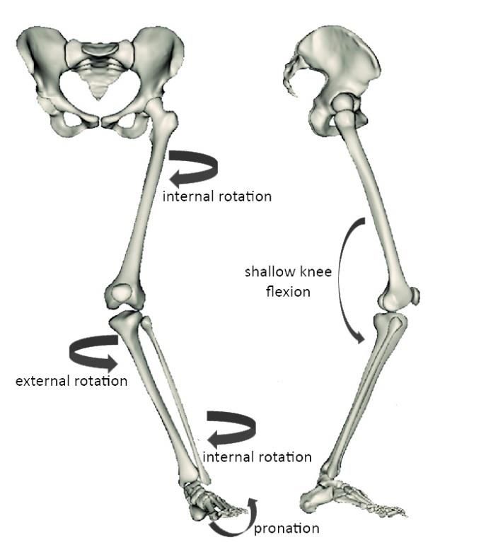
aspect of the tibial attachment of the ACL, further avascular and impacting the healing process post

injury to warrant surgical intervention to prevent joint and tissue degeneration (Duthon et al., 2006).

3.1.2 Function

 The ACL does not function as a simple band of fibers under constant tension as the knee moves

through its full range of motion (Amis & Dawkins, 1991; Zantop et al., 2006). This ligament acts as the

primary restraint to anterior tibial translation (Petersen & Zantop, 2007) and the secondary stabilizer to

rotatory instabilities (Norwood & Cross, 1979) and constraint to excessive joint space between the tibia

and femur in the medial and lateral compartments of the knee (Grood et al., 1981). In the frontal plane,

the major motion that the ACL restricts is varus movement, where this ligament carries a substantial

role in restraining varus-valgus rotation of an intact knee (Ohori et al., 2017).

3.1.3 Mechanism of Injury – A Perspective into Joint Degeneration

 The complete rupture of the ACL can result in pathological knee conditions that include knee

instability, meniscal damage, damage to the chondral surfaces, and predisposition to knee osteoarthritis

(Yu & Garrett, 2007). Ligament rupture can result from two mechanisms of injury, contact and non-

contact ACL tears (Salem et al., 2018). Contact ACL tears are a result of a direct external force to the

knee by another person or object (Salem et al., 2018). Contact tears have a higher incidence of collateral

ligament and articular cartilage injuries with an association of higher injury severity (Salem et al., 2018).

Lateral femoral condyles are especially prevalent to chondral injury upon arthroscopy (Salem et al.,

2018).

 Non-contact ACL tears are the most common mechanisms of ligament rupture, occurring when

individuals generate excessive force, moments, and loading on the ACL than it is capable of withstanding

(Yu & Garrett, 2007). This mechanism of injury is in the absence of external forces other than ground

reaction forces and result in multiplanar knee loading (Shimokochi & Shultz, 2008). The “position of no

 7
return” (Figure 3.2) is defined as the combined motions of hip adduction and internal rotation, external

rotation of the tibia relative to the femur, internal rotation of the tibia on the foot, and forefoot

pronation all occurring concurrently (Alentorn-Geli et al., 2009; Shimokochi & Shultz, 2008). In this

position, there is high likelihood of ACL injury.

Figure 3.2: Mechanism of non-contact ACL injury defined as “position of no return”.

 Regardless of the mechanism of injury, a torn ACL has limited repair capabilities (McLean et al.,

2015) due to the native tissue’s poor vascularity. Pathologically, an ACL rupture can lead to significant

instability and secondary damage including meniscus tears and articular cartilage injuries within the

knee joint (Zantop et al., 2006). ACL deficient knees that are left untreated have been shown to develop

increased mean contact stress on the posterior medial and lateral compartments of the joint due to an

increased anterior tibial translation and internal tibial rotation without restraint (Simon et al., 2015).

Excessive anterior tibial displacement results in shearing forces that are applied primarily on the medial

compartment, with the posterior horn of the medial meniscus splitting between the tibia and posterior

condyle of the femur (Louboutin et al., 2009). The degeneration of the posterior horn of the medial

 8
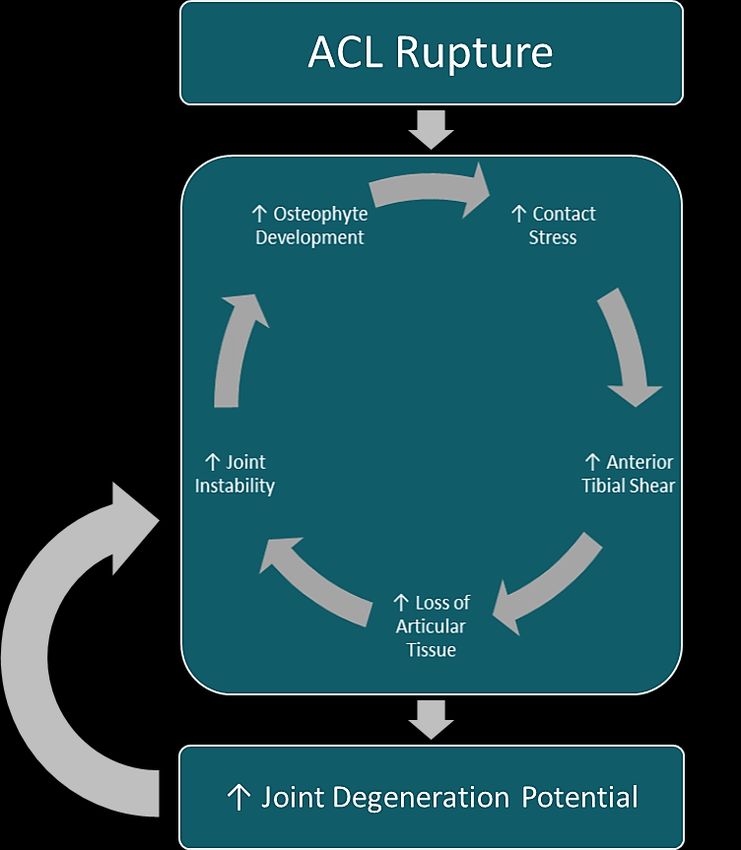
meniscus contributes to increased anterior tibial displacement on the femur as shear forces increase on

the articular surfaces of the joint, leading to a loss of articular cartilage (Louboutin et al., 2009) . These

series of events almost act as a positive feedback loop (Figure 3.3) to expose bone and accelerate

osteophyte development (Louboutin et al., 2009) . The overall lack of a primary restraint to anterior

tibial translation results in greater knee joint loading and increases the susceptibility to degeneration

(Simon et al., 2015). Radiographic signs of osteoarthritis and limitations in activities of daily living are

greatest in patients with conservative treatment without surgical interventions and combined

cumulative knee injuries (Simon et al., 2015). This often requires the complete surgical replacement of

the tissue in order to return to a moderate level of physical activity (McLean et al., 2015). Patients who

are symptomatic with meniscal or cartilage damage following ligament rupture, or experience joint

instability, are at an increased risk of developing osteoarthritis if surgical reconstruction is delayed as

cumulative loading worsens the state of the joint (Louboutin et al., 2009).

 9
Figure 3.3: Summary of the positive feedback loop that increases the potential of knee joint degeneration following a complete
ACL rupture.

 ACL reconstructions aim to surgically restore joint stability, re-establish optimal knee kinematics,

and protect the knee from further developing severe meniscal tears, cartilage damage, and

osteoarthritis (Xie et al., 2015; Zantop et al., 2006). However, the grafts used for surgical reconstruction

can be too stiff and can over-constrain the joint, restricting the range of motion (Dargel et al., 2007; Mae

et al., 2010), or can be too elastic and can result in residual joint laxity (Smeets et al., 2017). These are

two examples of potential initiators of the cascade of events that lead to degenerative joint disease.

Increased residual joint laxity increases joint instability, which has been reported as a risk factor for the

development of osteoarthritis (Øiestad et al., 2009). Identification of residual joint laxity is important

 10
when trying to ensure optimal knee kinematics of the lower limb and decreasing the risk of

osteoarthritic development.

 Females are 2.4 to 4.1 times more likely to rupture their ACL compared to their male

counterparts in a 5-year study period (Arendt & Dick, 1995). Subsequently, the greater ratio of females

to males sustaining an ACL rupture increases the risk of mechanical stress to the knee, cartilage damage

to the surrounding tissue, risk of joint instability, and risk of developing osteoarthritis (Louboutin et al.,

2009) if left untreated with this particular sex. With a minimum follow-up of 10 years from receiving an

ACL reconstruction, patients are overall 1.63 times more likely to rupture their contralateral ACL than

that of their reconstructed ligament (Magnussen et al., 2015), where females are more likely to

experience a contralateral rupture than males (Sutton & Bullock, 2013). The overall greater ACL rupture

ratio in females also creates an increased ratio females to males receiving surgical reconstructions,

where surgical reconstructions themselves increase the risk of post-traumatic osteoarthritic

development (Taruc-Uy & Lynch, 2013). Sex differences must be considered when addressing risk

factors associated with ACL reconstructed knees, such as residual joint laxity, and the development of

osteoarthritis.

 11
3.2 Knee Osteoarthritis Following an ACL Tear

3.2.1 Knee Osteoarthritis

 Osteoarthritis is a joint disorder, characterized by cell stress and extracellular matrix

degradation that is initiated by micro- and macro-injury, activating maladaptive repair responses and

pro-inflammatory pathways (March et al., 2016). First manifesting as abnormal joint tissue metabolism,

it is followed by anatomic and/or physiologic imbalances to cartilage, bone remodelling, osteophyte

formation, joint inflammation, and loss of joint function, that can culminate in illness (March et al.,

2016). It is the most common degenerative joint disorder that remains challenging to treat due to

evolving risk factors and pathophysiology that could lead to worsening of disease severity and

progression over time (Martel-Pelletier et al., 2016). Osteoarthritis can be classified into primary

(idiopathic) or secondary forms (Martel-Pelletier et al., 2016; Taruc-Uy & Lynch, 2013). Primary

osteoarthritis can result from a combination of risk factors such as the wear and tear on the cartilage

with increasing age (Taruc-Uy & Lynch, 2013) and obesity (Martel-Pelletier et al., 2016). Secondary

osteoarthritis can be attributed to predisposing causative factors such as trauma (Martel-Pelletier et al.,

2016; Taruc-Uy & Lynch, 2013). Mechanical stress, repeated trauma to soft tissue, and surgery, are

some examples of factors that can lead to secondary or post-traumatic osteoarthritis (Taruc-Uy & Lynch,

2013). The complexity of an ACL tear can result in increased mechanical stress to the knee and damage

to the surrounding tissue (Louboutin et al., 2009), where surgical reconstruction aims to reduce these

risk factors (Xie et al., 2015; Zantop et al., 2006). However, surgical intervention itself is an additional

predisposing risk factor to the development of osteoarthritis, resulting in joint trauma but also residual

joint laxity (Smeets et al., 2017). The grafts used for surgical reconstruction can be too elastic and can

result in residual joint laxity (Smeets et al., 2017), where increased residual joint laxity increases joint

instability, which has been reported as a risk factor for the development of osteoarthritis (Øiestad et al.,

2009).

 12
3.2.2 Instability and Laxity

 Joint instability and increased joint laxity have been reported as a risk factor for the

development of osteoarthritis (Øiestad et al., 2009). Acute instability following rupture can become a

chronic instability, resulting in greater knee joint loading and increasing the susceptibility to

osteoarthritic degeneration (Simon et al., 2015). One important factor in restoring knee joint stability is

correcting the increased knee joint laxity that results from an ACL tear (Xie et al., 2015; Zantop et al.,

2006).

AP Laxity

 AP laxity is clinically assessed the most, measured as either the AP translation or the angular

range of motion of the tibia with respect to the femur (Aït Si Selmi et al., 2006; Salmon et al., 2006). Pre-

operatively following rupture, it has been shown that AP laxity increased as a higher injury-to-surgery

time was presented (Signorelli et al., 2016). Post-operatively, it has been shown that AP laxity persists 6

months to 6 years following an ACL reconstruction (Shimizu et al., 2019). Increased AP laxity (assessed as

AP translation via Lachman test) has been identified as having a statistically significant relationship with

degenerative radiographic knee changes (Aït Si Selmi et al., 2006; Salmon et al., 2006). Sustaining an ACL

rupture increases the anterior tibial translation and internal tibial rotation (Simon et al., 2015), resulting

in an increase of knee joint instability and laxity.

 13
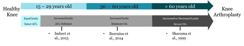
3.3 Frontal Plane Laxity

 Frontal plane laxity can be defined as the passive, frontal plane angular range of motion with an

application of a varus-valgus moment (Sharma, Lou, et al., 1999; van der Esch et al., 2005). In the frontal

plane, the ACL acts as a constraint to excessive joint space between the tibia and femur in the medial

and lateral compartments of the knee (Grood et al., 1981). Frontal plane (varus-valgus) knee laxity can

manifest due to the damage or impairment in the passive restraint of the system of ligamentous tissue

(Sharma, Lou, et al., 1999) such as an ACL rupture. It has been speculated that a portion of increased

frontal plane laxity measured in osteoarthritic patients may precede osteoarthritic development and

raises the question on whether increased frontal plane laxity may contribute to osteoarthritis

progression (Sharma, Lou, et al., 1999).

 ACL ruptured knees were found to have an increased frontal plane laxity as rupture-to-surgery

time increased between a duration of 2 to 220 months (Signorelli et al., 2016). When comparing the ACL

ruptured knee to the contralateral knees of patients, it was found that there was a significantly larger

laxity in the ACL deficient knees (Figure 3.4) (Imbert et al., 2015). It was suggested that performing early

surgical reconstruction might prevent the deterioration of knee stability over time, prior to and

following surgical intervention (Signorelli et al., 2016). Immediately following surgical reconstruction, it

was found that the ACL reconstruction did not restore frontal plane laxity to the level of the

undiagnosed contralateral knees of patients, indicating residual laxity in the reconstructed knee (Imbert

et al., 2015). Unfortunately, there was no further follow-up to determine if, or for how long this residual

laxity persisted post-operatively (Imbert et al., 2015).

 Fourteen years following an ACL reconstruction there was a higher prevalence of moderate

osteoarthritic development in the ACL reconstructed knee compared to the contralateral knees without

an osteoarthritic diagnosis for a cohort ranging between 29-57 years of age (Barenius et al., 2014).

 14
Comparing frontal plane knee joint laxity between osteoarthritic patients and healthy controls has

reported that frontal plane laxity was greater in knees with mild osteoarthritis and in the contralateral

knees (that did not have an osteoarthritic diagnosis) compared to the control (Sharma, Lou, et al., 1999).

In that study, controls were not age matched, and they were older than the osteoarthritic cohort (mean

71.4 versus 62.6 years of age), where controls had a sample size of N=25 and osteoarthritic had N = 164

(Sharma, Lou, et al., 1999). As the Kellgren and Lawrence grade increased for the severity of knee

osteoarthritic changes, the mean frontal plane laxity increased (Sharma, Lou, et al., 1999). Both joint

space narrowing, and malalignment of the knee have been associated with higher frontal plane knee

joint laxity values in those diagnosed with knee osteoarthritis for an mean time of 10 years for patients

66.5 ± 10.3 years of age (van der Esch et al., 2005). When stress radiographs were taken following the

application of a 15N force at the knee to create a varus and valgus motion, it was found that patients

(50.3 ± 7.4 years of age) with medial compartmental osteoarthritis and genu varum had greater frontal

plane laxity and instability compared to age- and gender-matched controls without osteoarthritis (Lewek

et al., 2004).

Figure 3.4: A summary of previously published literature examining frontal plane laxity and osteoarthritic development through
a timeline of aging. Imbert et al. (2015) examined frontal plane laxity prior to and immediately following an ACL reconstruction.
Barenius et al. (2014) found moderate osteoarthritic development in knees 14-years following an ACL reconstruction. Sharma et
al. (1999) examined increasing frontal plane laxity as osteoarthritis severity increased as defined by the Kellgren and Lawrence
grading system.

 Across most studies above, authors reported samples sizes with a greater number of females

than males but neglected to include sex comparisons (Barenius et al., 2014; Sharma, Lou, et al., 1999;

van der Esch et al., 2005). There was also limited follow-up after ACL reconstruction to determine if, or

 15
for how long residual laxity persisted (Imbert et al., 2015). Furthermore, most patients undergoing an

ACL reconstruction were 14-21 years of age for females or 18-25 years for males (Csintalan et al., 2008),

whereas previous frontal plane laxity studies examined patients that were approaching middle age or

older (participant ages were ≥33 years) and had already developed osteoarthritis (Lewek et al., 2004;

Pottenger et al., 1990; Sharma, Lou, et al., 1999; van der Esch et al., 2005). This age range omits the

examination of young adults who most frequently sustain an ACL tear and subsequent surgical

reconstruction, a population that have tissue joint trauma that place them at a greater susceptibility to

osteoarthritic development with sex differences (Louboutin et al., 2009; Martel-Pelletier et al., 2016;

Taruc-Uy & Lynch, 2013).

 16
3.4 Frontal Plane Laxity Measurement System Designs

 Physician examinations have been reported to have poor interobserver reliability (Cushnaghan

et al., 1990), where instrumented frontal plane laxity measurement devices were required to measure

repeatable angles. Frontal plane measurement systems have been designed to assess laxity of the knee

and minimize major sources of variation commonly seen during physical examination tests (Sharma,

Lou, et al., 1999). The thigh is immobilized while the shank is supported and rotated along the

transverse axis about the knee joint with the application of a fixed moment from the knee (Sharma, Lou,

et al., 1999; Shultz et al., 2007; van der Esch et al., 2005). The application of this fixed moment occurs

both in the abduction and adduction directions separately, along the frontal plane to create varus and

valgus angular deviations, and then combined to get the total frontal plane laxity (Sharma, Lou, et al.,

1999; van der Esch et al., 2005). However, the key attribution to the variance in reported measures was

the use of different measurement system parameters across study devices (Freisinger et al., 2017).

 Previous literature has used varying laxity system designs for the examination of passive frontal

plane laxity in the knee joint (Table 3.1) of young adults using the right lower limb. The SEM has been

used to measure how repeatable the measurement scores where, whereas the MDD was used to

identify the smallest difference or change that the system could detect.

Table 3.1: Comparison of standard error of measurement (SEM) and minimal detectable difference (MDD) for frontal plane laxity
measurement systems in the literature with young adults.

 Number of Sex Measurement Laxity (°) Session SEM (°) MDD
 Participants Method Design for (°)
 (N) Determining
 MDD and
 SEM
 (Mines, 2016) 10 5M/5F Motion 7.67 (2.4) Same day 0.44 1.22
 Tracking
 (Shultz et al., 10 5M/5F Motion 9.6 (3.0) Between-day 0.67 1.86
 2007) Tracking
 (van der Esch, 20 10M/10F Electrical 5.92 (2.6) Between-day 1.55 4.30
 Steultjens, Goniometer
 Ostelo, et al.,
 2006)

 17
Measurement of frontal plane laxity using an electrical goniometer resulted in a MDD that was up to

approximately 73% of the of the average frontal plane laxity values recorded (van der Esch, Steultjens,

Ostelo, et al., 2006). When the error of the system is a high proportion of the measurements

themselves, it can obscure differences in laxity measurements recorded by the system. However, when

using a motion capture system and same day testing, improved repeatability and sensitivity occurs as

reflected by a smaller SEM and MDD (Mines, 2016).

 Previous studies also report using varying knee flexion angles from when measurements were

taken (Freisinger et al., 2017), where 20° of knee flexion was seen to be the most common (Sharma, Lou,

et al., 1999; Shultz et al., 2007; van der Esch et al., 2005). Designs combined gravitational force and

applied loads as a result of the shank not being orientated horizontally, where individuals would

experience different total applied loads at their end range of motion (Sharma, Lou, et al., 1999; Shultz et

al., 2007; van der Esch, Steultjens, Ostelo, et al., 2006). These previous designs also forced the shank to

rotate about a fixed mechanical axis rather than the knee’s natural joint center (Shultz et al., 2007),

potentially limiting the range of rotation. Finally, in previous work, stabilizing muscles that cross the

knee joint were not monitored to confirm that they remained passive during the laxity measurement

and thus that passive laxity was being measured (Sharma, Lou, et al., 1999; Shultz et al., 2007; van der

Esch, Steultjens, Ostelo, et al., 2006). Only visual cues and palpation of the muscles supporting the lower

limb was completed to assess muscle contraction and passive laxity measurement (Sharma, Lou, et al.,

1999).

 Overall, when examining frontal plane laxity there was a lack of focus on sex differences

(Barenius et al., 2014; Sharma, Lou, et al., 1999; van der Esch et al., 2005) and young adults who most

frequently sustain an ACL tear and subsequent surgical repair (Csintalan et al., 2008). Investigations of

frontal plane laxity as a risk factor for the development of osteoarthritis have only been carried out

when osteoarthritic development has already occurred at long-term follow-up (Lewek et al., 2004;

 18
Pottenger et al., 1990; Sharma, Lou, et al., 1999; van der Esch et al., 2005), and not under short-term

examination with an ACL reconstructed population that has a greater susceptibility to osteoarthritic

degeneration (Simon et al., 2015). Addressing frontal plane measurement system design limitations, by

ensuring a consistent knee flexion angle and total applied moment, rotation about the knee’s natural

joint center, and examination of muscle activation for passive laxity, would further improve the

repeatability and detectability of laxity values. Adopting motion tracking into the measurement system

used to record these values would also be more repeatable with a higher sensitivity in detecting

measurement differences (Shultz et al., 2007) to examine populations at greater susceptibility of

osteoarthritic degeneration.

 In summary, both increased frontal plane laxity and ACL rupture/reconstruction are linked to

osteoarthritis development. Increased frontal plane laxity has been observed in ACL deficient knees,

reconstructed knees immediately following reconstruction, and in knees with (not necessarily post-

traumatic) osteoarthritis. However, it’s unclear if the laxity observed immediately following surgery

persists, therefore potentially preceding and contributing as a factor for knee OA development in ACL

reconstructed knees. This study addresses the short term follow up time frame after surgery to

determine if frontal plane laxity persists and uses methodology for measuring frontal plane laxity that

addresses some of the limitations of previous work.

 19
Chapter 4: Methods
4.1 Study Population

 Twenty-four university aged participants (twelve females mean age 20.5 ± 1.8 and twelve males

mean age 21.7 ± 2.3) were recruited for this cohort study with approval from the institutional ethics

board and provided informed consent. There were two groups: twelve participants with one ACL

reconstructed knee and one contralateral knee (that had no previous ACL tear to repair) and twelve age-

and sex-matched controls. ACL reconstructed participants were eligible to participate had they

sustained a complete ACL rupture isolated to one knee, with at least six months since reconstructive

surgery (Ajuied et al., 2014; Barenius et al., 2014; Hoffelner et al., 2012; Imbert et al., 2015; Sharma,

Lou, et al., 1999; van der Hart et al., 2008). ACL reconstructed participants were required to self-report

that their contralateral knee was free of injury (Imbert et al., 2015) had no previous surgery (van der

Hart et al., 2008), and was asymptomatic (Hoffelner et al., 2012); showing no symptoms of pain,

swelling, stiffness, or evidence of knee osteoarthritis (Signorelli et al., 2016). Evidence of osteoarthritis

was assessed using the American College of Rheumatology Clinical Criteria (Wolfe et al., 1990). The

contralateral knee with no osteoarthritis diagnosis represents the best possible reference for

comparison purposes and has been used for comparison with both ACL deficient and ACL reconstructed

knees (Hoffelner et al., 2012). Control participants knees were required to meet the same criteria as the

contralateral knees for those with an ACLR knee. Participants were excluded if they had a history of

unpredictable clicking, locking, or buckling, known meniscal tears, known joint fractures, known

cartilage injury or Baker’s cyst, or if they had additional surgical procedures in either knee aside from

one ACLR (Imbert et al., 2015; Sharma, Lou, et al., 1999). All ACLR participants were varsity athletes,

sustaining their sports related ACL tear in soccer, basketball, or rugby. One participant chose not to

report the specific date of when their operation was held, however they confirmed that it was at least

six months prior to study participation.

 20
4.2 Instrumentation and Experimental Design

4.2.1 Instrumentation

 All participants were fitted with rigid body clusters (Figure 4.1) that attached bilaterally to the

thigh, shank, and foot using an optoelectronic system (Certus, NDI, Waterloo, Canada), used to collect

kinematic data sets recorded at 64Hz.

Figure 4.1: Optoelectronic system (Certus, NDI, Waterloo, Canada) rigid body clusters locations, attached to the lateral aspect of
the thigh, shank, and foot.

Electromyographic (EMG) activity was monitored bilaterally from the vastus lateralis and vastus medialis

using bipolar Ag/AgCl electrodes (BlueSensor N, Ambu Inc., Glen Burnie, MD, USA) that were affixed

following SENIAM guidelines (Hermens et al., 1999). Raw EMG signals were sampled at 2048 Hz.

4.2.2 Experimental Design

 Maximal voluntary isometric contractions (MVICs) for the vastus lateralis and vastus medialis

were recorded with participants attempting seated leg extensions (two per leg) against resistance at

approximately 60° of knee flexion (measured from full extension) (Becker & Awiszus, 2001) using a leg

extension machine. ACL reconstructed participants with patellar tendon autografts were advised by

surgeons to refrain from leg extension machine use, therefore they attempted leg extensions in a leg

 21
press machine at approximately 90° of knee flexion instead, as minimal ACL forces have been reported

with this exercise (Escamilla et al., 2012).

 The frontal plane laxity device (Figure 4.2) consisted of a chair and backrest (Chang et al., 2014;

van der Esch et al., 2005) with the seat tilted to allow the tibia to be fixated horizontally to a free-

moving sled (van der Esch et al., 2005) as designed by Mines (2016). The horizontal orientation of the

tibia allowed gravity to act perpendicular to the frontal plane, eliminating a gravitational moment that

would be inconsistent in magnitude between subjects (Chang et al., 2014). The tilted seat combined

with a horizontally fixed tibia produced 20° (Sharma, Lou, et al., 1999; Shultz et al., 2007; van der Esch et

al., 2005) of knee flexion and the sled allowed frontal plane rotation of the tibia without the fixation to a

specific mechanical axis, that has been seen in previous laxity device designs (Sharma, Lou, et al., 1999;

van der Esch et al., 2005). The sled that hosted the tibia was constructed with a LEXANTM base, that

could slide over a LEXANTM surfaced table, covered in 4mm zinc-plated ball bearings. The ball bearings

between the sled and table minimized friction (Sharma, Lou, et al., 1999; van der Esch et al., 2005)

during sled motion. Femoral condylar clamps were located at the end of the seat to secure the femur

while a low-friction cable-pulley system located 0.45m distal to the clamps was fixated to the table.

 22
Figure 4.2: Participant seated in free-moving laxity measurement apparatus to record passive frontal plane laxity in response to
a 10Nm varus/valgus moment designed by Mines (2016). A) Infrared light emitting electrodes for motion capture (Certus, NDI,
Waterloo, Canada). B) Thigh strap. C) EMG electrodes for vastus lateralis (or vastus medialis, not shown). D) Femoral condylar
clamps. E) Free-moving sled with Velcro attached shank cradle that can move (left to right in photograph) to support shank
regardless of leg length F) Shank strap. G) Fixed low-friction cable-pulley system with a cable attached to the free-moving sled to
maintain same distance of force application to create a 10N/m moment.

Figure 4.3 Frontal plane view of the shank cradled on the free-moving sled. Movement of the sled in the valgus or varus
directions to measure valgus or varus laxity of the shank in respect to the femur.

 23
Participants sat in the chair with their tibia fixed horizontally to the free-moving sled. The thigh

was fastened to the chair and the femoral condylar clamps were positioned on the medial and lateral

sides of the knee and engaged to minimize internal-external rotation of the femur (Chang et al., 2014). A

2.28kg load application to the low-friction pulley system occurred in two steps (1.14kg each) to create a

maximum moment of 10Nm. Previous frontal plane laxity studies have applied a moment between

7.7Nm (van der Esch et al., 2005) - 12Nm (Sharma, Lou, et al., 1999). This 2.28kg load was applied to the

medial and then lateral aspects of the tibial sled to exert a moment in each of the varus and valgus

directions. Three laxity measurement tests were performed on each leg by the same single examiner

across all participants.

4.3 Data Processing

 Knee joint angles were calculated using previously defined (Chong et al., 2017) femoral and

tibial coordinate systems and a Z-X-Y Euler sequence (flexion/extension- abduction/adduction-internal

rotation/external rotation) (Visual3D, v6.01.07, C-motion, Germantown, MD) to link passive laxity to

active laxity seen in gait and other lower limb activities (Mines, 2016). In a given pair of trials (one

medial and one lateral load test), frontal plane laxity was defined as the sum of the maximum absolute

varus and valgus deviations (in degrees) of the knee following application of the loads (Sharma, Lou, et

al., 1999; van der Esch et al., 2005).

 EMG was used only to confirm that the vastus lateralis and the vastus medialis in the measured

leg were indeed passive (activity
You can also read