Prussian Blue Technique is Prone To Yield False Negative Results In Magnetoreception Research
←
→
Page content transcription
If your browser does not render page correctly, please read the page content below
Prussian Blue Technique is Prone To Yield False
Negative Results In Magnetoreception Research
Franziska Curdt
Institute of Biology and Environmental Sciences, University Oldenburg
Katrin Haase
Institute of Biology and Environmental Sciences, University Oldenburg
Laura Ziegenbalg
Institute of Biology and Environmental Sciences, University Oldenburg
Helena Greb
Institute of Biology and Environmental Sciences, University Oldenburg
Domink Heyers
Institute of Biology and Environmental Sciences, University Oldenburg
Michael Winklhofer ( michael.winklhofer@uol.de )
Institute of Biology and Environmental Sciences, University Oldenburg
Research Article
Keywords: Biogenic magnetite, fine-dust contamination, magnetosomes, Magnetospirillum, rainbow trout,
songbirds, Trigeminal system
Posted Date: February 14th, 2022
DOI: https://doi.org/10.21203/rs.3.rs-1324083/v1
License: This work is licensed under a Creative Commons Attribution 4.0 International License.
Read Full License
Page 1/25
Abstract
Perls’s Prussian blue staining technique has been used in magnetoreception research to screen tissues
for iron-rich structures as proxies for putative magnetoreceptor structures based on magnetic particles.
However, seemingly promising structural candidates in the upper beak of birds detected with Prussian
Blue turned out to be either irreproducible or located in non-neuronal cells, which has spurred a
controversy that has not been settled yet. Here we identify possible pitfalls in the previous works and
apply the Prussian Blue technique to tissues implicated in magnetic-particle-based magnetoreception, in
an effort to reassess its suitability for staining single-domain magnetite, i.e., the proposed magnetic
substrate for the interaction with the external magnetic field. In the upper beak of night-migratory
songbirds, we found staining products in great numbers, but not remotely associated with fiber terminals
of the traced ophthalmic branch of the trigeminal nerve. Surprisingly, staining products were absent from
the lamina propria in the olfactory rosette of rainbow trout where candidate magnetoreceptor structures
were identified with different techniques earlier. Critically, magnetosome chains in whole cells of
magnetotactic bacteria remained unstained. The failure to label single-domain magnetite in positive
control samples is a serious limitation of the technique and suggests that two most influential but
antipodal studies conducted previously stood little chances of obtaining correct positive results under the
assumption that magnetosome-like particles were present in the tissues. Nonetheless, the staining
technique appears suitable to identify tissue contamination with iron-rich fine dust trapped in epithelia
already in vivo.

1. Introduction
Numerous behavioral experiments have demonstrated the ability of migratory animals to orient by the
Earth’s magnetic field, but the nature of the underlying magnetic sensory structures remains one of the
greatest mysteries in sensory biology (Mouritsen 2018). The magnetic sense has been studied most
thoroughly in night-migratory songbirds, where various lines of evidence point towards the existence of at
least two fundamentally different magnetoreception mechanisms. One is based on radical pairs, with the
flavoprotein cryptochrome 4 as the potential magnetic sensory molecule, located in cone photoreceptor
cells in the retina (Hore & Mouritsen, 2016; Günther et al., 2018; Wu et al., 2020; Xu et al., 2021), and
suggested to provide directional, i.e., “compass” information (Zapka et al., 2009). The radical-pair
mechanism is consistent with the observation that songbirds tested in Emlen funnels were magnetically
disoriented during exposure to weak radiofrequency magnetic fields aimed at interfering with the
mechanism (Ritz et al., 2004; Engels et al., 2014; Schwarze et al., 2016). The second magnetoreception
mechanism is suggested to be based on ferrimagnetic particles within the upper beak innervated by the
ophthalmic branch of the trigeminal nerve (V1; Williams & Wild, 2000; Fleissner et al., 2003, 2007;
Falkenberg et al., 2010), and which seem to provide magnetic information for a navigational map
(Kishkinev et al., 2013; Pakhomov et al., 2018). This mechanism is consistent with the observation that
birds pre-exposed to a brief but strong magnetic pulse (as a tool to perturb a magnetic particle-based
magnetoreceptor) had shifted orientations compared to untreated control birds (Wiltschko et al., 1994,

                                                  Page 2/25
2007; Beason et al., 1997; Holland & Helm, 2013). The observation that pulse effects were restricted to
experienced migrants which had already successfully finished at least one migratory journey has led to
the notion that adults use magnetic-particle based receptors to acquire magnetic map information.
Further evidence in support of the so-called magnetic map theory has come from both physical
displacement and virtual magnetic displacement studies, where animals readjusted their orientation
according to a different place when tested under magnetic field parameters mimicking the displacement
site (Kishkinev et al., 2013, 2015; Pakhomov et al., 2018).
From ablation studies, V1 has been identified as necessary for conveying magnetic map information in
migratory songbirds (Kishkinev et al., 2013; Pakhomov et al., 2018). In addition, behavioral molecular
mapping has shown that birds exposed to a strongly changing magnetic field stimulus display
significantly increased expression levels of immediate early genes in subcompartments of the principle
and spinal sensory trigeminal brainstem nuclei which receive V1 input (Heyers et al., 2010; Lefeldt et al.,
2014; Elbers et al., 2017; but see Kishkinev et al., 2016). The magnetically activated neurons in the
principal sensory trigeminal brainstem nucleus were recently shown to define a morphologically distinct
neuronal subpopulation, likely to form the origin of a neuronal processing stream exclusively dedicated to
transmitting trigeminally perceived magnetic information to higher telencephalic integration centers
(Kobylkov et al., 2020).
Given that magnetic map information is conveyed by V1 but misinterpreted after magnetic pulsing, it is
sensible to postulate that magnetic particles form the basis of trigeminal magnetoreception. With V1
responsible for sensory innervation of the upper beak, iron-rich structures found in the upper beak of
homing pigeons and songbirds (Williams and Wild, 2001; Fleissner et al., 2003, 2007; but see Winklhofer
& Kirschink, 2008; Tian et al. 2007; Falkenberg et al., 2010) were considered to represent the long-sought
trigeminal magnetic sensor, particularly since the structures were found to contain magnetite
nanocrystals in large numbers (Hanzlik et al., 2000; Winklhofer et al., 2001) and suggested to colocalize
with nervous tissue (Williams and Wild, 2001; Fleissner et al., 2003). Critically, however, the association
between iron-rich structures and nervous tissue turned out irreproducible in independent follow-up
studies, who found iron-rich structures not associated with nervous tissue but highly colocalized with
cells presenting MHC class II, i.e. probably macrophages (Treiber et al., 2012, 2013; Engels et al., 2018).
The majority of these studies screened tissue sections for non-hemin iron-rich structure as possible hints
towards magnetosensory structures, using the Prussian blue (PB) staining technique. However, upon
closer comparison of earlier PB studies on the upper beak of birds, we realized potential methodological
pitfalls, which might have been the cause for the contradictory results of previous studies:
(1) Fleissner et al. (2003) and Treiber et al. (2012, 2013) relied on antibodies against generic neuronal
markers such as neurofilaments to find possible colocalizations of PB and nervous tissue. It is not clear if
this approach is suitable to also label free nerve endings, where primary sensory receptors are expected to
occur.
Page 3/25(2) Apart from false negatives, such a generic labelling approach may also yield false positives in the
form of PB positive sites colocalizing with nerves of the autonomous system, e.g., for regulation of blood
vessels.
(3) None of the previous works used a suitable positive control for intracellular magnetite. Although the
PB method detects ultrafine iron oxide nanoparticles (< 10 nm) in tissue when present as dense
accumulations measuring several hundreds of microns in diameter (Hanzlik et al., 2000; Nimpf et al.,
2017), such nanoparticulate structures are far from representing an optimized solution for realizing a
sensor that should be capable of detecting the small magnetic field differences which were implied from
the magnetic map experiments mentioned above. For instance, cuticulosomes, which are iron-rich
vesicles found in the cuticular plate of hair cells in the Avian inner ear (Lauwers et al., 2013; Nimpf et al.,
2017; Malkemper et al. 2019), are so weakly magnetic that they would not even qualify for a compass
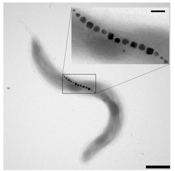
sense to begin with (de Gille et al., 2021). In contrast, magnetite crystals with particle-sizes between 40
and 100 nm have superior magnetic properties, forming single-domain magnets, which makes them
much more suitable as magnetic field sensitive structures (Kirschink et al., 2010), as can be best seen in
the example of magnetotactic bacteria (Fig. 1).
These microorganisms biomineralize chains of membrane-enclosed sub-100 nm magnetite crystals,
termed magnetosomes, which impart a permanent magnetic dipole moment to the bacterial cell body and
keep it thus aligned with the magnetic field (Frankel et al., 1979). Notably, similar crystals were found in
structural magnetoreceptor candidates associated with trigeminally innervated regions of the olfactory
epithelium of rainbow trout (Walker et al., 1997; Diebel et al., 2000), using reflectance confocal laser
scanning microcopy for screening in combination with electron microscopy or magnetic scanning probe
microscopy for validation of iron chemistry or of magnetic properties, respectively. In contrast to these
methods sensitive to physical properties of single-domain magnetite, it is not known if the PB method is
capable of detecting just a dozen of magnetosome-like crystals, which suffice for a magnetoreceptor
according to theoretical models (Winklhofer & Kirschvink, 2010). We therefore embarked on a
reexamination of the topic, where we studied beak tissues of a night-migratory songbird, the Eurasian
blackcap (Sylvia atricapilla), in which we specifically labelled V1 fiber terminals within the beak using
neuronal tract tracing. As in previous studies (Williams & Wild, 2001; Fleissner et al., 2003, 2007; Tian et
al. 2007; Treiber et al., 2012, 2013), we used the PB technique, for the lack of a better histochemical stain
with specificity for the magnetic iron compounds magnetite (or its oxidized form, maghemite) – two
components which we would ultimately expect to find in a magnetic-particle based magnetoreceptor
structure. None of the previous studies applied the modified PB protocol with diaminobenzidine (DAB)
postenhancement of PB (Nguyen-Legros et al. 1980; Moos & Møllgård 1993; Meguro et al. 2003), since
DAB was already used as chromogen in immunohistochemistry, which is why we did without the
modified protocol, too.
To approach point 3) also independently, we applied the PB technique to the olfactory epithelium of
rainbow trout where we expect to find candidate magnetoreceptor structures (Walker et al., 1997; Diebel et
al., 2000), however in sparse occurrence, and therefore, for ground-truthing, to cells of a magnetotactic
Page 4/25bacterium (Magnetospirillum magnetotacticum strain MS-1, see Fig. 1), where we know with certainty of
the occurrence of intracellular iron-rich target structures.
Since the PB technique is essential for our study, we commence with a brief summary of the chemical
reactions involved in PB staining, followed by theoretical considerations exploring the potential sensitivity
of the PB stain in relation to various iron oxide minerals found in tissues.
The Prussian Blue staining reactions
The PB stain for ferric ion was introduced by Perls in 1867 and has since been employed widely in
biology and pathology. Despite variations in the protocol, all ferric iron staining kits use the
[
hexacyanoferrate anion, FeII(CN) 6 ] 4−
, as reagent to bind free Fe3+ in the form a blue pigment
referred to as a Prussian Blue. The reagent is applied in acid solution, typically hydrochloric acid (HCl)
solution, to liberate Fe3+ from its bound forms in the tissue, because ferric iron requires pH < 2 to exist as
free Fe3+ ion, see Pourbaix diagram for the iron-water system in Delahay et al. (1950); a pH value of 2
corresponds to 10mM HCl solution or approx. 0.04 wt% HCl. When a crystalline iron oxide is present, e.g.,
Fe2O3, the low pH also affords the protons to dissolve the oxide, i.e.,
Fe2O 3 + 6H + − > 2Fe3 + + 3H 2O. (1)
Thus, unlike a conventional histochemical stain, which would label a target molecule directly, the PB
staining technique requires the (acidic) dissolution of the target compound to then label the liberated
target ion. Depending on the mobility of the free Fe3+, the PB stain may not mark the original site of the
target compound, but rather its diffusion trace. Depending on the ease with which the target compound
can be dissolved, it may also disappear altogether. In any case, the target compound will be irreversibly
altered.
The actual PB staining reaction is thought to occur in two steps, with the following reaction proceeding
first:
[
Fe3 + + FeII(CN) 6 ] 4−
[
+ K + − > KFeIII FeII(CN) 6 , (2) ]
[
where the potassium ions are delivered with the dissolved reagent, K 4 FeII(CN) 6 . The product in ]
Eq. (2) is referred to as (water-)soluble PB (Keggin & Miles, 1936). In the presence of excess Fe3+,
insoluble PB precipitates according to:
[
4Fe3 + + 3 FeII(CN) 6 ] 4−
+ xH 2O − > Fe4
III
[Fe II(CN)
]
6 3 ∙ xH 2O, (3)
with variable amounts x (14 … 16) of unbound water of crystallization in the crystal structure of insoluble
PB (Buser et al., 1977, for structure visualization see also Kraft, 2021). Combining Eq. (1) with (3), the
Page 5/25complete reaction writes
[
12H + + 2Fe2O 3 + 3 FeII(CN) 6 ] 4−
+ (x − 6)H 2O − > Fe4
III
[Fe II(CN)
]
6 3 ∙ xH 2O. (4)
Some of the hexacyanoferrate on the left of Eqs. (3,4) is derived from the soluble PB forming first (Eq. 2),
so that also mixed phase crystals containing both soluble and insoluble PB can be expected. Either way,
all these forms of PB containing iron in mixed valence have blue color. However, after incubation of the
tissue with the PB reagent, at least one rinsing step with distilled water ensues, which does not affect the
insoluble forms of PB but is likely to wash away the soluble form of PB forming first, which then
diminishes the sensitivity and accuracy with which a cellular target can be marked with the technique
(Hirose et al., 1970).
Theoretical considerations on sensitivity of Prussian Blue staining
Despite the vagaries with soluble PB, we here point out that the PB reaction in principle can enhance the
apparent amount of iron present. To understand the multiplication effect, it is necessary to compare the
volumetric concentration of ferric iron in the iron source material with that in the PB product (Table 1).
Strikingly, both soluble and insoluble PB have approximately ten times lower concentration in ferric iron
compared to the possible iron oxide source mineral. This means that the ferric iron liberated from a
completely dissolved iron-oxide crystal of initial volume v0 is equivalent to a PB volume of approximately
10 times v0 (with reagent ad libitum), hence the enhancement.
Next, we ask if the PB method is theoretically sensitive enough to amplify a chain of magnetosomes
(Fig. 1) such that it can be visualized in a normal wide-field, transmitted-light micrograph taken with a
high numerical aperture objective. We assume that all magnetite in a volume v0 can be dissolved
completely. We then compare that volume with known cellular iron-containing structures that were
marked with the PB technique in tissue sections and verified independently with transmission electron
microscopy in unstained ultrathin sections. A good example here are cuticulosomes in hair cells of birds,
iron-rich structures with diameter of 0.4 µm, densely packed with ferritin nanoparticles (Lauwers et al.,
2013; Nimpf et al., 2017). Assuming a packing density of 0.5, a ferritin protein shell diameter of 12.5 nm
and a mineral core containing 1000 – 3000 ferric iron atoms (typical iron load in human-liver or horse-
spleen ferritin; Harrison & Arosio, 1996), we obtain the total number of Fe3+ ions in a cuticulosome as 1.6
– 4.9 x 10⁷. A typical magnetosome chain in a magnetotactic spirillum consists of 15 magnetite crystals
(Fig. 1), with 45 nm edge length each, amounting to an equivalent of 3.7 x 10⁷ Fe3+ ions. Judging from
this close numerical agreement in ferric iron loads between magnetosomes and cuticulosomes, the PB
technique is deemed capable of resolving a magnetosome chain under normal transmitted light, which
without staining is short of impossible unless special optical contrast enhancement techniques are used.
However, there are two important unknowns that remain to be constrained from experiments. First, while
magnetite nanocrystals are dissolved completely within 5 minutes of exposure to a 4% HCl solution
(Fleissner et al., 2003), it may take significantly longer to dissolve a 50 nm sized crystal, and yet longer
Page 6/25when the crystal is enveloped by a membrane vesicle. Second, it is not known if the acid treatment is
sufficient to perforate membranes to a point where the PB reagent can enter the iron source region. We
address these questions by applying the PB technique to whole cells of magnetotactic bacteria.
Table 1
Ferric iron content in Prussian Blue in relation to other iron-oxide minerals and iron-oxyhydroxides found
in tissues, as calculated from chemical formulae and volumetric mass density.
Compound Chemical formula Density [g/cm3] FeIII conc. [mol/cm3]
Prussian Blue FeIII4[FeII(CN)6]3 · x H2O (x = 14-16) 1.45 4.71
soluble1 1.72 - 1.78 6.35
KFeIII[FeII(CN)6] „soluble“
insoluble2
Magnetite3 Fe3O4 5.21 45.0 (total Fe: 67)
FeIII2O3 FeIIO
Maghemite4 γ-FeIII2O3 4.80 61.5
FeIII2O3 FeIII2/3O
Hematite5 α-FeIII2O3 5.27 66.0
Ferrihydrite6,7 FeIII8.2O8.5 • 7.4 OH •3 H2O 4.0 46.7
4.9 60.0
FeIII10O14(OH)2
1
Keggin & Miles 1936, 2Buser et al. 1977;
3,4
Magnetite and its fully oxidized form, maghemite, are strongly ferrimagnetic minerals. The
oxidation of magnetite to maghemite produces one vacancy at every third of the former iron(II) sites
in the lattice, hence the lower density compared to magnetite. To emphasize the relationship between
maghemite and magnetite, the chemical formula of maghemite can also be written as FeIII2O3
FeIII2/3O or in sum, Fe8/3O4.
5,6,7
Hematite and ferrihydrite (for structural details see Michel et al., 2007, 2010) are weakly
ferrimagnetic minerals. In the biological iron storage protein ferritin, iron is typically stored in the form
of a nanocrystalline core of ferrihydrite or hematite, sometimes of a magnetite-like phase (magnetite
or maghemite), at least in human brains (Quintana et al., 2004). Magnetic data of horse spleen ferritin
can be explained by a combination of ferrihydrite and a small, variable amount of strongly magnetic
phase like magnetite or maghemite (Brem et al., 2006).
2. Methods
2.1 Eurasian blackcaps (Sylvia atricapilla)
Page 7/252.1.1. Animals and housing
Two male adult Eurasian blackcaps (Sylvia atricapilla) were wild-caught using mist nets near the
University Oldenburg. The birds were kept as pair in indoor wire cages (102 x 50 x 40 cm) at the institute´s
animal facility at around 21°C and were exposed to a circannual and circadian light-dark cycle simulating
the natural light-dark cycle of Oldenburg. Food and water were provided ad libitum. All animal procedures
were approved by the Animal Care and Use Committees of the Lower Saxonian State Office for Consumer
Protection and Food Safety (LAVES, Oldenburg, Germany, Az.: 33.19-42502-04-15/1865; 33.19-42502-04-
20/3492).
2.1.2. Neuronal tract tracing
Distal fibre terminals of V1 within the upper beak were visualized by neuronal tract tracing. Birds were
anaesthetized with Isoflurane CP® (~1-1.5% Vol. dissolved in oxygen; 1 ml/ml; cp-pharma, Burgdorf,
Germany). V1 was accessed unilaterally through an incision along the dorsal rim of the orbit and careful
retraction of the eyeball and oculomotor muscles. This procedure was identical to previous studies
(Zapka et al., 2009; Heyers et al., 2010; Kishkinev et al., 2013; Lefeldt et al., 2014; Elbers et al., 2017;
Pakhomov et al., 2018; Kobylkov et al., 2020). Ca. 250 nl of the neuronal tracer substance Cholera toxin
subunit B (CtB; 1% in distilled water; C9903, Sigma-Aldrich, St. Louis, MO, USA) was administered by
pressure injection into the nerve using a microinjector (WPI-2000, World Precision Instruments, Sarasota,
FL, USA) and bevelled glass capillaries. After the injection, all tissues were repositioned and resealed
using cyanoacrylate surgical glue (Histoacryl®, BRAUN, Rubi, Spain). For post-surgical analgesia, each
bird was administered meloxicam (Metacam®, Boehringer Ingelheim, Ingelheim, Germany), 0.1 ml/kg
body weight dissolved in 0.9% sodium chloride (NaCl), intramuscular 24 and 48 hours post-surgery. Each
bird was given three to six days to recover from the surgery and to let the tracer transport.
2.1.3. Immunohistochemistry and Prussian blue staining
Birds were deeply anaesthetized with pentobarbital (Narcoren®, Boehringer Ingelheim, Ingelheim,
Germany; 2.5 ml/kg body weight) and transcardially perfused using 0.9% NaCl followed by 4%
paraformaldehyde (PFA) dissolved in phosphate buffered saline (PBS; pH 7.4). Beaks were postfixed in
4% PFA in PBS for 24 hours and cryoprotected in 30% D(+)-saccharose dissolved in PBS for at least 48
hours. Beaks were cut in 25 µm thick slices in the frontal plane in ten parallel series using a freezing
microtome (Leica CM 1860, Wetzlar, Germany) and a polytetrafluoroethylene coated knife (Thermo Fisher
Scientific, Waltham, MA, USA). Slices were dried on gelatinized glass slides (Menzel SuperFrost® Plus,
Thermo Fisher Scientific, Waltham, MA, USA), and stored at -20°C until being subjected to
immunohistochemistry.
Each of the parallel series of beak slices were stained in one run. To visualize CtB, slices were washed in
Tris-buffered saline (TBS; pH 7.6). Endogenous peroxidases were saturated with 0.3% hydrogen peroxide
for 30 minutes and washed three times for 5 minutes each. Unspecific binding sites were blocked with
10% normal donkey serum (NDS; Antibodies-online, Aachen, Germany) dissolved in TBS containing 0.3%
Page 8/25Triton-X100 (TBS-T; Carl Roth, Karlsruhe, Germany) for 30 minutes. Slices were incubated with a
polyclonal rabbit anti-CtB antibody (working dilution 1:1000 in 5% NDS in TBS-T; Sigma-Aldrich, St. Louis,
MO, USA, cat. #C3062, lot. #045M4864V, RRID: AB_258833) overnight at 4°C. After washing three times
10 minutes each in TBS, slices were incubated with a biotinylated antibody (working dilution 1:200 in
TBS-T; PK-6101, Vector Laboratories, Burlingame, CA, USA) for 120 minutes followed by three washing
steps in TBS-T for 10 minutes each and incubation in an avidin-coupled peroxidase complex (according
to the manufacturer’s instructions, PK-6101, Vector Laboratories, Burlingame, CA, USA) for 60 minutes.
Two washing steps in TBS for 5 minutes each followed. The immunosignal was visualized using a 3’3-
diamino-benzidine (DAB) reaction according to the manufacturer’s instructions (SK-4105, Vector
Laboratories, Burlingame, CA, USA).
For Prussian blue staining, after three washing steps of 5 minutes each in distilled water, the slices were
incubated in 5% potassium hexacyanoferrate and 5% hydrochloric acid for 20 minutes according to the
manufacturer’s instructions (Hematognost Fe®, 112084, Sigma-Aldrich, St. Louis, MO, USA). After another
three washing steps of 5 minutes each with distilled water, slices were counterstained with 0.1% nuclear
fast red (working dilution 1:12 in distilled water). Beak slices were rinsed in distilled water, dehydrated in a
graded alcohol series (70% ethanol, 96% ethanol, isopropanol, twice xylene) and cover-slipped with Eukitt
(Sigma-Aldrich, St. Louis, MO, USA). Negative controls were done on parallel beak slices by omitting the
primary antibody. Slices were imaged using light microscopy (Nikon Eclipse Ni-Ei, Nikon, Minato, Tokyo,
Japan) using an 40x CFI Plan-Fluor, 0.75 NA objective. Contrast was adjusted with identical settings
using ImageJ (NIH, Bethesda, MD, USA; Schindelin et al., 2012).
2.1.4. Antibody characterization
The used anti-Cholera toxin subunit B (CtB) antibody (Sigma-Aldrich, St. Louis, MO, USA, Cat. #C3062,
Lot. #045M4864V, RRID: AB_258833, working dilution: 1:1000) is a polyclonal antibody of the host
species rabbit. It was obtained after immunization from a virulent strain (CTXΦ+) of Vibrio cholerae. CtB
(KEGG Entry K10929) is physically absent in any animal unless harboring toxigenic Vibrio cholerae, which
we can rule out here, thus making any unspecific binding highly unlikely.
2.1.5. Analysis of Prussian blue positive structures
To visualize the location of Prussian blue positive structures in the upper beak of Eurasian blackcaps, the
Prussian blue labelled structures were identified in 50 stained slices against CtB, Prussian blue, and
Nuclear Fast Red in 250 µm increments. The identified locations were transferred to a schematic drawing
outlining the upper beak at a dorso-ventral axis and medio-lateral axis.
2.2. Rainbow trout (Oncorhynchus mykiss)
2.2.1. Animals and housing
Three juvenile rainbow trout (Oncorhynchus mykiss) were used for this study. Fish were obtained from a
local breeder (Fischfarm Schubert, Wildeshausen, Germany) and kept in a 150 l aquarium at 12°C under a
Page 9/2514:10 hours light:dark cycle. Fish were fed once daily with commercial fish food pellets. All animal
procedures were approved by the Animal Care and Use Committees of the Lower Saxonian State Office
for Consumer Protection and Food Safety (LAVES, Oldenburg, Germany, Az.: 33.19-42502-04-17/2450).
All experiments were carried out in accordance with the approved guidelines.
2.2.2. Prussian blue staining
Rainbow trout were deeply anaesthetized with clove oil, transcardially perfused with 0.9% NaCl followed
by 4% PFA in PBS (pH 7.4) and decapitated. The heads were post-fixed by immersion in 4% PFA for 24
hours. After thorough washing with PBS (pH 7.4), the olfactory rosettes were dissected using titanium
alloyed forceps and micro spring scissors. The nostrils were covered with PBS (pH 7.4) to prevent them
from drying out during this process. The isolated rosettes were cryoprotected with 30% D(+)-saccharose
in PBS for 72 hours at 4°C, embedded in Tissue-Tek® O.C.T.™ Compound (Sakura Finetek Europe B.V.,
Alphen aan den Rijn, Netherlands) and horizontally sectioned (20 µm) using a cryostat (Leica CM 1860,
Wetzlar, Germany) with polytetrafluoroethylene-coated broadband blades (Thermo Fisher Scientific,
Waltham, MA, USA). The sections were mounted onto SuperFrost Plus™ GOLD glass slides (Thermo
Fisher Scientific, Waltham, MA, USA) and stored at -20°C until subjected to the staining procedure.
For PB staining, the sections were placed on a heating plate (37°C) for 2 hours. The rest of the procedure
was conducted as per the manufacturer´s instructions (Hematognost Fe®, 112084, Sigma-Aldrich, St.
Louis, MO, USA), as already described in section 2.1.3. above. Subsequently, the sections were
counterstained with 0.1% nuclear fast red (1:3 in distilled water), rinsed with Milli-Q water and dehydrated
in ascending alcohol series. Finally, the sections were cover-slipped with Eukitt (Sigma-Aldrich, St. Louis,
MO, USA) and imaged under transmitted light (Zeiss Axio Scan.Z1, Oberkochen, Germany), using a 10x
Plan-Apochromat, 0.45 NA objective.
2.3 Magnetotactic bacteria (Magnetospirillum
magnetotacticum)
2.3.1. Cultivation of Magnetotactic bacteria
Cells of Magnetospirillum magnetotacticum, strain MS-1 (No. 3856, DSMZ, Braunschweig, Germany)
were cultivated at 30°C under microaerophilic conditions in Magnetospirillum culture medium (No. 380,
DSMZ, Baunschweig, Germany) containing succinate and tartrate as main electron donors and ferric
quinate as iron source; sodium-thioglycolate, the reducing agent from the original recipe, was replaced by
Na2S. After cultivation, cells were stored at 8°C before harvesting magnetically enriched samples of cells.
For this purpose, magnetic cells were separated with a NdFeB magnet and centrifuged. The pellet was re-
suspended in 4% PFA, then washed in PBS and distilled water.
2.3.2. Prussian blue staining
The PB staining solution for bacterial cells consisted of equal volumes of 2% potassium
hexacyanoferrate in distilled water and 5% hydrochloric acid in distilled water (pH=0.7 in final solution,
Page 10/250.7 N HCl). A drop of 10 µl magnetically enriched bacteria solution was pipetted onto an objective slide
on a position marked with a diamond engraving pen and allowed to air dry, yielding a film of cells. The
film was fixed to the glass surface by immersing the slide in methyl alcohol 99.6% for 15 minutes
followed by washing three times for 5 minutes with distilled water. To start dissolving the iron from the
magnetosomes, we immersed the slide in 5% hydrochloric acid (1.4 N HCl solution, pH=0) for 10 minutes.
Slides were incubated in the PB staining solution for 2 hours. Variations of the protocol with shorter
incubation times did not result in qualitatively different results. After incubation, residues were washed
off with distilled water, three times for 5 minutes each. The samples were dehydrated with a graded
alcohol series of 70% ethanol, 96% ethanol, and isopropanol. Glycerol was used to cover the slide-
mounted cells for imaging.
Some samples of magnetotactic bacteria cells were also stained free-floating for detailed investigation
under the transmission electron microscope, where we adapted the protocol as follows: All triple washing
steps were reduced to one, as a means to limit the loss of cells. After each step the bacteria were
centrifuged at 4500 rpm for 20 minutes.
2.3.3 Bright field, Confocal reflectance, and Enhanced
Darkfield Microscopy
Bright field transmitted light images were taken on a Leica SP2 with a 63x ApoPlan objective NA 1.32 oil
(Leica, Wetzlar, Germany) with an eyepiece CMOS RGB camera (MikroCam SP 5.0, Bresser, Germany).
Confocal reflectance scans were acquired with 488 nm illumination, a 70/30 beamsplitter, and the same
objective lens.
Enhanced Darkfield images were taken on an upright DMLB microscope (Leica, Wetzlar, Germany)
equipped with an Enhanced Darkfield Illuminator (Cytoviva, Auburn, AL, USA), a 63x ApoPlan objective,
NA 1.4-0.6 oil (Leica) and a dark field RGB camera (KA-DF-12S, MikroAge, Germany).
2.3.4. Electron microscopy
A 10 µl drop of bacteria solution, either unstained or free-floating stained, was placed on parafilm and a
copper grid was placed on top. After 5 minutes, access liquid was removed by blotting with filter paper.
The samples were dried before images were taken on a EM 900N transmission electron microscope
(Zeiss, Oberkochen, Germany)
Further electron micrographs, selected area electron diffraction (SAED) patterns, and energy-dispersive X-
ray analysis (EDX) maps, were obtained on a JEM2100F TEM at 200kV (JEOL Ltd., Tokyo, Japan)
equipped with two Orius SC200D cameras (Gatan-Ametek, Pleasanton, CA, USA). EDX was performed on
an Oxford INCA energy TEM250 EDX system with SDD detector X-Max80 (Oxford Instruments Inc., High
Wycombe, UK).
3. Results
Page 11/25To investigate if iron-containing structures are located in close proximity to nervous structures in the
upper beak, nerve fiber terminals of V1 and PB labelled structures were analyzed for possible
colocalization, using the night-migratory songbird Eurasian blackcap as a model species (Fig. 2A-H). V1
nerve fiber terminals were mainly located in outgrowths of the nasal septum of the upper beak (Fig. 2A,
C), in bulges in the ventral part of the upper beak (Fig. 2A, D), and in the ventral subepidermis of the upper
beak (Fig. 2A, E). We found no PB positive structures located in close proximity to these nerve fiber
terminals (Fig. 2C-F). Punctiform structures of PB appeared partially nucleated shown by a colocalization
of PB and the nucleic acid marker nuclear fast red (NFR; Fig. 2B, F).
In general, the overall spatial distribution of PB positive structures in the upper beak strongly varied
between the two individuals analysed and did not appear to follow an ordered pattern (Fig. 3). These
findings were overall consistent with previous studies (Treiber et al., 2012, 2013; Engels et al., 2018),
which also failed to observe the six distinct PB sites reported in the original study (Fleissner et al., 2003;
2007). The only approximate systematic tendency we noticed was a difference between caudal and
rostral areas in terms of PB site clustering and anatomical locations: in caudal regions of the beak, PB
positive structures were mainly located in the dorso-lateral parts and formed larger clusters (Fig. 2B, F, 3),
while PB sites in more rostral parts were sparse and occured in lateral and ventro-medial parts (Fig. 3).
Having found no PB positive sites associated with V1 in the upper beak of Eurasian blackcaps, we next
applied the PB protocol to olfactory rosettes of rainbow trout, where candidate magnetoreceptor
structures have been reported earlier as cells containing highly reflective particles (Walker et al., 1997)
with magnetic properties (Diebel et al., 2000; Eder et al., 2012, Bellinger et al., 2022). According to the
tracings of Walker et al. (1997), some processes of the superficial ophthalmic branch of the trigeminal
nerve (referred to as rosV) enter into the olfactory lamellae from their tips down to the lamina propria,
where candidate magnetoreceptor structures have been consistently found, albeit in low numbers. In our
PB-treated sections of olfactory rosettes (Fig. 4), however, we did not observe any meaningful PB staining
patterns. The vast majority of PB sites were found at the luminal side of the epithelium, which at the
same time forms the interface with the aqueous environment through the nasal openings. It is thus well
conceivable that iron-rich fine-dust particles suspended in water flowing though the olfactory organ were
trapped at the external surface of the epithelium. Therefore, no matter how much precautions regarding
clean lab environments are taken (Kirschvink 1989; Kobayashi et al., 1995), external particles already
incorporated in vivo, remain a constant source of concern when dealing with olfactory, gustatory, and
other sensory epithelia bathed in environmental water, such as lateral line canals. This also needs to be
borne in mind when assessing non-luminal PB sites, such as the one shown in Fig. 4C. Simply by varying
the focal plane under the microscope, we found this particular PB site and similar ones in a different
focal plane than in other sections to overlay the tissue which means they are situated on top of it. These
non-luminal PB sites therefore are likely derived from entrained external particles, which were sheared
across during the sectioning process.
With regard to the NFR counterstain, we often found blueish color seams around NFR stained nuclei,
which may be mistaken as PB stains. These color artifacts are a clear manifestation of chromatic
Page 12/25aberration, which despite apochromatic objectives, often emerge when imaging curved objects with
slightly higher density compared to their surroundings, such as nuclei against cytoplasmic background.
The curvatures may act as additional microlenses in the transmitted light path.
Having failed to find promising PB sites in Eurasian blackcap beaks and trout olfactory rosettes, we
engaged in some ground truthing work on bacteria, where we know that magnetite is present. When
applied to cells of M. magnetotacticum (Fig. 5A-I), the PB method does not label the magnetite chains but
instead yields extracellular staining products, which can be clearly seen as blue spots in bright-field
transmitted light images (Fig. 5B, C) and as orange or pink spots in enhanced-darkfield imaging, which
collects scattered light and thus inverts the color seen in transmitted light (Fig. 5H, I). The magnetosome
chains remain in place, as shown in the confocal reflectance images (Fig. 5D-F) as well as in the
enhanced darkfield images (Fig. 5G-I). In bright-field transmitted light, the magnetosome chains would
appear black, but do not produce enough contrast to be visible. We found very similar results when
applying the PB reagent in HCl solution immediately to slide mounted cells without the prior HCl
incubation intended to prolong the iron leaching time.
Our observation that PB reaction products are extracellular prompts the question as to the source region
of PB stained ferric ion. Magnetosomes provide, by far, the highest local concentrations of ferric ion (ca.
45 M, see Table 1), which however is chemically locked in the magnetite host crystals and therefore
needs to be leached from them to be accessible to the PB reagent. The necessary leaching process is
afforded by the strong acid solution applied with the PB reagent, which in dead cells (i.e., in absence of
active pH homeostasis mechanisms) is sufficient to increase the intracellular proton concentration to the
point of dissolving magnetite at least partially, which should leave behind etching pits on magnetosomes
and which is supported by our electron micrographs showing irregularities on the surface of the
magnetosomes (Fig. 6), similar to the corrosion features observed in fossil magnetosomes extracted
from deep-sea and lake sediments undergoing changes in pH-redox conditions (Vali et al. 1987; Vali &
Kirschvink, 1989). However, compared to small protons, the PB reagent hexacyanoferrate is obviously too
hydrophilic to permeate both bacterial cell wall and magnetosome membrane during the incubation time,
so that it does not label intracellular magnetosomes slide mounted but instead stains magnetosome-
derived ferric iron leaving the cells by passive or facilitated diffusion. The electron dense extracellular
crystals with square faces, as seen in Fig. 6A, can be identified as PB crystals by their characteristic
electron diffraction pattern and lattice spacings (Fig. 7).
To rule out that the PB reagent did not stain ferric iron leftovers from the culture medium, despite several
washing steps of the cells prior to PB treatment, we applied the PB procedure to cells of a different type
of Gram-negative bacterium (Aromatoleum aromaticum, strain EbN1, see Rabus et al., 2014) that lacks
the machinery to produce magnetosome chains, but otherwise, has similar cell-wall structure, which
implies transport properties largely similar to those of Magnetospirillum. We did not observe any PB
staining products in this negative control sample (Fig. S1), which supports our proposition that
magnetosomes are the single-most significant source region of PB stainable ferric iron in the
magnetotactic bacteria samples.
Page 13/254. Discussion
In contrast to the PB study on V1-traced pigeon beaks by Williams and Wild (2000), we did not find any
instance of PB colocalization with V1 in the upper beak of Eurasian blackcaps. This should not be taken
as evidence against V1-mediated magnetoreception in this songbird given all the independent lines of
evidence for trigeminal-based magnetoreception in night migratory songbirds mentioned earlier. However,
our findings strongly suggest the absence of clusters of superparamagnetic magnetite-maghemite
nanoparticles in or at V1 fiber terminals in the upper beak of Eurasian blackcaps. Such clusters were
identified earlier in the upper beak skin of homing pigeons with the aid of electron microscopy techniques
and did produce an intense PB stain in microtome sections (Hanzlik et al. 2000; Winklhofer et al., 2001),
regardless of the fact that their spatial association with antibody-labelled neurofilaments (Fleissner et al.,
2003) turned out to be irreproducible in an independent follow up study by Treiber et al. (2012). The
absence of colocalization between PB-staining and V1 fiber terminals in Eurasian blackcaps therefore
confirms the negative result on homing pigeons by Treiber et al. (2012), who used antibodies against
neuronal cytoskeletal proteins. Likewise, the PB stained structures seen in Figure 2F resemble the ones in
the nucleated cells shown in Treiber et al. (2012), who proposed macrophages as host cells of PB-stained
iron-rich structures, representing hemosiderin deposits, i.e., proteolyzed ferritin aggregates.
PB-stainable iron structures certainly comprise hemosiderin aggregates, but iron sources of external
origin need to be considered too. The problem of contamination with externally sourced iron trapped
already in vivo is most obvious in the olfactory rosettes of rainbow trout, where we found many instances
of PB-stained particles adhering to the lumen-side of the olfactory epithelium (Fig. 4). These particles are
obviously iron-rich nanoparticles suspended in the water flow through the olfactory capsules. Similarly,
the breathing passage of birds and other land animals is exposed to ultrafine dust suspended in the
incoming air flow. This would not matter if the dust particles were staying on the surface of the
epithelium, where they can be easily distinguished as external contaminants. Worryingly, ultrafine
particles deposited on the olfactory mucosa of rats were found 24 hours later in the olfactory bulb
(Oberdörster et al., 2004), which suggests that the particles infiltrate olfactory sensory neurons and
diffuse anterogradely in the axoplasm into the brain, even across the synaptic cleft. It therefore comes as
no surprise that combustion-derived magnetite nanoparticles have also been detected in the olfactory
bulb of humans exposed to heavy air pollution (Maher et al., 2016). The precise mechanisms by which
nanoparticles infiltrate cells are not known, but given their small size, they may enter, suspended in
colloidal form, by way of pinocytosis, an unspecific uptake mechanism of extracellular fluid. In the case
of olfactory sensory neurons, we propose that odorant receptor internalization via a clathrin-dependent
endocytotis mechanism (Mashukova et al., 2006) can provide an entry portal for nanoparticles, allowing
them to slip in with the odorant-bound receptor. Since G-protein coupled receptors (GPCR) are found in
other types of cells, too, notably in nociceptive somatosensory terminals (e.g. Basbaum et al., 2009) and
on the apical membrane of airway epithelia (e.g., Kreda et al., 2000), clathrin-dependent GPCR-receptor
internalization could generally act as a collateral uptake mechanism for dust nanoparticles in the nasal
passage. Last, macrophages may take in PB-stainable nanoparticles either directly, because
Page 14/25macrophages are known to engulf iron-oxide nanoparticles (Wang et al., 2001; Raynal et al., 2004;
Schroeter et al., 2004), or indirectly by phagocytosing tissue cells with infiltrated nanoparticles.
In the context of nanoparticle infiltration, we note that Williams & Wild (2000) detected PB-structures
associated with V1 mainly in the bony cavities of rostral concha, which is the first anatomical structure in
the nasal passage, and that Treiber et al. (2012) found approximately half of all PB-stained structures in
the respiratory epithelium. To us, this indicates that a non-negligible fraction of PB detectable iron must
have been incorporated from an external source, already in the live animal. Thus, given the apparent ease
with which nanoparticles find their way into nerve tissue, the presence of PB stainable magnetic
nanoparticles in nerves should be considered with caution. We suggest to also include the olfactory nerve
or bulb as control tissue to assess the potential contamination in future studies.
To our surprise, we did not obtain any meaningful PB stainings in the subepithelial layers of trout
olfactory rosettes, where a candidate magnetoreceptor structure based on single-domain magnetite have
been detected by their pronounced confocal reflectance contrast (Walker et al., 1997, Diebel et al., 2000,
Bellinger et al., 2022). This absence of PB products is consistent with our observations on cells of
magnetotactic bacteria, where the PB method failed to label intracellular membrane-enclosed magnetite
crystals (Fig. 5B,E,H) and instead produced PB pigments in low numbers, at a clear distance to the cells. It
has been speculated repeatedly that a magnetosome chain might be too small to be detectable by the PB
stain. On the other hand, our theoretical considerations suggest that the amount of ferric iron present in
magnetosomes is sufficient to produce a PB pigment large enough to be visible under the light
microscope, provided that magnetite dissolves completely. For the PB technique, the problem with
magnetosomes rather is their large size in comparison to nanoparticles and therefore the small surface-
to-volume ratio, which limits the dissolution kinetics. Indeed, intracellular membrane-enclosed magnetite
crystals appeared to remain largely unaffected, even under HCl pre-treatment, except for developing some
etching pits at the surface (Fig. 6), with the result of supplying only a fraction of the ferric iron pool stored
in magnetosomes. While this mechanism readily explains the low amount of PB pigments observed in
the PB-treated slide-mounted cells (Fig. 5B, E, H), it does not account for the second observation that PB
products here do not colocalize with the ferric iron source region (allochtonous PB). Obviously, the PB
reagent is too hydrophilic to permeate the lipid bilayer membranes and instead stains dissolved iron
passively transported out of the cells. Both mechanisms together severely debilitate the reliability of the
PB staining technique in terms of sensitivity and site fidelity, which would also apply to the modified PB
technique with DAB post-enhancement of PB reaction products. In contrast, clustered nanoparticles, with
crystalline cores below 10 nm, have a large surface-to-volume ratio that allows for efficient dissolution by
the acidic carrier liquid of the PB reagent and concomitant formation of a PB reaction product. Strictly
speaking, the PB technique here does not exactly mark the original compound either but stains the
liberated iron diffusing away from the acid-dissolved source. Due to the smallness of the nanoparticles,
this parautochtonous staining would go unnoticed under the light microscope.
From our negative PB results on samples known to contain single-domain magnetite, we suggest that the
aforementioned PB studies on the pigeon beak were unable to label magnetosome chains even if present
Page 15/25and thus prone to miss out candidate magnetoreceptor structures based on single domain magnetite.
The only case we are aware of where the PB technique was able to stain single-domain magnetite in a
tissue are gills of a bivalve containing endosymbiontic magnetotactic bacteria, where intense PB
products were obtained in eukaryotic host cells digesting the endosymbiont (Dufour et al., 2014). Here it
was most likely the advancing lysosomal degradation of the magnetosome membranes which was
conducive to the PB staining method. A lipase/protease pretreatment mimicking lysosomal processes
may be necessary for detecting membrane-enclosed magnetite crystals with the PB technique, but given
the likely side effects on the tissue (disintegration, denaturation), we instead suggest to use an altogether
different combination of techniques that do not require acid dissolution and instead are sensitive to
distinct properties of magnetite crystals, such as confocal reflectance combined with Raman-
spectroscopy (Eder et al., 2014) and nitrogen-vacancy based magnetometry, which has recently been
applied to measuring in situ magnetic properties of cuticulosomes (de Gille et al., 2021). Ironically, the
Prussian Blue technique may be most useful in magnetoreception research for assessing the level of
background contamination with iron-rich fine dust.
Declarations
Ethics declarations:
Reporting in the manuscript follows the recommendations in the ARRIVE guidelines.
We declare not conflict of interest.
Data availability statement
Data generated and analysed during the current study are included in this submission.
Fundings
Our research is generously funded by Deutsche Forschungsgemeinschaft (DFG; SFB 1372
“Magnetoreception and Navigation in Vertebrates”; project number: 395940726 to DH and MW employing
FC and KH, and GRK 1885 “Molecular basis of sensory biology” to MW employing LZ), Human Frontier
Science Program (RGP2013/13 to MW employing HG).
Acknowledgments
The authors gratefully acknowledge the University´s animal keeping facility for taking care of the birds
and the University´s Fluorescence Microscopy and Electron Microscopy Service Unit for providing and
maintaining imaging facilities. The authors would also like to thank Prof. Dr. Christine Köppl and Dr.
Ulrike Sienknecht for microscopic support, Dr. Vita Solovyeva for electron microscopic support, Ramona
Buschen and Prof. Dr. Ralf Rabus for providing cells of EbN1, and Frank Meyerjürgens for help with
preparing Magnetospirillum cultivation media. Our research is generously funded by Deutsche
Forschungsgemeinschaft (DFG; SFB 1372 “Magnetoreception and Navigation in Vertebrates”; project
Page 16/25number: 395940726 to DH and MW employing FC and KH, and GRK 1885 “Molecular basis of sensory
biology” to MW employing LZ), Human Frontier Science Programme (RGP2013/13 to MW employing
HG).
Author contributions:
D.H. and M.W. designed the study. F.C. coordinated the study. D.H. and K.H. performed surgery. F.C., K.H.,
H.G., and L.Z. performed stainings and imaging. F.C., K.H., and M.W. performed image analysis. M.W.
performed theoretical considerations. All authors contributed substantially to the interpretation and
discussion of the results as well as to the writing of the manuscript, led by M.W.. All authors approved the
submitted version.
References
1. Basbaum, A. I., Bautista, D. M., Scherrer, G. & Julius, D. Cellular and molecular mechanisms of pain.
Cell 139, 267–284 (2009). DOI: 10.1016/j.cell.2009.09.028
2. Beason, R.C., Wiltschko, R. & Wiltschko, W. Pigeon homing: effects of magnetic pulses on initial
orientation. Auk 114, 405–415 (1997). DOI: 10.2307/4089242
3. Bellinger, M. R. et al. Conservation of magnetite biomineralization genes in all domains of life:
possible implications in magnetic sensing? Proc. Natl. Acad. Sci. USA 119, e2108655119 (2022).
DOI: 10.1073/pnas.2108655119
4. Brem, F., Tiefenauer, L., Fink, A., Dobson, J. & Hirt, A. M. A mixture of ferritin and magnetite
nanoparticles mimics the magnetic properties of human brain tissue. Phys. Rev. B 73, 224427–
224427 (2006). DOI: 10.1103/physrevb.73.224427
5. Buser, H. J., Schwarzenbach, D., Petter, W., & Ludi, A. The crystal structure of Prussian Blue:
Fe4[Fe(CN)6]3.xH2O. Inorg. Chem. 16, 2704–2710 (1977). DOI: 10.1021/ic50177a008
6. Delahay, P., Pourbaix, M. & Rysselberghe P. V. Potential-pH diagrams. J. Chem. Edu. 27, 683–683
(1950). DOI: 10.1021/ed027p683
7. Diebel, C. E., Proksch, R., Green, C. R., Neilson, P., & Walker, M. M. Magnetite defines a vertebrate
magnetoreceptor. Nature 406, 299–302 (2000). DOI: 10.1038/35018561
8. Dufour, S. C. et al. Magnetosome-containing bacteria living as symbionts of bivalves. ISME J. 8,
2453–2462 (2014). DOI: 10.1038/ismej.2014.93
9. Eder, S. H. K. et al. Magnetic characterization of isolated candidate vertebrate magnetoreceptor cells.
Proc. Nat. Acad. Sci. U.S.A. 109, 12022–12027 (2012). DOI: 10.1073/pnas.1205653109
10. Eder, S. H. K., Gigler, A. M., Hanzlik, M. & Winklhofer, M. Sub-micrometer-scale mapping of magnetite
crystals and sulfur globules in magnetotactic bacteria using confocal Raman micro-spectrometry.
PLoS ONE 9, e107356 (2014). DOI: 10.1371/journal.pone.0107356
11. Elbers, D., Bulte, M., Bairlein, F., Mouritsen, H. & Heyers, D. Magnetic activation in the brain of the
migratory northern wheatear (Oenanthe oenanthe). J. Comp. Physiol. A 203, 591–600 (2017). DOI:
Page 17/2510.1007/s00359-017-1167-7
12. Engels, S. et al. Anthropogenic electromagnetic noise disrupts magnetic compass orientation in a
migratory bird. Nature 509, 353–356 (2014). DOI: 10.1038/nature13290
13. Engels, S. et al. Lidocaine is a nocebo treatment for trigeminally mediated magnetic orientation in
birds. J. R. Soc. Interface 15, 20180124 (2018). DOI: 10.1098/rsif.2018.0124
14. Falkenberg, G. et al. Avian magnetoreception: elaborate iron mineral containing dendrites in the
upper beak seem to be a common feature of birds. PLoS ONE 5, e9231 (2010). DOI:
10.1371/journal.pone.0009231
15. Fleissner, G. et al. Ultrastructural analysis of a putative magnetoreceptor in the beak of homing
pigeons. J. Comp. Neurol. 458, 350–360 (2003). DOI: 10.1002/cne.10579
16. Fleissner, G., Stahl, B., Thalau, P., Falkenberg, G. & Fleissner, G. A novel concept of Fe-mineral-based
magnetoreception: histological and physicochemical data from the upper beak of homing pigeons.
Naturwissensch. 94, 631–642 (2007). DOI: 10.1007/s00114-007-0236-0
17. Frankel, R. B., Blakemore, R. P. & Wolfe, R. S. Magnetite in freshwater magnetotactic bacteria. Science
203, 1355–1356 (1979). DOI: 10.1126/science.203.4387.1355
18. Gille, R. W. de et al. Quantum magnetic imaging of iron organelles within the pigeon cochlea. Proc.
Nat. Acad. Sci. U.S.A. 118, e2112749118 (2021). DOI: 10.1073/pnas.2112749118
19. Günther, A. et al. Double-cone localization and seasonal expression pattern suggest a role in
magnetoreception for european robin cryptochrome 4. Curr. Biol. 28, 211–223 (2018). DOI:
10.1016/j.cub.2017.12.003
20. Hanzlik, M. et al. Superparamagnetic magnetite in the upper beak tissue of homing pigeons.
Biometals 13, 325–331 (2000). DOI: 10.1023/A:1009214526685
21. Harrison, P. M. & Arosio, P. The ferritins: molecular properties, iron storage function and cellular
regulation. Biochim. Biophys. Acta - Bioenergetics 1275, 161–203 (1996). DOI: 10.1016/0005-
2728(96)00022-9
22. Heyers, D., Zapka, M., Hoffmeister, M., Wild, J. M. & Mouritsen, H. Magnetic field changes activate the
trigeminal brainstem complex in a migratory bird. Proc. Nat. Acad. Sci. U.S.A. 107, 9394–9399
(2010). DOI: 10.1073/pnas.0907068107
23. Hirose, S. et al. A modified method of the Prussian blue reaction for the histochemical demonstration
of iron, and its application to the colloidal iron reaction of acid mucopolysaccharides. Acta
Histochem. Cytochem. 3, 18–27 (1970). DOI: 10.1267/ahc.3.18
24. Holland, R. A. & Helm, B. A strong magnetic pulse affects the precision of departure direction of
naturally migrating adult but not juvenile birds. J. R. Soc. Interface 10, 20121047 (2013). DOI:
10.1098/rsif.2012.1047
25. Hore, P. J. & Mouritsen, H. The radical-pair mechanism of magnetoreception. Ann. Rev. Biophys. 45,
299–344 (2016). DOI: 10.1146/annurev-biophys-032116-094545
Page 18/2526. Keggin, J. F. & Miles, F. D. Structures and formulæ of the Prussian Blues and related compounds.
Nature 137, 577–578 (1936). DOI: 10.1038/137577a0
27. Kirschvink, J.L. Magnetite biomineralization and geomagnetic sensitivity in higher animals: An
update and recommendations for future study. Bioelectromagn. 10, 239–259 (1989). DOI:
10.1002/bem.2250100304
28. Kirschvink, J. L., Winklhofer, M. & Walker, M. M. Biophysics of magnetic orientation: strengthening the
interface between theory and experimental design. J. R. Soc. Interface 7, S179–S191 (2010). DOI:
10.1098/rsif.2009.0491.focus
29. Kishkinev, D., Chernetsov, N., Heyers, D. & Mouritsen, H. Migratory reed warblers need intact
Trigeminal nerves to correct for a 1,000 km Eastward displacement. PLoS ONE 8, e65847 (2013).
DOI: 10.1371/journal.pone.0065847
30. Kishkinev, D., Chernetsov, N., Pakhomov, S., Heyers, D. & Mouritsen, H. Eurasian reed warblers
compensate for virtual magnetic displacement. Curr. Biol. 25, R822-4 (2015). DOI:
10.1016/j.cub.2015.08.012
31. Kishkinev, D. et al. Experienced migratory songbirds do not display goal-ward orientation after release
following a cross-continental displacement: an automated telemetry study. Sci. Rep. 6, 37326 (2016).
DOI: 10.1038/srep37326
32. Kobylkov, D. et al. A newly identified trigeminal brain pathway in a night-migratory bird could be
dedicated to transmitting magnetic map information. Proc. R. Soc. B 287, 20192788 (2020). DOI:
10.1098/rspb.2019.2788
33. Kobayashi, A. K, Kirschvink, J. L. & Nesson, M. H. Ferromagnetism and EMF. Nature 374, 123 (1995).
DOI: 10.1038/374123a0
34. Kraft, A. Some considerations on the structure, composition, and properties of Prussian Blue: a
contribution to the current discussion. Ionics 27, 2289–2305 (2021). DOI: 10.1007/s11581-021-
04013-0
35. Kreda, S. M., Pickles, R. J., Lazarowski, E. R. & Boucher, R. C. G-protein-coupled receptors as targets
for gene transfer vectors using natural small-molecule ligands. Nat. Biotechnol. 18, 635–640 (2000).
DOI: 10.1038/76479
36. Lauwers, M. et al. An iron-rich organelle in the cuticular plate of Avian hair cells. Curr. Biol. 23, 924–
929 (2013). DOI: 10.1016/j.cub.2013.04.025
37. Lefeldt, N. et al. Magnetic field-driven induction of ZENK in the trigeminal system of pigeons
(Columba livia). J. R. Soc. Interface 11, 20140777 (2014). DOI: 10.1098/rsif.2014.0777
38. Maher, B. A. et al. Magnetite pollution nanoparticles in the human brain. Proc. Nat. Acad. Sci. U.S.A.
113, 10797–10801 (2016). DOI: 10.1073/pnas.1605941113
39. Malkemper, E. P., et al. No evidence for a magnetite-based magnetoreceptor in the lagena of pigeons.
Current Biology, 29, R14-R15 (2019). DOI:10.1016/j.cub.2018.11.032.
40. Mashukova, A., Spehr, M., Hatt, H. & Neuhaus, E. M. β-Arrestin2-mediated internalization of
mammalian odorant receptors. J. Neurosci., 26, 9902–9912 (2006). DOI: 10.1523/JNEUROSCI.2897-
Page 19/25You can also read