Temporal Dynamics and Neuronal Specificity of Grin3a Expression in the Mouse Forebrain
←
→
Page content transcription
If your browser does not render page correctly, please read the page content below
Cerebral Cortex, April 2021;31: 1914–1926

                                                                              doi: 10.1093/cercor/bhaa330
                                                                              Advance Access Publication Date: 9 December 2020
                                                                              Original Article

ORIGINAL ARTICLE

Temporal Dynamics and Neuronal Specificity
of Grin3a Expression in the Mouse Forebrain

                                                                                                                                                         Downloaded from https://academic.oup.com/cercor/article/31/4/1914/6027877 by guest on 14 April 2021
Alvaro Murillo1,2 , Ana I. Navarro1 , Eduardo Puelles1 , Yajun Zhang3 ,
Timothy J. Petros3 and Isabel Pérez-Otaño1
1 Instituto
          de Neurociencias, Consejo Superior de Investigaciones Científicas – Universidad Miguel Hernández,
03550 Sant Joan d’Alacant, Spain, 2 UK Dementia Research Institute at Cardiff University, CF24 4HQ Cardiff,
UK and 3 Eunice Kennedy Shriver National Institute of Child Health and Human Development, Bethesda, MD,
USA
Address correspondence to Isabel Pérez-Otaño, Instituto de Neurociencias, CSIC-UMH, Av. Ramón y Cajal s/n, 03550 Sant Joan d’Alacant,
Spain. Email: otano@umh.es.

Abstract
GluN3A subunits endow N-Methyl-D-Aspartate receptors (NMDARs) with unique biophysical, trafficking, and signaling
properties. GluN3A-NMDARs are typically expressed during postnatal development, when they are thought to gate the
refinement of neural circuits by inhibiting synapse maturation, and stabilization. Recent work suggests that GluN3A also
operates in adult brains to control a variety of behaviors, yet a full spatiotemporal characterization of GluN3A expression is
lacking. Here, we conducted a systematic analysis of Grin3a (gene encoding mouse GluN3A) mRNA expression in the mouse
brain by combining high-sensitivity colorimetric and f luorescence in situ hybridization with labeling for neuronal subtypes.
We find that, while Grin3a mRNA expression peaks postnatally, significant levels are retained into adulthood in specific
brain regions such as the amygdala, medial habenula, association cortices, and high-order thalamic nuclei. The time-course
of emergence and down-regulation of Grin3a expression varies across brain region, cortical layer of residence, and sensory
modality, in a pattern that correlates with previously reported hierarchical gradients of brain maturation and functional
specialization. Grin3a is expressed in both excitatory and inhibitory neurons, with strong mRNA levels being a
distinguishing feature of somatostatin interneurons. Our study provides a comprehensive map of Grin3a distribution across
the murine lifespan and paves the way for dissecting the diverse functions of GluN3A in health and disease.

Key words: circuit refinement, excitatory glycine receptors, high-order thalamus, neocortical maturation, somatostatin
interneurons

Introduction
N-Methyl-D-Aspartate receptors (NMDARs) are a family of                       are rarer and predominate during early postnatal stages.
excitatory glutamate-gated ion channels with central roles in                 Heteromeric complexes composed of GluN1/GluN2/GluN3A
brain plasticity, development, learning, and memory (Paoletti                 respond to glutamate and NMDA, and are thus bona-fide
et al. 2013). Each receptor consists of four subunits, an obligatory          NMDARs. However, they have nonconventional properties
GluN1 subunit and combinations of GluN2 (A-D) and GluN3 (A,                   that distinguish them from classical NMDARs (GluN1/GluN2
B) subunits, which generates a variety of subtypes with distinct              heteromers) including lower calcium permeability, reduced
biophysical and signaling properties, distribution, and functions.            voltage-dependent block by magnesium, and lesser attachment
Whereas GluN1 and GluN2 are expressed in the central nervous                  to postsynaptic densities. Because of these properties, they
system throughout life in high concentrations, GluN3A subunits                can work as dominant-negative regulators of NMDAR-mediated

© The Author(s) 2020. Published by Oxford University Press.
This is an Open Access article distributed under the terms of the Creative Commons Attribution-NonCommercial License (http://creativecommons.org/
licenses/by-nc/4.0/), which permits non-commercial re-use, distribution, and reproduction in any medium, provided the original work is properly cited.
For commercial re-use, please contact journals.permissions@oup.com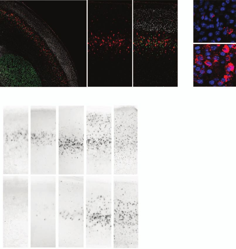
Timing and Neuronal Specificity of Grin3a Expression      Murillo et al.       1915

plasticity and have been proposed to fine-tune the refinement of       Materials and Methods
neural circuits during critical postnatal periods by inhibiting the
                                                                       Animals and Tissue Collection
stabilization of nonused synapses and promoting their pruning
(Pachernegg et al. 2012; Perez-Otano et al. 2016).                     Several strains of C57BL/6 J background mice were used:
    Recent studies point towards broader roles of GluN3A               WT, GAD67GFP (B6.Cg-Tg(Gad1-EGFP) (Tamamaki et al. 2003)
subunits in the mammalian brain. First, GluN3A can form                and Nkx2.1-Cre::Ai9tdTomato . The latter was obtained crossing
GluN1/GluN3 complexes that do not bind glutamate and                   Tg(Nkx2.1-Cre)2S and/J (JAX 008661; (Xu et al. 2008) and
instead generate excitatory currents activated by glycine              Ai9(B6.Cg-Gt(ROSA)26Sortm9(CAG-tdTomato)Hze/J (JAX 007909)
(Perez-Otano et al. 2001; Chatterton et al. 2002). Although            (Madisen et al. 2010). Mice were housed 4–6 per cage with
initially studied in recombinant systems, native GluN1/GluN3A          ad libitum access to food and water and maintained in a
receptors have now been identified in juvenile mouse hip-              temperature-controlled environment on a 12-h light/dark cycle
pocampus and adult medial habenula (Grand et al. 2018;                 at humidity between 40 and 60% in a standard pathogen free
Otsu et al. 2019). Rather than modulating plasticity, their            environment. All procedures were conducted in accordance with
activation potentiates basal firing rates and could have               the European and Spanish regulations (2010/63/UE; RD53/2013)

                                                                                                                                            Downloaded from https://academic.oup.com/cercor/article/31/4/1914/6027877 by guest on 14 April 2021
a major influence on neuronal excitability. Second, while              and were approved by the Ethical Committee of the Government
GluN3A protein and mRNA levels are largely down-regulated              of the Generalitat Valenciana (2017/VSC/PEA/00196).
after critical developmental periods, the Allen Brain Atlas                After deep anesthesia with isoflurane, mice were tran-
documents remaining Grin3a mRNA expression in adult                    scardially perfused with 4% paraformaldehyde in phosphate
brains and a variety of phenotypes of cognition, social, and           buffered saline (PBS) pH 7.4. Brains were removed and postfixed
aversive behavior have been reported in adult mice lacking             overnight at 4◦ C. From this step on, tissue treatment was
GluN3A, suggesting that functional relevance may continue              different depending on the later technique. For colorimetric and
throughout life (Mohamad et al. 2013; Lee et al. 2016; Otsu            fluorescence ISH, brains were then embedded in 4% agarose,
et al. 2019). Third, genetic mutations or alterations in human         sectioned coronally in 100 or 80 μm-thick slices respectively with
GRIN3A expression have been linked to disorders of cognition,          a vibrating microtome and stored in PBS at 4◦ C. For RNAscope
mood, and emotion such as schizophrenia, bipolar disorder,             assays, tissue was immersed in 30% sucrose at 4◦ C and frozen
or substance abuse, suggesting alterations in the control of           in OCT. Frozen brains were sectioned at 14 μm with a cryostat
affective, impulsive traits or executive functions (Mueller and        and stored at –80◦ C.
Meador-Woodruff 2004; Yang et al. 2015; Lee et al. 2016; Huang
et al. 2017). Importantly, preclinical mouse studies provided
                                                                       Cloning and In Situ Hybridization
causal relationships between age-inappropriate reactivation of
functional GluN3A expression and pathological spine pruning            Templates for synthesizing Grin3a riboprobes were obtained
in Huntington’s disease, or maladaptive forms of plasticity that       by PCR from cDNA libraries of hippocampus of C57BL/6 using
predispose to drug relapse (Marco et al. 2013, 2018; Yuan et al.       the following primers: forward GAACACATAGTGTACAGACTGC;
2013), making GluN3A an attractive candidate for therapeutic           reverse CTAGGATTCACAAGTCCGGTT. The resulting amplicon
intervention.                                                          was purified with QIAquick gel extraction kit- (QIAGEN) and
    At present it is unclear how GluN3A subunits subserve              cloned into pBlueScript SK plasmid. Plasmids for riboprobes
such diverse functions and whether links to behavior or                against Gad1 mRNA were a gift from Dr Jordi Guimer. The
disease endophenotypes reflect adult roles of GluN3A or                riboprobes were: Grin3a-ctd (spanning 495 bp of the C-terminal
faults in the developmental trajectories of specific neuronal          domain, accession number NM_001033351.2) and Gad1 (bp
circuits. A limitation for dissecting GluN3A functions has been        934–1786 of the Gad1 cDNA, accession number NM_008077).
the lack of a comprehensive understanding of the timing                Plasmids were linearized and riboprobes labeled with UTP-
and regional variations in GluN3A expression throughout                digoxigenin or UTP-fluorescein during in vitro transcrip-
development and in the adult brain, and of the cellular                tion using polymerase T3 or T7 (for antisense and sense
populations where GluN3A is expressed. To address this                 riboprobes, respectively). Riboprobes were then purified by
limitation, we have conducted a systematic analysis of Grin3a          RNeasy Mini Kit Qiagen and concentrations measured with
mRNA expression in the mouse brain from embryonic to early             NanoDrop.
postnatal and adult stages using colorimetric and fluorescence             Free-floating 100 μm-thick brain sections were permeabi-
in situ hybridization (ISH). We find that, while subject to a          lized with detergent mix (1% NP-40, 1% SDS, 0.5% sodium
prominent down-regulation, substantial Grin3a expression               deoxycholate, 50 mM Tris pH 8.0, 1 mM EDTA, and 150 mM
is retained in a subset of brain regions. Specifically, adult          NaCl) for 1 h at room temperature, and incubated with
Grin3a expression was highest in multimodal or associative             digoxigenin-labeled riboprobes at 63◦ C in hybridization buffer
areas such as the insular, prefrontal, and anterior cingulate          (50% formamide; 2x saline-sodium citrate pH 5.3; 50 μg/mL
cortices or the claustrum, and in areas encoding or integrating        heparin; 50 μg/mL tRNA; 50 μg/mL salmon sperm DNA; 0.1%
internal emotional or arousal/awareness states such as the             Tween-20) overnight. Hybridized probes were detected using
amygdala, medial habenula, or “high-order” thalamic nuclei.            an alkaline phosphatase-conjugated antidigoxigenin (Roche
Throughout postnatal development, Grin3a expression exhibits           11 093 274 910, 1:2000) in blocking solution (2% Roche Blocking
precise temporal dynamics that differ in neocortical areas across      reagent and 20% sheep serum in MABT buffer) overnight at
sensory modalities and cortical layer. At the cellular level, Grin3a   4◦ C, followed by several washes in MABT buffer (500 mM
is expressed by both excitatory and inhibitory neurons, with           maleic acid, 750 mM NaCl, 0.95 M NaOH, and 0.1% Tween-
high expression a distinguishing feature of somatostatin (Sst)         20, pH 7.5). For visualization, sections were then incubated at
interneurons.                                                          room temperature during 6 or 22 h in a solution containing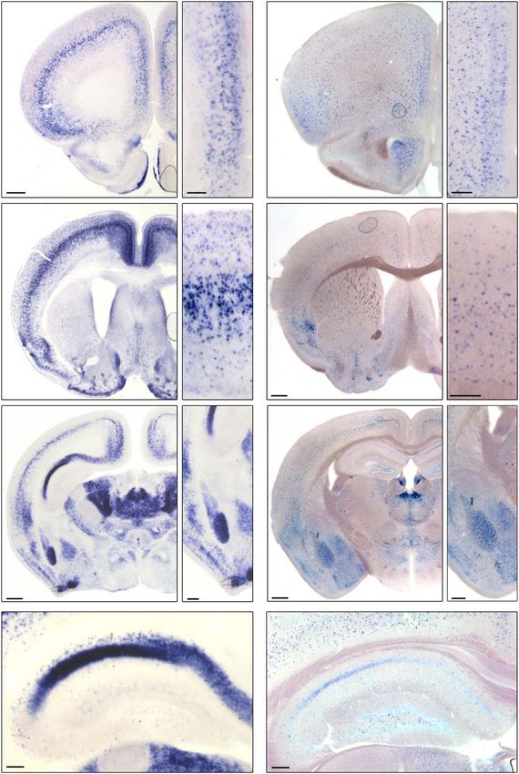
1916     Cerebral Cortex, 2021, Vol. 31, No. 4

nitroblue tetrazolium chloride (NBT) and 5-bromo-4-chloro-3 -      coordinates (Paxinos and Franklin, 2019 Edition), the Mouse
indolyphosphate p-toluidine salt (BCIP) in NTMT buffer (100 mM      nervous system (Watson, Paxinos, Puelles, 2012 Edition) and the
NaCl; 100 mM Tris-HCl pH 9.5; 50 mM MgCl2; 1% Tween-20)             Atlas of the developing mouse brain (Paxinos, Halliday, Watson,
that produces a colored precipitate due to the reaction with        Koutcherov, Wang, 2007 Edition) were used.
alkaline phosphatase. After several washes in PBS 1x, sections
were mounted onto Superfrost Plus slides (Thermo Fisher             RNAscope In Situ Hybridization Assay
Scientific™), dehydrated, and coverslipped with DPX mounting
solution.                                                           RNAscope® Multiplex Fluorescent Kit v2 (Advanced Cell
                                                                    Diagnostics) was used to perform ISH with the following
                                                                    probes: Grin3A-C1 (#551371), Gad1-C2 (#400951-C2), Slc17a7-
Fluorescence In Situ Hybridization                                  C3 (vGlut1)(#416 631-C3), Sst-C3 (#404631-C3), and tdTomato-C2
Identical protocol to the described above was followed, except      (#317041-C2).
80 μm-thick brain sections were used. Sections were incubated           Briefly, slides were dried for 60 min at –20◦ C, washed in
with digoxigenin- and fluorescein-labeled riboprobes (against       PBS for 5 min, then baked for 10 min at 60◦ C. The slides were
                                                                    postfixed by immersing them in prechilled 4% PFA for 10 min at

                                                                                                                                                Downloaded from https://academic.oup.com/cercor/article/31/4/1914/6027877 by guest on 14 April 2021
Grin3a and Gad1 mRNA, respectively) in hybridization buffer,
followed by incubation with peroxidase-conjugated antidigoxi-       ice, followed by two rinses in diH2 O. Slides were dehydrated for
genin or antifluorescein antibodies (Roche 11 207 733 910; Roche    5 min each in 50%, 70%, and 100% ethanol, then dried for 5 min at
11 426 346 910, 1:2000). A TSA plus fluorescence kit was used       RT. About 5–8 drops of RNAscope hydrogen peroxide was applied
for signal amplification and detection according to the manu-       to the slides for 10 min followed by diH2 O washing. Then, slides
facturer protocol (Thermo Fisher, NEL744001KT). To avoid non-       were transferred to a container with RNAscope target retrieval
specific signal, endogenous peroxidases were inactivated by         reagent and incubated at >95◦ C for 5 min, then incubated in
incubation with MABT-1% H2 O2 for 1 h in the dark. Sense probes     100% alcohol for 3 min. After drying the slides at RT, they were
used as control yielded no signal. Double fluorescence ISH was      treated with Protease III for 20 min at 40◦ C. After rinsing two
performed with dual hybridization and duplication of detec-         times in distilled water, 1X target probe mixes were applied to
tion and amplification steps. Sections were counterstained with     the brain sections and incubated at 40◦ C for 2 h in the HybEZ™
DAPI, mounted onto Superfrost Plus slides (Thermo Fisher), air-     oven (Advanced Cell Diagnostics). Sections were then incubated
dried, and cover-slipped with fluorescence mounting medium          with preamplifier and amplifier probes and develop HRP-C1,
(DAKO).                                                             C2, and C3 signals with TSA Plus fluorophores (PerkinElmer)
                                                                    by following the RNAscope® Multiplex Fluorescent Kit v2 user
                                                                    manual. After washing, sections were stained for 30 s with DAPI.
Immunohistochemistry
After the fluorescence ISH procedure, free-floating brain sec-      Statistical Analysis
tions were incubated with primary antibody in 1% BSA, 0.1%
                                                                    Statistical analysis was performed using GraphPad Prism 7. For
Triton X-100, and 1% NS in PBS overnight at 4◦ C. The following
                                                                    qualitative and semi-quantitative analysis of colorimetric ISH
primary antibodies were used: rabbit anti-GFP (Synaptic systems
                                                                    data in Figures 1–4 and Supplementary Table 1, the following
132 002, 1:1000), rabbit anti-Cux1 (Santa Cruz Biotechnology Sc-
                                                                    number of mice were analyzed: E17.5 (n = 3), P0 (n = 6), P3 (n = 8), P6
13 024, 1:500) and rat anti-Ctip2 (Abcam Ab18465, 1:500). After
                                                                    (n = 16, short and long exposure), P9 (n = 6), P12 (n = 4), adult (n = 3,
washing with PBS, sections were incubated with fluorophore-
                                                                    long exposure). Two-way ANOVA followed by Bonferroni’s mul-
conjugated secondary antibody in 1% BSA, 0.1% Triton X-100
                                                                    tiple comparisons posthoc test was used to assess differences
in PBS for 1 h at room temperature. The following secondary
                                                                    between groups in Figure 5D. In all figures, data are presented
antibodies were used: anti-rabbit-Cy3 (Jackson Immunoresearch
                                                                    as means ± SEM.
111 175 144, 1:500), anti-rat-488 (Invitrogen A11006, 1:500) and
anti-rabbit-488 (Invitrogen A11008, 1:500). After several washes
in PBS, sections were counterstained with DAPI, mounted onto        Results
Superfrost Plus slides, air-dried, and coverslipped with fluores-
                                                                    Predominant Postnatal Grin3a mRNA Expression
cence mounting medium (DAKO).
                                                                    and Down-Regulation into Adulthood
                                                                    We began by comparing the relative levels and regional pat-
Image Acquisition and Analysis
                                                                    terns of Grin3a mRNA expression in young (postnatal day (P)
Bright field images were acquired using a Leica DM6000B micro-      6) and adult (3–4 months) mouse brains using colorimetric ISH.
scope with 5X and 10X objectives or a MZ16FA stereomicroscope       Here and for most other experiments, sections were processed
equipped with a DC500 digital camera, and processed with Leica      using two different exposures to the developing solution. This
AF6000 software. Quantification of Grin3a expression across cor-    allowed us to achieve the high sensitivity needed for detecting
tical layers and sensory modalities was performed using Fiji        adult expression (22 h, high exposure, Fig. 1), while preserving a
for automatic detection of positive labeling after background       dynamic range for semi-quantitative assessment of expression
subtraction. Fluorescence confocal images were captured on an       levels at earlier postnatal stages (6 h, low exposure, Figs 2–4). P6
Olympus FV1200 with a 20X objective or a ZEISS AxioImager M2        was chosen based on previous quantitative immunoblot anal-
microscope with an Apochromat 20X objective, and processed          yses in rodent brain that reported maximal Grin3a expression
with FV10-ASW_Viewer and Zen Blue software, respectively. The       over the P5–P9 time window (Al-Hallaq et al. 2002; Wong et al.
colocalization of Grin3a-positive cells with neuronal subtype-      2002; Roberts et al. 2009).
specific markers was quantified using Fiji software.                    A first inspection confirmed that highest Grin3a mRNA
    For anatomical analysis, identification of structures and       levels are found postnatally and decline dramatically in most
functional interpretation, the Mouse brain in stereotaxic           brain regions into adulthood (Fig. 1). However, there were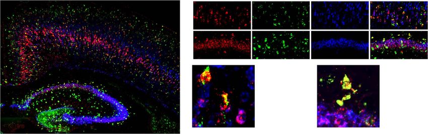
Timing and Neuronal Specificity of Grin3a Expression           Murillo et al.      1917

                                                                                      islands of Calleja), in nuclei of the contiguous cortical or
                                                                                      olfactory amygdala (anterior, ACo; and posteromedial, PMCo);
                                                                                      and in basolateral and central amygdala (Fig. 1B, D, F, see
                                                                                      Supplementary Fig. 1). Adult expression in the thalamus was
                                                                                      lower and more restricted than at P6, but high levels were
                                                                                      retained in some of the so-called “high-order” thalamic nuclei
                                                                                      including midline (paraventricular, intermediodorsal, reuniens,
                                                                                      rhomboid) and intralaminar nuclei (centromedial, paracentral,
                                                                                      centrolateral) (Fig. 1F, see Supplementary Fig. 1). It is worth
                                                                                      noting the exception of the medial habenula that displayed
                                                                                      an opposite pattern of Grin3a expression, with high adult levels
                                                                                      but low expression in young brains (Fig. 1E vs. F).
                                                                                          Our comparative analysis additionally revealed a clear dorso-
                                                                                      ventral gradient of Grin3a expression in principal neurons of

                                                                                                                                                          Downloaded from https://academic.oup.com/cercor/article/31/4/1914/6027877 by guest on 14 April 2021
                                                                                      adult CA1 hippocampus (see Supplementary Fig. 2). At P6, Grin3a
                                                                                      was highly expressed in CA1 pyramidal neurons throughout
                                                                                      the entire rostrocaudal extent of the hippocampus in both dor-
                                                                                      sal and ventral divisions (Fig. 1G, see Supplementary Fig. 2A,
                                                                                      B, D, E). In contrast, expression in dorsal CA1 was virtually
                                                                                      absent in adults but high levels were retained in ventral CA1
                                                                                      pyramidal neurons (Fig. 1H, see Supplementary Fig. 2C vs. F).
                                                                                      As previously reported (Sucher et al. 1995), no expression was
                                                                                      detected in CA2, CA3, or dentate gyrus at either age. However,
                                                                                      we noted a previously unappreciated strong Grin3a mRNA sig-
                                                                                      nal in a small subset of neurons in the CA1 stratum oriens
                                                                                      which was most prominent at P6 (see Supplementary Fig. 2,
                                                                                      insets).
                                                                                          As introduced above, shorter exposure to the developing
                                                                                      solution allowed a semi-quantitative analysis of postnatal
                                                                                      Grin3a expression levels across the rostro-caudal brain axis
                                                                                      (Fig. 2 and see Supplementary Table 1). The analysis revealed
                                                                                      a unique pattern of Grin3a expression with several remarkable
                                                                                      features. First, high Grin3a levels were observed in most
                                                                                      neocortical areas with predominance in deeper layers at P6
                                                                                      (Fig. 2A-H, see extended laminar analysis below). Expression
Figure 1. Grin3a expression is down-regulated into adulthood. Colorimetric
                                                                                      was also high throughout the entire rostro-caudal axis of the
ISH showing Grin3a mRNA expression in young (postnatal day (P)6; A, C, E,
                                                                                      olfactory cortex including the rostral anterior olfactory nucleus
G) and adult mouse brain (B, D, F, H). Sections were exposed for 22 h to the
developing solution to enhance sensitivity. Representative coronal sections at        and tenia tecta, the dorsal endopiriform nucleus and piriform
different rostro-caudal levels are displayed, and higher magnification images         cortex (Fig. 2B-H); in ACo and PMCo nuclei of the cortical
of the boxed areas are shown on the right. AI, agranular insular cortex; AON,         amygdala (Fig. 2E-H); and in the interposed cortex amygdala
anterior olfactory nucleus; BLA, basolateral amygdala; CA1, Cornu Ammonis 1;          transition zone (Fig. 2F, G). Second, high levels were found in
CeM, central amygdala; Cg, cingulate cortex; CL, centrolateral thalamic nucleus;
                                                                                      the basolateral, basomedial, and basoposterior divisions of the
Cla, claustrum; CM, centromedial thalamic nucleus; CoA, cortical amygdala;
                                                                                      amygdala. Lower but significant levels were detected in central
DEn, dorsal endopiriform nucleus; DG, dentate gyrus; DLG, dorsal lateral genic-
ulate nucleus; DP, dorsal peduncular cortex; Ect, ectorhinal cortex; ICj, islands     and lateral amygdala (Fig. 2F, G, H). Third, Grin3a expression
of Calleja; IL, infralimbic cortex; IMD, intermediodorsal thalamic nucleus; La,       was especially strong in the thalamus where it prevailed
lateral amygdala; MHb, medial habenula nucleus; Mo, motor cortex; OT, olfactory       in high-order thalamic nuclei including midline thalamic
tubercule; PC, paracentral thalamic nucleus, PFC, prefrontal cortex; Pir, piriform    nuclei (paraventricular, reuniens, rhomboid), intralaminar,
cortex; PrL, prelimbic cortex; PV, paraventricular thalamic nucleus; RS, retrosple-
                                                                                      mediodorsal, posterior, and lateroposterior nucleus (analogous
nial cortex; SS1, somatosensory cortex 1; TeA, temporal association cortex; TT,
                                                                                      to the primate pulvinar). By contrast, Grin3a was absent or
tenia tecta. Scale bars: 500 μm (A-F); 200 μm (G-H and insets).
                                                                                      expressed at very low levels in first-order, primary sensory
                                                                                      thalamic nuclei including the dorsolateral geniculate, ventral
                                                                                      division of the medial geniculate, or ventral posterior nuclei
notable exceptions. Substantial Grin3a levels were retained in                        (Fig. 2D-H).
specific cortical areas including the prefrontal cortex (limbic
and infralimbic divisions), anterior cingulate, retrosplenial,
                                                                                      Emergence and Down-regulation of Grin3a Expression
agranular insular, temporal association, ectorhinal, and perirhi-
                                                                                      Vary Across Brain Areas
nal cortices (Fig. 1B, D, F, see Supplementary Fig. 1). This
adult pattern of Grin3a expression correlates with a cortical                         While maximal Grin3a expression was previously reported in
hierarchy of functional specialization established based on                           P6–P9 brains and assumed to homogeneously decline across
connectivity patterns or structural properties (Fulcher et al.                        brain regions into adulthood, earlier embryonic expression has
2019; Harris et al. 2019). High adult levels of Grin3a mRNA                           been reported in human brains (Eriksson et al. 2002; Mueller
were also observed in areas of the olfactory cortex (tenia                            and Meador-Woodruff 2003). Furthermore, GluN3A-NMDARs
tecta, anterior olfactory nucleus, dorsoendopiriform nucleus,                         have been shown to control the propensity for precisely timed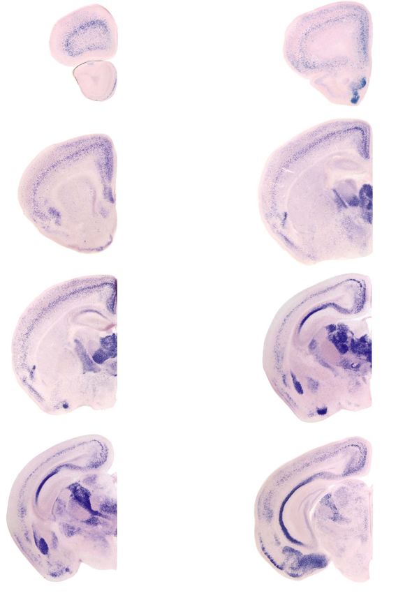
1918        Cerebral Cortex, 2021, Vol. 31, No. 4

                                                                                                                                                                           Downloaded from https://academic.oup.com/cercor/article/31/4/1914/6027877 by guest on 14 April 2021
Figure 2. Regional distribution of Grin3a expression in early postnatal development. (A-H) Representative images of colorimetric ISH for Grin3a mRNA in coronal sections
from P6 to P9 mouse brains. Here and in Figures 2–4, sections were exposed for 6 h to the developing solution. Corresponding schemes modified from the Paxinos’s
Atlas of the developing mouse brain are shown in the right, and mRNA expression levels are color-coded as indicated in the heatmap. The images were acquired at
different rostro-caudal levels. Abbreviations are listed in Supplementary Table 1. Scale bars: 500 μm.

forms of cortical plasticity such as the developmental onset                           but abundant in high-order nuclei (Fig 3C-E). Grin3a was also
of long-term potentiation in the hippocampus (Roberts et al.                           expressed at this early time-point in the CA1 hippocampus,
2009) or spike-timing dependent long-term depression in visual                         amygdala, cortical amygdala (Fig. 3C-E), and in the claustrum
cortex (Larsen et al. 2014) and could be predicted to display a                        (Fig. 3B), a nucleus highly interconnected with nearly all cortical
precise timing in specific brain regions. We thus investigated                         areas and involved in multimodal sensory processing (Kim et al.
the developmental time-course of Grin3a expression from                                2016).
embryonic mouse brain through multiple stages of postnatal                                 Grin3a expression in the neocortex was low at E17.5
development (P0, P3, P6, P9, P12).                                                     (Fig. 3A-E), increased postnatally, and down-regulated by
   We found that the age of emergence and down-regulation                              P12 (Fig. 4). Yet again differences in the timing of expres-
of Grin3a expression varies significantly across brain regions.                        sion were noted across neocortical areas. Grin3a expres-
By embryonic day (E) 17.5, high levels of Grin3a mRNA were                             sion appeared earliest (by P0) in cingulate, retrosplenial,
detected in the thalamus following a similar pattern to the                            and piriform cortices (Fig. 4F, K, P). By P3, expression was
observed postnatally, that is, absent/low in sensory thalamus                          observed in most of the neocortex including prefrontal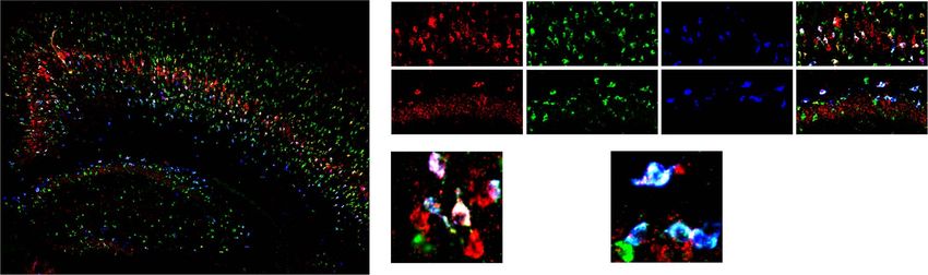
Timing and Neuronal Specificity of Grin3a Expression           Murillo et al.      1919

                                                                                   reached (Fig. 4E, J, O, T). Expression in CA1 became restricted to
                                                                                   its ventral division (Figure 4O, T, see Supplementary Fig. 2).

                                                                                   Temporal Dynamics of Cortical Grin3a Expression
                                                                                   Across Layers and Sensory Modalities
                                                                                   A major feature of developmental Grin3a expression in the neo-
                                                                                   cortex was the initial predominance as an intense band in
                                                                                   deeper cortical layers with later spread to other cortical lam-
                                                                                   inae. To establish the laminar specificity of Grin3a expression,
                                                                                   we combined fluorescence ISH with immunohistochemistry for
                                                                                   the layer-specific markers Ctip2 and Cux1 that label pyramidal
                                                                                   neurons in layer V and layers II–IV, respectively. Focused on P6
                                                                                   primary somatosensory cortex (SS1), we found that the highest

                                                                                                                                                        Downloaded from https://academic.oup.com/cercor/article/31/4/1914/6027877 by guest on 14 April 2021
                                                                                   number of Grin3a-positive fluorescent cells populate layer V as
                                                                                   determined by colocalization with Ctip2 (Fig. 5A). A subset of
                                                                                   layer V cells could be distinguished by their strong Grin3a mRNA
                                                                                   labeling (Fig. 5B, bottom panel). Only a few Grin3a-positive cells
                                                                                   with much lower fluorescence signal were detected in layers
                                                                                   II/III (Fig. 5B, upper panel).
                                                                                       A finer temporal analysis covering earlier to late postnatal
                                                                                   stages revealed a highly reproducible pattern of sequential
                                                                                   Grin3a expression, which extends from deep to superficial
                                                                                   layers, but which timing varied across sensory modalities.
                                                                                   For instance: in SS1 Grin3a expression begins early in layer V,
                                                                                   with Grin3a-positive cells already detected in newborns (P0).
                                                                                   A gradual increase in the number of Grin3a-positive cells and
                                                                                   Grin3a intensity levels in layer V was observed through P3–P6.
                                                                                   Laminar specificity became less apparent at P9 and was lost by
                                                                                   P12 (Fig. 5C, upper panels). Quantification of signal intensities
                                                                                   across SS1 confirmed a sharp peak of Grin3a expression in
                                                                                   layer V which reached a maximum at P6, and appearance
                                                                                   of a second peak in layers II/III by P9 (Fig. 5D, upper graph).
                                                                                   Expression in layer V declined significantly by P9-P12 (Fig. 5D,
                                                                                   red vs. blue and green traces; see Supplementary Table 2). A
                                                                                   comparison with primary visual cortex (V1) demonstrated a
                                                                                   delayed onset of Grin3a expression relative to SS1 (Fig. 5C,
                                                                                   bottom panels) or motor cortex (not shown). By P6 only a
Figure 3. Embryonic Grin3a expression. (A-E) Representative images of colorimet-
                                                                                   few, weakly labeled, Grin3a-positive cells were observed in
ric ISH showing Grin3a mRNA expression in E17.5 mouse embryos at various
rostro-caudal levels. Abbreviations: ACo, anterior cortical amygdaloid nucleus;
                                                                                   layer V of V1 and expression peaked around P9–P12 (Fig. 5D,
AM, anteromedial thalamic nucleus; BLA, basolateral amygdala, anterior part;       bottom graph). The spread to upper layers had not reached
BLP, basolateral amygdala, posterior part; BMP, basomedial amygdala, poste-        statistical significance by P12 and layer V down-regulation was
rior part; BST, bed nucleus of the stria terminalis; CA1, Cornu Ammonis 1;         also delayed relative to SS1 (blue vs. green lines in Fig. 5D, see
CeM, central amygdaloid nucleus; Cg, cingulate cortex; Cla, claustrum; CM,         Supplementary Table 2). The analysis demonstrates a precise
centromedial thalamic nucleus; CPu, caudate-putamen; CxA, cortex amygdala
                                                                                   laminar distribution of Grin3a expression, which changes over
transition zone; DEn, dorsal endopiriform nucleus; fr, fasciculus retrof lexus;
La, lateral amygdala; LP, lateral posterior thalamic nucleus; MD, medio dorsal
                                                                                   postnatal development and varies across sensory modalities,
thalamic nucleus; mt, mammillothalamic tract; OT, olfactory tubercle; PFC,         as exemplified by the shift in emergence and down-regulation
prefrontal cortex; Pir, piriform cortex; PMCo, posteromedial cortical amygdaloid   between SS1 and V1.
nucleus; Po, posterior thalamic nuclear group; PV, paraventricular nucleus; Rt,
reticular thalamic nucleus; VEn, ventral endopiriform nucleus; VG, ventrolateral
geniculate nucleus; VM, ventromedial thalamic nucleus; VMH, ventromedial           Grin3a is Expressed in Excitatory and Inhibitory
hypothalamic nucleus; VP, ventroposterior thalamic nucleus; ZI, zona incerta.      Neurons, with Predominance in Sst-Expressing
Scale bar: 500 μm.
                                                                                   Interneurons
                                                                                   We next aimed to achieve cellular resolution and define which
                                                                                   neuronal subpopulations express Grin3a, focusing on SS1 cortex
and motor cortex and primary sensory cortices such as                              and dorsal hippocampus at P6–P9. The neocortex is composed
the somatosensory and auditory (Fig. 4B, G, L, Q). Express-                        of a majority of excitatory glutamatergic neurons (∼80%) and a
ion in visual cortex was delayed and appeared around P6                            lower number of inhibitory GABAergic neurons (∼20%). Thus, we
(see Fig. 4P-R).                                                                   conducted multiplex FISH assays using RNAscope probes for the
   By P12, down-regulation had taken place in most regions                         vesicular glutamate transporter vGlut1, the GABA-synthesizing
and the characteristic adult pattern illustrated in Figure 1, with                 enzyme Gad1 (glutamate decarboxylase 1), and Grin3a (Fig 6A).
restriction to the amygdala, olfactory cortex, cortical amyg-                      In SS1, Grin3a mRNA was expressed in both excitatory and
dala, claustrum, and selected cortical and thalamic regions, was                   inhibitory neurons as demonstrated by colocalization of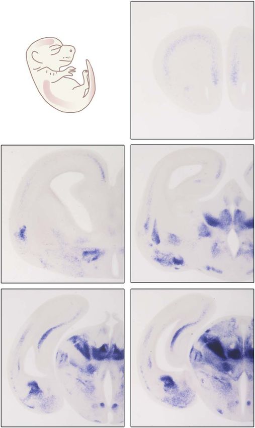
1920        Cerebral Cortex, 2021, Vol. 31, No. 4

                                                                                                                                                                           Downloaded from https://academic.oup.com/cercor/article/31/4/1914/6027877 by guest on 14 April 2021
Figure 4. Emergence and down-regulation of Grin3a expression throughout postnatal developmental stages. (A-T) Colorimetric ISH for Grin3a mRNA in mouse brain at
different postnatal (P) stages. Representative images of coronal sections at different rostro-caudal levels and indicated ages are shown. Abbreviations: ACo, anterior
cortical amygdaloid nucleus; AI, agranular insular cortex; AON, anterior olfactory nucleus; Au, auditory cortex; BLA, basolateral amygdala; BLP, basolateral amygdaloid
nucleus, posterior part; CA1, cornu ammonis 1; Cg, cingulate cortex; Cla, claustrum; CPu, caudate-putamen; CxA, cortex amygdala transition zone; DEn, dorsal
endopiriform nucleus; Ect, ectorhinal cortex; ICj, islands of Calleja; La, lateral amygdaloid nucleus; Mo, motor cortex; PFC, prefrontal cortex; OT, olfactory tubercle;
Pir, piriform cortex; PMCo, posteromedial cortical amygdaloid nucleus; RS, retrosplenial cortex; SS1, somatosensory cortex 1; TeA, temporal association cortex; Thal,
thalamus; TT, tenia tecta; Vis, visual cortex; VMH, ventromedial hypothalamic nucleus. Scale bar: 500 μm.

punctate nuclear labeling with vGlut1 or Gad1 mRNA (Fig. 6B).                          of inhibitory Gad1-positive neurons that displayed strong
Quantitatively, about half of Grin3a-positive neurons were Gad1-                       Grin3a labeling in the stratum oriens, whereas inhibitory
positive (51.57% ± 1.63) and the other half were vGlut1-positive                       interneurons intercalated in the CA1 pyramidal layer or in the
(48.29% ± 1.66) (Fig. 6D). Strongest expression was typically                          stratum radiatum lacked Grin3a expression (Fig. 6C, E). Similar
found in GABAergic interneurons (Fig. 6D). In the hippocampus,                         results were obtained when Grin3a localization in inhibitory
a majority of CA1 pyramidal excitatory neurons expressed                               interneurons was examined by conventional double FISH
Grin3a (Fig. 6C). We additionally identified a subpopulation                           with riboprobes against Grin3a and Gad1 (see Supplementary
Timing and Neuronal Specificity of Grin3a Expression Murillo et al. 1921
Downloaded from https://academic.oup.com/cercor/article/31/4/1914/6027877 by guest on 14 April 2021
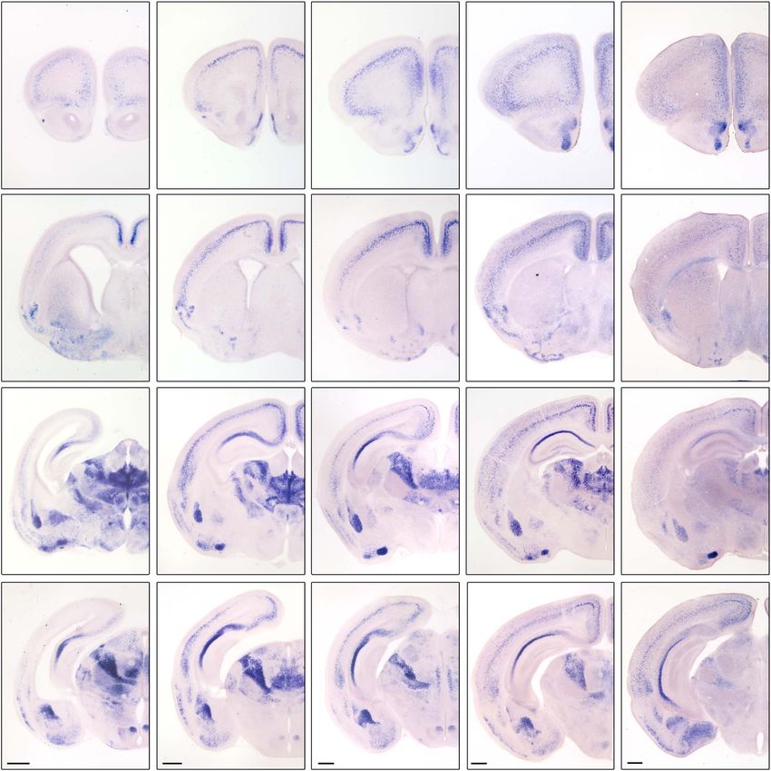
Figure 5. Dynamic temporal patterns of Grin3a expression across cortical layers and sensory modalities. (A) Left, mosaic of single confocal images of somatosensory
cortex 1 (SS1) at P6 showing f luorescence ISH labeling of Grin3a mRNA (red) combined with immunof luorescence staining for Citp2 (green) and Cux1 (gray). Middle,
right: higher magnification images of the boxed area on the left. Dotted lines mark boundaries between areas (left panel) or cortical layers (middle-right panels). (B)
High-magnification images of boxed areas in layers II–III and V of the middle panel in A. Grin3a mRNA (red) and DAPI-stained nuclei (blue) are shown. Solid arrows
indicate cells with high Grin3a mRNA levels; empty arrows show cells with low Grin3a mRNA levels. (C, D) Comparative analysis of the time-courses of Grin3a emergence
and down-regulation in SS1 and primary visual cortex (V1). (C) Representative images show colorimetric ISH for Grin3a mRNA in SS1 and V1 at indicated postnatal
ages. Dotted lines mark layer V boundaries. (D) Graphs of Grin3a mRNA intensity levels across layers in SS1 (top) and V1 (bottom) at P0 (gray), P3 (dashed red), P6 (red),
P9 (blue), P12 (green). Data are mean ± S.E.M (n = 3 mice per postnatal age; 2 cortical fields taken at different levels were analyzed per mouse). Shown are statistical
significances between P0 and P6 (∗∗∗∗ P < 0.0001) and between P0 and P9 (### P < 0.001, #### P < 0.0001) calculated using two-way ANOVA followed by Bonferroni’s multiple
comparisons test. All others can be found in Supplementary Table 2. CPu, caudate-putamen; SS1, primary somatosensory cortex 1; V1, primary visual cortex 1. Scale
bars: 100 μm (A), 20 μm (B), 100 μm (C).1922 Cerebral Cortex, 2021, Vol. 31, No. 4
Downloaded from https://academic.oup.com/cercor/article/31/4/1914/6027877 by guest on 14 April 2021
Figure 6. Grin3a expression in excitatory and inhibitory neurons. (A) Representative coronal section from a P6 wild-type mouse brain labeled by RNAscope for Grin3a
mRNA (red) and markers of inhibitory (green: Gad1 mRNA) and excitatory neurons (blue: vGlut1 mRNA). (B, C) High-magnification images of the boxed areas in A
corresponding to SS1 and CA1 hippocampal subfield. In (C), dotted lines mark the boundary between the different hippocampal layers. (D) Higher magnification of the
boxed area in B and quantification of percentage of Grin3a-positive cells in SS1 layer V which express inhibitory or excitatory markers at P6–P9. (E) Higher magnification
of the boxed area in C and quantification of percentage of Grin3a mRNA positive cells in the SOr of CA1 that colocalize with Gad1. Solid arrows: Grin3a inhibitory neurons
as shown by colocalization with Gad1 mRNA. Empty arrowheads: Grin3a excitatory neurons as shown by colocalization with vGlut1 mRNA. Data are mean ± S.E.M (n = 4
animals, 6 SS1 fields, and 5 SOr of CA1 at different antero-posterior levels were analyzed). Ex, excitatory neurons; In, inhibitory neurons; SOr, stratum oriens; SP, stratum
pyramidale. Scale bars: 100 μm (A); 25 μm (B-C); 5 μm (D-E).
Fig. 3A) or using GAD67GFP mice (Tamamaki et al. 2003) (see major postnatal down-regulation known a priori, and addition-
Supplementary Fig. 3B, C). ally reveal highly dynamic and differential Grin3a regulation
The two most prevalent types of interneurons are fast- across different brain regions. We further show that Grin3a is
spiking parvalbumin-containing interneurons and low-threshold expressed in both excitatory and inhibitory neurons, and iden-
spiking Sst interneurons, which are generated in the medial tify a previously underappreciated enrichment of Grin3a mRNA
ganglionic eminence (MGE) and account for ∼70% of all in Sst interneurons. The work provides a roadmap for dissecting
interneurons (Tricoire et al. 2011; Lim et al. 2018). To determine the diverse functions attributed to GluN3A-NMDARs in brain
which interneuron populations express Grin3a, we used Nkx2– physiology, behavior, and disease states.
1-cre:Ai9tdTomato transgenic mice where the tdTomato signal is During early postnatal life, neuronal circuits in the brain
restricted to MGE-derived interneurons (Madisen et al. 2010) are massively remodeled by experience via long-lasting plastic-
(Fig. 7A-C). Quantitatively, 52.14% ± 2.44 of Grin3a-expressing ity mechanisms that stabilize subsets of synapses but weaken
cells colocalized with tdTomato mRNA in SS1 layer V (Fig. 7D), or eliminate others (Katz and Shatz 1996). GluN3A-NMDARs
a figure almost identical to the 51.57% of Grin3a-positive cells have been proposed to control the timing and magnitude of
that colocalize with Gad1 (Fig. 6D), indicating that essentially all this remodeling by limiting classical NMDAR signaling until
Grin3a-expressing interneurons derive from MGE progenitors. the arrival of sensory experience, and/or fine-tuning synapse
Of these, the majority expressed Sst mRNA at high levels plasticity and maturation at later stages (Roberts et al. 2009;
(34.88% ± 2.45 of the Grin3a-expressing population that is ∼70% Fiuza et al. 2013; Perez-Otano et al. 2016). Our anatomical work
of all Grin3a-positive interneurons, Fig. 7D). At present, it is lends support for this idea by showing early, sequential, and
unclear if the remaining tdTomato-positive/Sst-negative cells transient (or protracted) expression of Grin3a in cortical areas
belong to a population of immature Sst-fated interneurons that over postnatal development.
have not yet upregulated Sst mRNA levels, a different MGE- The variations in the timing of Grin3a expression and down-
derived population such as parvalbumin cells, or a subset of regulation across cortical regions correlate with differences in
MGE-derived cells that do not express either parvalbumin or time-scales of cortical maturation (Guillery 2005) and degree of
Sst (Petros et al. 2015). Since parvalbumin is not upregulated in functional specialization (Wang 2020). For instance, transient
cortical interneurons until the second postnatal week, we are waves of Grin3a expression are a characteristic of primary
unable to directly address this. Similar results were obtained sensory cortices (see Figure 5), whereas expression appears
in the stratum oriens where virtually all Grin3a-expressing early and is retained in association and multimodal areas.
cells expressed tdTomato and Sst (Fig. 7E). Conversely, nearly Primary sensory cortices provide modality-specific information,
all Sst-positive interneurons expressed Grin3a (96.39% ± 0.98 of and early maturation guided by experience is needed to sculpt
the total Sst population in SS1 layer V, 91.98% ± 3.10 in stratum receptive fields and preserve the fidelity of the information. By
oriens, Fig. 7F). contrast, associative and multimodal cortices play an integrative
role, have long, protracted maturation periods, and maintain
a potential for plastic responses that primary sensory areas
lose early (Guillery 2005). Adult Grin3a expression was also
Discussion observed in noncortical areas with strong plasticity needs
We provide a comprehensive description of Grin3a expression in into adulthood or a functional requirement for association of
the mouse brain from embryonic to postnatal and adult stages. multiple inputs such are the olfactory system, the amygdala, or
Our results recapitulate and expand with anatomical detail the the claustrum (see Figure 1). Within primary sensory cortices,Timing and Neuronal Specificity of Grin3a Expression Murillo et al. 1923
Downloaded from https://academic.oup.com/cercor/article/31/4/1914/6027877 by guest on 14 April 2021
Figure 7. Grin3a expression in molecularly defined interneuron populations. RNAscope was used to assess colocalization of Grin3a and Sst mRNA in Nkx2–1-
cre:Ai9tdTomato mice that report MGE-derived interneurons. (A) Mosaic image of a P6 Nkx2–1-cre:Ai9tdTomato mouse brain labeled for Grin3a mRNA (red) and specific
cell-types (green: tdTomato mRNA; blue: Sst mRNA, somatostatin interneurons). Images of the corresponding boxes from SS1 (B) and CA1 area (C). Dotted lines mark
the boundary between the different hippocampal layers. (D) Image of the boxed area in B (left panel) and quantification of Grin3a-positive cells that colocalize with
tdTomato or Sst mRNA in SS1 layer V of P6–P9 mice (right panel). (E) Image of the boxed area in C (left panel) and quantification of Grin3a-positive cells that colocalize
with tdTomato or Sst mRNA in the CA1 stratum oriens of P6–P9 mice (right panel). (F) Quantification of Sst mRNA positive cells that express Grin3a mRNA in SS1 layer
V and stratum oriens. Solid arrows: Grin3a MGE-derived SSt interneurons as shown by colocalization with tdTomato and Sst mRNA. Empty arrowheads do not show
colocalization and are likely Grin3a-positive excitatory neurons. Data are mean ± S.E.M (n = 4 animals, 5–6 fields at different rostro-caudal levels were analyzed). SOr,
stratum oriens; SP, stratum pyramidale. Scale bars: 100 μm (A); 25 μm (B-C); 5 μm (D-E).
Grin3a expression follows a stereotyped sequential pattern with also been shown to control the developmental onset of long-
initial restriction to layer V followed by expression in outer layers term forms of plasticity such as hippocampal long-term poten-
and overall down-regulation (see Figure 5), which resembles tiation (Roberts et al. 2009), spike-timing dependent long-term
the inside-out patterning model of cortical maturation. While depression in V1 (Larsen et al. 2014), or the synaptic incorpo-
this stereotyped pattern is conserved across motor and sensory ration of GluN2A subunits and AMPA receptors (Henson et al.
cortices, its timing varies and is coupled to the arrival of sensory 2012). Other properties such as the maintenance of persistent
experience as exemplified by the delayed Grin3a expression or recurrent activity in the prefrontal cortex could reflect roles
in visual cortex, an area of the neocortex that matures later of GluN3A-NMDARs or excitatory GluN1/GluN3A receptors on
coinciding with eyelid opening (Yoshii et al. 2003). NMDA spiking activity or neuronal excitability (Mahfooz et al.
Our findings confirm and expand a recent report that found 2016; Otsu et al. 2019). Finally, association and frontal areas
Grin3a expression in adult mouse brain to be one of the strongest contain a greater proportion of Sst interneurons that innervate
correlates with a hierarchical gradient of functional integra- dendrites and have an “input-modulating function,” while sen-
tion—from primary sensory to transmodal association cortices, sory areas contain more parvalbumin interneurons that inner-
established using the T1w/T2w magnetic resonance imaging vate the soma and axon initial segment and have an “output-
ratio (Fulcher et al. 2019). In primate and mouse cortex, the modulating” function.
T1w/T2w ratio inversely correlates with structural markers of Grin3a expression is very high in the thalamus where it
increased differentiation and stability across cortices (Garcia– prevails in higher order thalamic nucleus, while primary sensory
Cabezas et al. 2017; Wang 2020). Low T1w/T2w ratios and high thalamic areas lack Grin3a or express it at low levels. Infor-
Grin3a expression were found to be typical of functionally inte- mation from the sensory periphery is first transmitted to the
grative areas in mouse and human brains (Fulcher et al. 2019), cerebral cortex via the primary sensory, or first-order, thala-
as also observed here. This is the case of the agranular cortex mic nuclei including the lateral geniculate in visual thalamus,
that receives not only sensory inputs but integrates multimodal ventral division of the medial geniculate in auditory thalamus,
information, or of the temporal association and rhinal cortices and ventral posterior nuclei in somatosensory thalamus. By
and the prefrontal cortex (Gogolla 2017). It is worth noting that contrast, higher order thalamic nuclei do not receive substan-
genetic variations in human GRIN3A have been reported to tial input from the sensory periphery and instead form exten-
modulate prefrontal cerebral activity during selective attention sive cortico-thalamic-cortical circuits or connect functionally
tasks (Gallinat et al. 2007), supporting a role in higher cognitive related cortical and subcortical regions. They provide a route for
processing. cortico-cortical communication which integrates cortical and
A number of parameters that vary over postnatal develop- subcortical inputs, linking cognition and motivated behavior to
ment and along the cortical hierarchy to fulfill specific func- internal states and levels of consciousness (Sherman 2007). Of
tional requirements could be related to the Grin3a expression relevance here, highest adult Grin3a expression was observed
described here. For instance, GluN3A enhances spine turnover in midline and intralaminar nuclei such as the centromedial
(Kehoe et al. 2014), and high rates of disassembly and forma- and paraventricular, involved in arousal and wakefulness, and
tion of synapses and spines during postnatal critical periods in the medial habenula, an epithalamic structure that controls
are thought to be permissive for refinement while low spine negatively valued emotional associations (Van der Werf et al.
turnover leads to circuit stabilization. GluN3A subunits have 2002; Ren et al. 2018; Redinbaugh et al. 2020). Consistent with1924 Cerebral Cortex, 2021, Vol. 31, No. 4
these findings, unbiased mammalian reverse genetics detected Supplementary Material
alterations in wakefulness-sleep transitions in Grin3a knockout
Supplementary material can be found at Cerebral Cortex online.
mice (Sunagawa et al. 2016), and GluN1/GluN3A glycine recep-
tors in the medial habenula have been implicated in mediating
conditioned place-aversion (Otsu et al. 2019). Notes
Finally, our data revealed particularly high Grin3a mRNA
We are grateful to Francisca Almagro-García for expert technical
levels in Sst interneurons of SS1 and hippocampus from early
help, to Drs Francisco Clascá, Eloísa Herrera, Salvador Martínez,
postnatal stages. This is in-line with single-cell RNAseq analyses
Elisa Mengual, John Wesseling, and Óscar Elía-Zudaire for advice
of gene expression in adult mouse somatosensory and visual
on manuscript preparation, and Miguel Pérez-Otaño for help
cortex interneurons (Pfeffer et al. 2013; Paul et al. 2017) (T Pet-
with graphic design.
ros, et al, unpublished data). Publicly available transcriptome
We acknowledge support of the publication fee by the CSIC
datasets indicate that Grin3a expression might be a conserved
Open Access Publication Support Initiative through its Unit of
feature of Sst interneurons across species (http://interneuron.
Information Resources for Research (URICI). Conflict of Interest:
mccarrolllab.org/). Moreover, a recent genome-wide epigenomic
None declared.
Downloaded from https://academic.oup.com/cercor/article/31/4/1914/6027877 by guest on 14 April 2021
analysis identified the Grin3a locus as a site of open chromatin
and low DNA methylation (mCG) in Sst interneurons (Yao et al.
2020). Both epigenetic marks are typical of actively expressed
Funding
genes and are thought to promote and maintain cell-type spe-
cific expression and cell identity. Spanish Ministry of Education and Science (BES-2014-069359
Together these data provide strong validation of Grin3a fellowship to A.M., SAF2016-80895-R grant to I.P.O., Severo-
expression as a secondary marker for Sst interneurons. Ochoa Excellence Awards SEV-2013-0317 and SEV-2017-0723);
They further suggest that GluN3A subunits might play more the Generalitat Valenciana (PROMETEO 2019/020 grant to I.P.O);
general roles than currently envisioned in modulating the a NARSAD Independent Investigator Award (to I.P.O.); and NICHD
development of neural circuits. Along with the well-studied Intramural Funding (to Y.Z. and T.J.P.).
refinements of connections between excitatory principal
neurons, circuit formation requires the network integration of
local interneurons that also involves a refinement of excitatory
References
synaptic inputs onto these inhibitory cells (De Marco Garcia Al-Hallaq RA, Jarabek BR, Fu Z, Vicini S, Wolfe BB, Yasuda RP.
et al. 2015; Tuncdemir et al. 2016). For example, Sst interneurons 2002. Association of NR3A with the N-methyl-D-aspartate
in infragranular layers of SS1 receive dense but transient receptor NR1 and NR2 subunits. Mol Pharmacol. 62:1119–1127.
thalamocortical innervation during the first postnatal week. Chatterton JE, Awobuluyi M, Premkumar LS, Takahashi H, Talan-
This innervation is required for the later functional maturation tova M, Shin Y, Cui J, Tu S, Sevarino KA, Nakanishi N et al.
and synaptic integration of parvalbumin interneurons, but is 2002. Excitatory glycine receptors containing the NR3 family
reduced by 4-fold by P14 (Tuncdemir et al. 2016). Our data show of NMDA receptor subunits. Nature. 415:793–798.
that Grin3a is highly expressed between P3 and P9 in layer V Sst De Marco Garcia NV, Priya R, Tuncdemir SN, Fishell G, Karayan-
interneurons, providing a candidate mechanism for mediating nis T. 2015. Sensory inputs control the integration of neu-
the refinement of thalamocortical inputs. Excitatory synapses rogliaform interneurons into cortical circuits. Nat Neurosci.
onto MGE-derived interneurons undergo a defined program of 18:393–401.
synapse maturation, with predominance of GluN2B-NMDARs in Eriksson M, Nilsson A, Froelich-Fabre S, Akesson E, Dunker J,
neonates which are later replaced by GluN2A-NMDARs (Matta Seiger A, Folkesson R, Benedikz E, Sundstrom E. 2002. Cloning
et al. 2013; Perszyk et al. 2016). This developmental switch in and expression of the human N-methyl-D-aspartate receptor
subunit composition resembles the experienced by excitatory subunit NR3A. Neurosci Lett. 321:177–181.
neurons, in which GluN3A down-regulation has been implicated Fiuza M, Gonzalez-Gonzalez I, Perez-Otano I. 2013. GluN3A
(Perez-Otano and Ehlers 2004; Henson et al. 2012). expression restricts spine maturation via inhibition of
In sum, our study offers new insight onto the spatiotem- GIT1/Rac1 signaling. Proc Natl Acad Sci USA. 110:20807–20812.
poral patterns of Grin3a distribution in the brain and opens Fulcher BD, Murray JD, Zerbi V, Wang XJ. 2019. Multimodal
up important cell biological and functional questions. At the gradients across mouse cortex. Proc Natl Acad Sci USA.
cell biological level, further investigations on regulation will 116:4689–4695.
be needed to define the transcriptional and epigenetic mecha- Gallinat J, Gotz T, Kalus P, Bajbouj M, Sander T, Winterer G.
nisms that determine the area-to-area and temporal variations 2007. Genetic variations of the NR3A subunit of the NMDA
in Grin3a expression. Is experience a critical determinant factor, receptor modulate prefrontal cerebral activity in humans. J
as suggested by the observation that rearing rats in the dark Cogn Neurosci. 19:59–68.
prevents GluN3A down-regulation in visual cortex (Larsen et al. Garcia-Cabezas MA, Joyce MKP, John YJ, Zikopoulos B, Barbas H.
2014); or is regulation by the calcium-regulated transcription 2017. Mirror trends of plasticity and stability indicators in
factor CaRF (Lyons et al. 2016)? In terms of function, the transient primate prefrontal cortex. Eur J Neurosci. 46:2392–2405.
expression of Grin3a in cortical areas and temporal correlation Gogolla N. 2017. The insular cortex. Curr Biol. 27:R580–R586.
with hierarchical cortical maturation emphasizes the concept of Grand T, Abi Gerges S, David M, Diana MA, Paoletti P. 2018.
GluN3A as a key controller of the timing of brain maturation Unmasking GluN1/GluN3A excitatory glycine NMDA recep-
and its coupling to experience. Further advances will require tors. Nat Commun. 9:4769.
selective deletion of the Grin3a gene at specific times and in spe- Guillery RW. 2005. Is postnatal neocortical maturation hierarchi-
cific neuronal populations, combined with tools to distinguish cal? Trends Neurosci. 28:512–517.
roles of GluN3A-NMDARs from those of the yet more enigmatic Harris JA, Mihalas S, Hirokawa KE, Whitesell JD, Choi H, Bernard
excitatory GluN1/GluN3 receptors. A, Bohn P, Caldejon S, Casal L, Cho A et al. 2019. HierarchicalTiming and Neuronal Specificity of Grin3a Expression Murillo et al. 1925
organization of cortical and thalamic connectivity. Nature. Otsu Y, Darcq E, Pietrajtis K, Matyas F, Schwartz E, Bessaih
575:195–202. T, Abi Gerges S, Rousseau CV, Grand T, Dieudonne S et al.
Henson MA, Larsen RS, Lawson SN, Perez-Otano I, Nakanishi N, 2019. Control of aversion by glycine-gated GluN1/GluN3A
Lipton SA, Philpot BD. 2012. Genetic deletion of NR3A accel- NMDA receptors in the adult medial habenula. Science. 366:
erates glutamatergic synapse maturation. PLoS One. 7:e42327. 250–254.
Huang X, Chen YY, Shen Y, Cao X, Li A, Liu Q, Li Z, Zhang LB, Dai Pachernegg S, Strutz-Seebohm N, Hollmann M. 2012. GluN3
W, Tan T et al. 2017. Methamphetamine abuse impairs motor subunit-containing NMDA receptors: not just one-trick
cortical plasticity and function. Mol Psychiatry. 22:1274–1281. ponies. Trends Neurosci. 35:240–249.
Katz LC, Shatz CJ. 1996. Synaptic activity and the construction of Paoletti P, Bellone C, Zhou Q. 2013. NMDA receptor subunit
cortical circuits. Science. 274:1133–1138. diversity: impact on receptor properties, synaptic plasticity
Kehoe LA, Bellone C, De Roo M, Zandueta A, Dey PN, Perez-Otano and disease. Nat Rev Neurosci. 14:383–400.
I, Muller D. 2014. GluN3A promotes dendritic spine pruning Paul A, Crow M, Raudales R, He M, Gillis J, Huang ZJ.
and destabilization during postnatal development. J Neurosci. 2017. Transcriptional architecture of synaptic communica-
34:9213–9221. tion delineates GABAergic neuron identity. Cell. 171:522,
Downloaded from https://academic.oup.com/cercor/article/31/4/1914/6027877 by guest on 14 April 2021
Kim J, Matney CJ, Roth RH, Brown SP. 2016. Synaptic organi- e520–539.
zation of the neuronal circuits of the claustrum. J Neurosci. Paxinos G, Franklin K. 2019. Mouse brain in stereotaxic coordinates.
36:773–784. 5th ed. Amsterdam (Netherlands): Academic Press.
Larsen RS, Smith IT, Miriyala J, Han JE, Corlew RJ, Smith SL, Paxinos G, Halliday G, Watson C, Koutcherov Y, Wang H. 2007.
Philpot BD. 2014. Synapse-specific control of experience- Atlas of the developing mouse brain. 1st ed. Amsterdam (Nether-
dependent plasticity by presynaptic NMDA receptors. Neuron. lands): Academic Press.
83:879–893. Perez-Otano I, Ehlers MD. 2004. Learning from NMDA recep-
Lee JH, Wei L, Deveau TC, Gu X, Yu SP. 2016. Expression of the tor trafficking: clues to the development and maturation of
NMDA receptor subunit GluN3A (NR3A) in the olfactory sys- glutamatergic synapses. Neurosignals. 13:175–189.
tem and its regulatory role on olfaction in the adult mouse. Perez-Otano I, Larsen RS, Wesseling JF. 2016. Emerging roles
Brain Struct Funct. 221:3259–3273. of GluN3-containing NMDA receptors in the CNS. Nat Rev
Lim L, Mi D, Llorca A, Marin O. 2018. Development and functional Neurosci. 17:623–635.
diversification of cortical interneurons. Neuron. 100:294–313. Perez-Otano I, Schulteis CT, Contractor A, Lipton SA, Trimmer JS,
Lyons MR, Chen LF, Deng JV, Finn C, Pfenning AR, Sabhlok A, Sucher NJ, Heinemann SF. 2001. Assembly with the NR1 sub-
Wilson KM, West AE. 2016. The transcription factor calcium- unit is required for surface expression of NR3A-containing
response factor limits NMDA receptor-dependent transcrip- NMDA receptors. J Neurosci. 21:1228–1237.
tion in the developing brain. J Neurochem. 137:164–176. Perszyk RE, DiRaddo JO, Strong KL, Low CM, Ogden KK, Khatri
Madisen L, Zwingman TA, Sunkin SM, Oh SW, Zariwala HA, Gu A, Vargish GA, Pelkey KA, Tricoire L, Liotta DC et al. 2016.
H, Ng LL, Palmiter RD, Hawrylycz MJ, Jones AR et al. 2010. GluN2D-containing N-methyl-d-aspartate receptors mediate
A robust and high-throughput Cre reporting and charac- synaptic transmission in hippocampal interneurons and reg-
terization system for the whole mouse brain. Nat Neurosci. ulate interneuron activity. Mol Pharmacol. 90:689–702.
13:133–140. Petros TJ, Bultje RS, Ross ME, Fishell G, Anderson SA. 2015.
Mahfooz K, Marco S, Martinez-Turrillas R, Raja MK, Perez-Otano Apical versus basal neurogenesis directs cortical interneuron
I, Wesseling JF. 2016. GluN3A promotes NMDA spiking by subclass fate. Cell Rep. 13:1090–1095.
enhancing synaptic transmission in Huntington’s disease Pfeffer CK, Xue M, He M, Huang ZJ, Scanziani M. 2013. Inhi-
models. Neurobiol Dis. 93:47–56. bition of inhibition in visual cortex: the logic of connec-
Marco S, Giralt A, Petrovic MM, Pouladi MA, Martinez-Turrillas tions between molecularly distinct interneurons. Nat Neu-
R, Martinez-Hernandez J, Kaltenbach LS, Torres-Peraza J, rosci. 16:1068–1076.
Graham RK, Watanabe M et al. 2013. Suppressing aber- Redinbaugh MJ, Phillips JM, Kambi NA, Mohanta S, Andryk S,
rant GluN3A expression rescues synaptic and behavioral Dooley GL, Afrasiabi M, Raz A, Saalmann YB. 2020. Thalamus
impairments in Huntington’s disease models. Nat Med. modulates consciousness via layer-specific control of cortex.
19:1030–1038. Neuron. 106:66, e12–75.
Marco S, Murillo A, Perez-Otano I. 2018. RNAi-based GluN3A Ren S, Wang Y, Yue F, Cheng X, Dang R, Qiao Q, Sun X, Li X, Jiang
silencing prevents and reverses disease phenotypes induced Q, Yao J et al. 2018. The paraventricular thalamus is a critical
by mutant Huntington. Mol Ther. 26:1965–1972. thalamic area for wakefulness. Science. 362:429–434.
Matta JA, Pelkey KA, Craig MT, Chittajallu R, Jeffries BW, McBain Roberts AC, Diez-Garcia J, Rodriguiz RM, Lopez IP, Lujan R,
CJ. 2013. Developmental origin dictates interneuron AMPA Martinez-Turrillas R, Pico E, Henson MA, Bernardo DR, Jarrett
and NMDA receptor subunit composition and plasticity. Nat TM et al. 2009. Downregulation of NR3A-containing NMDARs
Neurosci. 16:1032–1041. is required for synapse maturation and memory consolida-
Mohamad O, Song M, Wei L, Yu SP. 2013. Regulatory roles of tion. Neuron. 63:342–356.
the NMDA receptor GluN3A subunit in locomotion, pain Sherman SM. 2007. The thalamus is more than just a relay. Curr
perception and cognitive functions in adult mice. J Physiol. Opin Neurobiol. 17:417–422.
591:149–168. Sucher NJ, Akbarian S, Chi CL, Leclerc CL, Awobuluyi M, Deitcher
Mueller HT, Meador-Woodruff JH. 2003. Expression of the NR3A DL, Wu MK, Yuan JP, Jones EG, Lipton SA. 1995. Develop-
subunit of the NMDA receptor in human fetal brain. Ann N Y mental and regional expression pattern of a novel NMDA
Acad Sci. 1003:448–451. receptor- like subunit (NMDAR-L) in the rodent brain. J Neu-
Mueller HT, Meador-Woodruff JH. 2004. NR3A NMDA receptor rosci. 15:6509–6520.
subunit mRNA expression in schizophrenia, depression and Sunagawa GA, Sumiyama K, Ukai-Tadenuma M, Perrin D,
bipolar disorder. Schizophr Res. 71:361–370. Fujishima H, Ukai H, Nishimura O, Shi S, Ohno RI, NarumiYou can also read