"CHARACTERIZATION OF CAMPYLOBACTER-SPECIFIC BACTERIOPHAGES FOR ASSESSMENT OF THEIR USE IN FOOD PRODUCTION AND PROCESSING." - TIHO ELIB
←
→
Page content transcription
If your browser does not render page correctly, please read the page content below
University of Veterinary Medicine Hannover
          Institute for Food quality and food safety (LMQS)

   „Characterization of Campylobacter-specific
 bacteriophages for assessment of their use in food
           production and processing.”

„Charakterisierung von Campylobacter-spezifischen
Bakteriophagen für eine Beurteilung ihrer Eignung in
           der Lebensmittelproduktion.“

  Submitted in partial fulfilment of the requirements for the degree of
                       Doctor of Natural Sciences

                     - Doctor rerum naturalium –
                            (Dr. rer. nat.)

                                  by
                        Severin Michael Steffan
                         Hannover, Germany

                       Hannover, Germany 2022
Wissenschaftliche Betreuung: Prof. Dr. Corinna Kehrenberg, PhD
Betreuungsgruppe : Prof. Dr. Corinna Kehrenberg, PhD
Institute for Veterinary Food Science (IfTN)
Justus-Liebig-University (JLU), Gießen,
Germany
Prof. Dr. George Herrler, Dr. rer. nat.
Institute for Virology
University of Veterinary Medicine Hannover
(TiHo), Hannover, Germany
Zweitgutachter: Prof. Dr. Martin Hasselmann
Specialist group population genomics of
livestock,
University of Hohenheim, Hohenheim, Germany
Tag der mündlichen Prüfung: 12.05.2022
iParts of this thesis have been presented to the public prior to publications at following
conferences:
“Characterisation of novel Campylobacter-specific Bacteriophages from Germany
Identification of promising candidates for use along the food production chain”
Severin Michael Steffan, Golshan Shakeri, Jens Andre Hammerl, Corinna Kehrenberg, Elisa
Peh, Manfred Rohde, Claudia Jäckel, Madeleine Plötz, Sophie Kittler
Oral presentation: “ 73. DGHM Jahrestagung”, 12. – 14. September 2021, online-meeting
“Characterization of group II and group III bacteriophage combinations for use along
the food production chain against Campylobacter coli and jejuni”
Severin Michael Steffan, Golshan Shakeri, Jens Andre Hammerl, Corinna Kehrenberg, Elisa
Peh, Manfred Rohde, Claudia Jäckel, Madeleine Plötz, Sophie Kittler
Poster presentation: “Zoonoses 2021 – International Symposium on Zoonoses Research”,
13 – 15 October 2021, online-meeting
iiAcknowledgments Throughout the preparation and writing of this dissertation I have received support and assistance by many individuals. Fist I would like to thank my supervisor Prof. Dr. Corinna Kehrenberg for the opportunity to write this dissertation and its topic. In equal parts I would like to thank both members of my supervision group, Prof. Dr. Corinna Kehrenberg and Prof. Dr. George Herrler, for being part my supervision group as well as supplying insightful feedback. In addition, I would like to thank Prof. Dr. Madeleine Plötz for allowing me to work at her institute (LMQS), including the opportunity to expand my horizon via the Junior Scientist Zoonoses Meeting. Moreover, I would like to thank our Cooperation partners Dr. Jens Andre Hammerl and Dr. Claudia Jäckel from the Department of Biological Safety situated at the German Federal Institute for Risk Assessment (BfR) for their efforts in trying to sequence and assemble bacteriophage genomes. Furthermore, I would like to thank Prof. Dr. Manfred Rohde and his technical staff for all the electron micrographs they prepared and sent my way. I would like to thank my involved scientist Dr. Sophie Kittler for a stable work relationship and help in preparing two publications. I would like thank my former colleagues Dr. Elisa Peh and Dr. Golshan Shakeri for a perfect work atmosphere, friendship, feedback and a sympathetic ear. In addition, I would like to thank the technical staff at the Research Center for Emerging Infections and Zoonoses (RIZ) and the Institute for Food quality and - safety (LMQS), especially Anke Bertling, Iris Oltrogge, Silke Ortaeri and Inna Pahl without whom I would have been unable to shoulder the high workloads. Furthermore, I would like to thank Inna Pahl for the PFGE and qPCR results I used to do phage group associations. Finally, I would like to thank Lebensmittel- und Veterinärlabor GmbH, Emstek, Germany for generous financial support of this study, including the supply of some Campylobacter field isolates and three Campylobacter phages. iii
Table of Contents

1. Introduction ..................................................................................................... 1
2. Einleitung ......................................................................................................... 3
3. Summary of results ......................................................................................... 5
3. Zusammenfassung der Ergebnisse ................................................................ 9
4. First Publication ............................................................................................ 14
5. Second Publication ........................................................................................ 16
6. Discussion ....................................................................................................... 18
  6.1 Introduction...................................................................................................................... 18
  6.2 General phage safety and current regulations .................................................................... 18
  6.3 Phage production .............................................................................................................. 19
  6.4 Phage applications ............................................................................................................ 19
  6.5 Overview over Campylobacter phages ................................................................................ 20
  6.6 General considerations...................................................................................................... 21
  6.7 Resistance and defense mechanisms .................................................................................. 21
  6.9 Host range ........................................................................................................................ 23
  7. Phage cocktails ................................................................................................................... 24
  7.1 Outlook............................................................................................................................. 25
References…………………………………………………………………..…27

                                                                                                                                          iv
List of abbreviations
- CBHI calcium supplemented brain heart infusion broth
- CFU colony forming units
- CPS capsule polysaccharides
- CsCl Cesium chloride
- DNA desoxyribonucleic acid
- DNS Desoxyribonukleinsäure
- DST direct spot test
- EU European Union
- EFSA European Food Safety Authority
- EOP Efficiency of Plating
- FDA Food and Drug Administration
- GIT gastro intestinal tract
- h hour
- ICTV International Committee on Taxonomy of Viruses
- kb kilobase pairs
- LPS lipooligigosaccharides
- NCTC National Collection of Type Cultures, United Kingdom
- OD600 optical density measured at 600 nm
- Omps outer membrane proteins
- phage short form of bacteriophage
- PKA planktonic killing assay
- PFGE Pulsed-field gel electrophoresis
- qPCR quantitative real time PCR
- RBP receptor binding protein
- R-M restriction modification system
- µl microliter or (10-6) liter
vList of figures
Figure 1 Distribution of defense systems in 500 Campylobacter jejuni predicted by the defenseFinder
program.. ................................................................................................................................................. 7
Figure 2 Distribution of defense systems in 109 Campylobacter coli predicted by the defenseFinder
program. .................................................................................................................................................. 7
Figure 3 Tests for remaining phage susceptible isolates after single phage applications during PKA. . 8
Abbildung 1 Verteilung von Verteidigungs Systemen gegen Phagen in 500 Camypylobacter jejuni;
hervorgesagt von dem defenseFinder Programm. ................................................................................. 11
Abbildung 2 Verteilung von Verteidigungs Systemen gegen Phagen in 109 Camypylobacter coli;
hervorgesagt von dem defenseFinder Programm. ................................................................................. 12
Abbildung 3 Tests um die verbleibende Anzahl an Phagen empfänglichen Isolaten nach Einzelphagen
Applikations PKAs festzustellen. .......................................................................................................... 13
viv
1. Introduction

Campylobacteriosis is a foodborne bacterial gastroenteritis. It is the most frequently reported
illness in the European Union (EU) since 2005 [1], as well as an important zoonosis worldwide
[2]. In general, consumption of raw or undercooked broiler meat is considered the most
common cause for human infections [3]. Symptoms of campylobacteriosis include watery to
hemorrhagic diarrhea and abdominal pain. In rare occasions severe but long-term sequelae can
occur such as Guillain-Barré syndrome, reactive arthritis, and erythema nodosum [2, 4, 5].
Campylobacter jejuni and coli are the most frequent species linked to campylobacteriosis cases
[1]. They are thermophilic, gram-negative Proteobacteria that thrive in microaerophilic
environments such as intestines of various wild, domestic, and farm animals [1, 6]. During
slaughter and meat processing, the pathogens can be spread form the gastro intestinal tract onto
the animal carcasses. Later occurring contaminations of other foodstuffs due to cross-
contamination during food preparation are possible [7-9].
Bacteriophages (phages) are bacterial viruses, whose bactericidal effects were first described
independently by Frederick Twort in 1915 and by Félix Hubert d’Hérelle in 1917, both were
credited with the discovery [10].
The ideal phage life cycle for commercial use would be a lytic, nontemperate one. It starts with
free floating virions that bind to their host cells via receptor-binding proteins (RBPs) in a two-
stage attachment process. First, they reversibly bind to an attachment site on the bacterial
surface, then bind irreversibly to the same receptor or shift to a secondary one, and finally inject
the phage’s genetic material into the host cell [11]. Then the host cell’s genomic DNA is
degraded and the cell’s metabolism is directed towards phage biosynthesis. After enough phage
DNA and proteins have been assembled into virions, host cells are lysed and the virions are
released [12]. Furthermore, there are phages with life cycles that are described as temperate,
which means, the phage’s DNA may be maintained in the host cell either as an episome or as a
prophage, that was integrated into the bacterial chromosome, and/or the life cycle is chronic,
which means that new virions are released without destruction of the host cell [12]. Therefore,
genomes of newly isolated phages have to be checked for genes that indicate a temperate and/or
chronic life style if they are intended for use in food production. Campylobacter phages are
non-enveloped, tailed bacteriophages, with icosahedral heads containing AT-rich double
stranded genomic DNA [13]. These phages belong to the order of Caudovirales, that includes
the families Myoviridae, Siphoviridae and Podoviridae. Most Campylobacter phages, including
all officially recognized by the International Committee on Taxonomy of Viruses (ICTV), are
Myoviridae. Siphoviridae have been described, but are few in numbers [14]. The ICTV
subdivides the Myoviridae based on DNA sequence analysis further into two genera,
Firehammerviruses and Fletcherviruses. A former typing scheme based on genome length
estimation by gel electrophoresis divides Campylobacter phages into three groups (group I
~320 kb, group II ~184 kb, and group III ~138 kb) [15, 16]. Currently, group II viruses overlap
with the Firehammerviruses and group III viruses overlap with the Fletcherviruses. The two
genera differ in lysis regulation [17], in the use of different noncanonical nucleotides [18], in
host range and primary attachment site [16]. Potential secondary receptors for Campylobacter
phages are still undiscovered. Group II phages infect Campylobacter coli and jejuni and
recognize their hosts via flagella proteins, while group III phages are restricted to infecting
Campylobacter jejuni and bind to their host’s capsular polysaccharides (CPS) [15, 16, 19-22].
The use of potential alternative receptors like lipopolysaccharides (LPS), outer membrane
proteins (Omps), and porins, have not yet been described for Campylobacter phages [14].
Intervention strategies geared towards reducing Campylobacter in poultry meat production are
still a work in progress. However, the use of phages in food production could become an integral
part of these strategies, as phages could be employed at the preharvest, while the animals are
                                                                                                  1
still alive, and/or postharvest level in processing plants or packaging [14]. Nevertheless, both scenarios differ widely in temperature and pH values or bacterial metabolic states [14]. Therefore, different phage cocktail compositions and application strategies will be required, and thus the fast characterization of Campylobacter-specific phages and the assessment of their use in food production and processing will become a much more important task in the future. Aims of the present study were to test the stability of phages at different temperatures and pH values comparable to food production settings, to investigate the effects of different phage concentrations on Campylobacter jejuni growth (1st publication), and to identify promising group II and group III phage combinations for use in a phage cocktail capable of minimizing Campylobacter jejuni or coli (2nd publication). 2
2. Einleitung

Campylobacter-Enteritis        ist    eine    lebensmittelassoziierte,   bakteriell verursachte
Schleimhautentzündung von Magen und Darm. Es handelt sich hierbei um eine bedeutende,
weltweit vorkommende Zoonose [2], die seit 2005 die meist gemeldete Krankheit in der
Europäischen Union (EU) ist [1]. Für gewöhnlich werden Infektionen des Menschen mit dem
Verzehr von rohem oder nicht durcherhitztem Hühnerfleisch in Zusammenhang gebracht [3].
Die Symptome einer Campylobacter-Enteritis können wässrigen bis blutigen Durchfall und
Unterleibsschmerzen umfassen. In seltenen Fällen können auch schwere Langzeitfolgen wie
das Guillain-Barré Syndrom, reaktive Arthritis und Erythema nodosum auftreten [2, 4, 5].
Campylobacter jejuni und coli sind die am häufigsten mit Campylobacter-Enteritis assoziierten
Spezies [1]. Es handelt sich um wärmeliebende, gram-negative Proteobakterien, die sich in
sauerstoffarmen Umgebungen wie dem Magen-Darmtrakt von verschiedenen Wild-, Haus- und
Nutztieren vermehren [1, 6]. Während des Schlachtprozesses und der anschließenden
Fleischverarbeitung kann der Krankheitserreger vom Magen-Darmtrakt auf den Schlachtkörper
übertragen werden. Im Anschluss sind Kreuzkontaminationen auf andere Lebensmittel
während des Zubereitungsprozesses möglich [7-9].
Bakteriophagen (Phagen) sind Bakterien-spezifische Viren, deren bakterizide Wirkung
unabhängig voneinander durch Frederick Twort in 1915 und Félix Hubert d’Hérelle in 1917
beschrieben wurden, beiden wird gleichermaßen ihre Entdeckung zugeschrieben [10].
Der lytische, nicht temperente Lebenszyklus gilt als ideal für kommerzielle Phagen
Anwendungen. Er beginnt mit freischwimmenden Viruspartikeln, deren Rezeptorbindeproteine
(RBPs) in einem zweistufigen Bindungsprozess an die Wirtszellen andocken. Zunächst binden
sie reversibel an eine Bindungsstelle auf der Bakterienoberfläche, um dann in einem zweiten
Schritt irreversibel an denselben Rezeptor zu binden oder zu einem Zweiten zu wechseln. Im
Anschluss wird das Phagen Genom in die Wirtszelle injiziert [11]. Danach wird das
Wirtsgenom abgebaut und der Metabolismus der Wirtszelle wird auf die Produktion von
Phagen umgestellt. Nachdem genügend Phagen DNA-Moleküle und Proteine zu Viruspartikeln
zusammengebaut wurden, werden die Wirtszellen lysiert und die neuen Viruspartikel
freigesetzt [12].
Darüber hinaus gibt es auch Phagen mit einem Lebenszyklus, der als temperent beschrieben
wird. Während dieses Zyklus kann die Phagen DNA in den Wirtszellen in Form eines Episomes
oder als Prophage, der ins Wirtsgenom integriert wurde, bewahrt werden und/oder der
Lebenszyklus ist chronisch, dies bedeutet, dass Viruspartikel freigesetzt werden, ohne die
Wirtszelle zu zerstören [12]. Dementsprechend müssen die Genome von neu isolierten Phagen
daraufhin überprüft werden, ob Gene vorhanden sind, die auf einen temperenten und/oder
chronischen Lebenszyklus hinweisen, wenn sie im Bereich der Lebensmittelproduktion
eingesetzt werden sollen. Campylobacter Phagen sind unbehüllte Bakteriophagen mit
schwanzförmigen Anhängseln, mit ikosaedrischen Köpfen gefüllt mit AT-reicher
doppelsträngiger DNA [13]. Diese Phagen gehören zur Ordnung Caudovirales, die die Familien
Myoviridae, Siphoviridae und Podoviridae einschließt. Die meisten Campylobacter Phagen,
einschließlich aller die offiziell durch das Internationale Komitee für die Taxonomie von Viren
(ICTV) anerkannt wurden, sind Myoviridae. Es gibt auch eine kleine Anzahl beschriebener
Siphoviridae [14]. Basierend auf DNA-Sequenz Analysen unterteilt die ICTV die Myoviridae
in zwei Gattungen, in die der Firehammerviren und die der Fletcherviren. Ein älteres
Typisierungschema auf Basis von Genomlängenabschätzungen per Gelelektrophorese unterteilt
Campylobacter Phagen in drei Gruppen (Gruppe I ~320 kb, Gruppe II ~184 kb und Gruppe III
~138 kb) [15, 16]. Im Moment kann man Gruppe II Viren mit den Firehammerviren und Gruppe
III Viren mit den Fletcherviren gleichsetzen. Die Gattungen unterscheiden sich in der
Regulation der Lyse [17], im Einsatz von nicht kanonischen Nukleotiden [18], in ihren
Wirtsspektren und ihrer primären Bindungsstelle [16]. Potentielle sekundäre Rezeptoren sind
                                                                                             3
für Campylobacter Phagen bis jetzt unbekannt. Gruppe II Phagen infizieren Campylobacter coli und jejuni und erkennen ihre Wirte auf Basis von Flagellen-Proteinen, währen Gruppe III Phagen auf Campylobacter jejuni beschränkt sind und an die Kapselpolysaccharide (CPS) ihrer Wirte binden [15, 16, 19-22]. Das Binden an alternative Rezeptoren wie Lipooligosaccharide (LPS), Membranproteinen (Omps) und Porine ist für Campylobacter Phagen noch nicht beschrieben worden [14]. Interventionsstrategien, die darauf abzielen Campylobacter in der Geflügelfleischproduktion zu minimieren, sind immer noch in der Entwicklung. Der Einsatz von Phagen in der Lebensmitteproduktion könnte jedoch ein integraler Bestandteil dieser Strategien werden, da Phagen sowohl vor der Schlachtung, also während der Mästung, und/oder nachdem Schlachtvorgang im Fleischverarbeitenden Betrieb oder während der Verpackung eingesetzt werden könnten [14]. Trotzdem unterscheiden sich die beiden Einsatzszenarien in der Temperatur, den pH-Wert Bereichen und dem zu erwartenden metabolischen Status der Bakterien [14]. Daher werden unterschiedliche Phagencocktail Zusammensetzungen und Applikationsstrategien nötig sein, die in Zukunft eine schnelle Charakterisierung von Campylobacter-spezifischen Phagen und eine Beurteilung ihrer Eignung in der Lebensmittelproduktion notwendig machen werden. Die Ziele der vorliegenden Studie waren die Stabilität von Phagen zu testen, wenn diese Temperaturen und pH-Werten ausgesetzt sind, die auch in der Lebensmittelproduktion anzutreffenden sind, sowie Untersuchungen der Effekte von unterschiedlichen Phagenkonzentrationen auf das Wachstum von Campylobacter jejuni (1. Veröffentlichung) und die Identifizierung von vielversprechende Kombinationen von Gruppe II und Gruppe III Phagen für einen Phagencocktail gerichtet gegen Campylobacter jejuni oder coli (2. Veröffentlichung). 4
Characterization of Campylobacter-specific bacteriophages for
assessment of their use in food production and processing
Severin Michael Steffan

3. Summary of results

In total, 401 samples form poultry farms (ncecal = 136, nfecal = 111, nneck-skin =54) form Lower
Saxony, Germany, were used to isolate new Campylobacter specific phages. Campylobacter
jejuni NCTC 12662 was used to isolate putative group III phages for the first publication (n =
19) and Campylobacter coli 084610 to isolate putative group II phages for the second
publication (n = 18) [15, 22]. The host range of the phages was determined by a direct spot test
(DST) assay with a newly characterized bacteria panel combined with phage dilution series [15,
23]. The phage dilution series allowed the calculation of the efficiency of plating (EOP). EOP
allows a relative comparison of phage susceptibility between different bacteria. EOP values are
obtained by comparing the plaque formation ability of a specific phage on one strain with the
plaque formation ability on a reference strain. For the group II phages, Campylobacter coli
084610, and for the group III phages, Campylobacter jejuni NCTC 12662, were chosen as
reference stains. The bacteria panel consisted of 28 Campylobacter isolates or strains. These
bacteria were chosen after characterization by flaA-typing [24] and SmaI-macrorestiction
analysis [25].
Based on the host ranges, eight phages were chosen, three group II and five group III phages,
of which the first group of phages was used for Publication 1 and the second for Publication 2.
Group 1 consisted of four group III phages (vB_CjM-LmqsCP1-4 (CP1-4), vB_CjM-
LmqsCP1-5 (CP1-5), vB_CjM-LmqsCP74-2c1 (CP74-2c1), and vB_CjM-LmqsCP132-3c
(CP132-3c)) while group 2 consisted of three group II phages (vB_CcM-LmqsCPL1/2
(CPL1/2), vB_CcM-LmqsCP218-2c2 (CP218-2c2), and LmqsCP288/3 (CP288/3)) and one
group III phage (vB_CjM-LmqsCP1-1 (CP1-1)). Selection of phage-bacteria combinations was
facilitated by comparison of EOP values.
Electron micrographs, done at HZI Braunschweig, allowed identifying these phages as
members of the Myoviridae family and to determine dimensional parameters like tail lengths,
head diameters and head lengths. The length of the Campylobacter phage genomes was
estimated by PFGE. Group II phage genomes had a length of 174 - 175 kb, while the group III
phage genomes were shorter with a length of 144 - 152 kb. Also, all group II phage genomes
were resistant to degradation by the restriction endonuclease HhaI (5’…GCGꜜC…3’) while
group III phage genomes were not. Further tests revealed that group II phage genomes were
susceptible to degradation by SwaI (5’…ATTTꜜAAAT…3’). The group identities were
confirmed by qPCR results [26].
DNA of all eight phages was extracted by a combination of virion purification via CsCl density
centrifugation with a kit-based DNA extraction protocol [15]. The DNA was then sent to our
cooperation partners at the German Federal Institute for Risk Assessment, Berlin Germany,
who subjected it to short read, paired-end sequencing on different Illumina devices [15].
However, sequencing or assembly of all eight phage genomes failed.
The four group III phages (CP1-4, CP1-5, CP74-2c1 and CP132-3c) were subjected to different
pH and temperature ranges to determine their stability under practical conditions. To test the
phages stability over long periods of storage (“shelf life”) and during applications, the four
group III phages were subjected to a series of tests. First, the average reduction rates of all
phages in a defined volume of 1 ml were determined. At 23.5 °C, the reduction rates stayed
between 1.29 to 1.82 PFU/month, while at 4 °C, they dropped to values between 1.02 to 1.20
PFU/month [15]. Afterwards these phages were subjected to a range of different pH values (pH
                                                                                              5
2 - 11) at two different temperatures for 24 hours. At 22 °C all phages were stable in a pH range from 3 to 11, while at 42 °C all phages were stable in a pH range from 4 to 11. Remarkable were the differences that occurred at pH 3 at 42 °C: only one phage (CP1-4) was stable for 24 hours under these conditions [15]. Subsequent to these results, an additional test with the other three group III phages (CP1-5, CP74-2c1, and CP132-3c) was conducted to investigate if they could remain stable for at least 2 hours (pH 3 at 42 °C), the minimum estimated time for upper intestinal track passage. All three phages appeared to be stable enough to fulfil this requirement [15]. Follow up tests were conducted to prove that the phages would lose activity after heat treatment in a kitchen stetting. Heat treatments were applied at 50, 60, 70 and 80 °C for 15 or 60 minutes. Results showed that heat inactivation of all phages could be achieved after 15 min at 70 °C [15]. To test the efficiency of phages to inhibit or hinder bacterial population growth, planktonic killing assays (PKAs) were performed. Liquid cultures of field isolates were incubated in the presence and absence of phages using a Tecan Spark Multiplate reader. Phages and bacteria were combined at defined ratios at the start of the experiments. Changes in cell density were recorded by optical density measurements at OD600 for 24 or 26 hours [15, 22]. For the first publication, the effects of four group III phages on two Campylobacter jejuni field isolates (Cj18 and LH83) were tested with single phage applications at five different multiplicity of infection ratios (MOIinput: 10 – 0.001) [15]. The MOIinput refers here to the ratio of virions per bacteria cell. Results showed that all phages were effective in inhibiting the growth of the two isolates. However, while for six phage bacteria combinations a significant inhibition of bacteria growth occurred at all MOIinput (Cj18 + CP1-4 or CP1-5; LH86 + CP1-4, CP1-5, CP74-2c1, or CP132-3c), for two combinations this only occurred at the highest MOIinput (Cj18 + CP74-2c1 or CP132-3c). Moreover, for these six phage bacteria combinations the highest MOIinput did not result in the significant differences in inhibition of bacterial growth compared to lower MOIinput [15]. For the second publication three group II (CPL1/2, CP218-2c2, and CP288/3) and one group III (CP1-1) phages were tested in combination with two Campylobacter jejuni (LH83 and Cj18) and one coli (Cc4) field strains. Time constrains restricted the number of MOIinput to the highest and the lowest (10 and 0.001) of the previous experiments. Phages were added as one, two or four phage applications [22]. The aim of this experiment was to evaluate the effect of phage combinations on bacterial population growth. As suggested by Zachary et al. the mean virulence index (meanvi) was used to determine the phage or phage combinations with the best bacterial inhibition at both high and low MOIinput [27]. Results showed that application of multiple phages could be more efficient at inhibiting bacterial population growth than single phage applications. In case of Cj18 all phage combinations outperformed single group II phage applications, while for LH83 all combinations with group II and group III phages outperformed single and double group II applications. The Campylobacter coli isolate Cc4 was resistant against the group III phage from the beginning, while all single and multiple group II phage applications were similar effective [22]. It was not possible to take samples during the experiments to determine the change in phage concentrations over time without significantly disrupting bacterial growth. The thermophily of Campylobacter bacteria was assumed to be the cause for this problem. Therefore, the phage concentrations at the end of the experiments were used as an indicator for successful phage replication. The mean adjusted phage concentrations (meanc24) were calculated to evaluate the overall phage replication at high and low MOIinput [22]. The host specificity of group II and group III phages allowed to determine phage concentrations separately. Results showed that certain combinations of group II and group III phages could increase the end phage concentration of one or both groups of phages. Reasons for this are however unknown [22]. To investigate the distribution of defense systems, protein faster files of 109 Campylobacter coli and 500 Campylobacter jejuni were uploaded to the online tool DefenseFinder [28, 29] 6
(Supplementary Table 1 and 2 / unpublished data). Results showed a similar distribution of
restriction modification systems (R-M) in Campylobacter coli and jejuni (see Figure 1 and 2).
Surprisingly, many Campylobacter jejuni contained PARIS, CAS, but no Zorya systems (see
Figure 1), while DRT and Hachiman systems were relative often found in Campylobacter coli
(see Figure 2).
Figure 1 Distribution of defense systems in 500 Campylobacter jejuni predicted by the defenseFinder program.
The histogram depicts the number of defense systems found in one strain dependent on the relative number of
strains containing a certain number of these systems (unpublished data).
Figure 2 Distribution of defense systems in 109 Campylobacter coli predicted by the defenseFinder program.
The histogram depicts the number of defense systems found in one strain dependent on the relative number of
strains containing a certain number of these systems (unpublished data).
Moreover, for all PKA experiments of the second publication potential phage resistant bacteria
colonies were collected [22]. At the end of each experiment 100 µl of well content was diluted
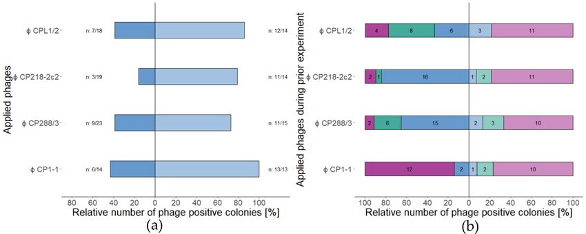
7by 3 log10 units with liquid media (CBHI) and 100 µl of the final dilution was plated on blood agar plates. After 48 to 72 h, at least five colonies were picked per plate and stored at -80 °C for further tests. Time constraints and problems with bacteria growth after phage exposure just allowed a limited number of phage resistance tests. Therefore, only colonies of Campylobacter jejuni LH83 from single phage application experiments were exposed to phage solutions that formed approximately 1000 plaques on soft-agar overlays with the original Campylobacter jejuni LH83. However, prior phage exposure led to reduced plaque sizes (“pinholes”) and reduced overall bacterial lawn densities. In consequence, it was concluded that experimental results did not reach publication quality and remained unpublished. However, certain trends became clear. Exposure to high MOIinput resulted in lower relative numbers of phage positive colonies than low MOIinput and cross-resistance formation between group II and group III phages appeared to be minimal. Figure 3 Tests for remaining phage susceptible isolates after single phage applications during PKA. C. jejuni LH83 was exposed to either one of three group II (CPL1/2, CP218-2c2, and CP288/3) or a group III phage at MOIinput 10 (dark colors), or 0.001 (light colors). After 24 h, bacteria isolates were recovered from microtiter plate wells by colony picking. Bacteriophage solutions were adjusted to 1000 PFU/ml on LH83 and bacteria isolates tested for phage susceptibility by soft-agar overlay technique. Isolates were either exposed to the same bacteriophage again (a) (n: total number of tested colonies vs. number of phage sensitive isolates; ■ MOIinput 10, ■ MOIinput 0.001), or to one of the remaining three phages (b) (colors represent number of phages, isolates were susceptible to: ■/■ one, ■/■ two and ■/■ three) (unpublished data). 8
Charakterisierung       von       Campylobacter-spezifischen
Bakteriophagen für eine Beurteilung ihrer Eignung in der
Lebensmittelproduktion.
Severin Michael Steffan

3. Zusammenfassung der Ergebnisse

Es wurden 401 Proben von Hühnerfarmen (nBlinddarm = 136, nfäkal = 111, nNackenhaut=54) aus
Niederachsen, Deutschland, benutzt um neue Campylobacter spezifische Phagen zu isolieren.
Für die erste Veröffentlichung wurde Campylobacter jejuni NCTC 12662 benutzt um
potentielle Gruppe III Phagen (n = 19) und für die zweite Veröffentlichung wurde
Campylobacter coli 084610 benutzt um potentielle Gruppe II Phagen zu isolieren (n = 18) [15,
22]. Das Wirt Spektrum (host range) der Phagen wurde mit einem direct spot test (DST) Assay
bestimmt. Dafür wurde ein Bakterien Panel, mit neu charakterisierten Bakterien, in Verbindung
mit Verdünnungsreihen von Phagen Lösungen verwendet [15, 23]. Der Einsatz der Phagen
Lösungen erlaubten die Bestimmung der efficiency of plating (EOP). EOP ermöglicht den
relativen Vergleich der Empfänglichkeit von verschieden Bakterien bezüglich ausgewählter
Phagen. EOP Werte werden errechnet, indem das Vermögen eines Phagen Plaques auf einem
Stamm zu erzeugen mit der Vermögen Plaques auf einem Referenzstamm zu erzeugen
verglichen wird. Für die Gruppe II Phagen wurde Campylobacter coli 084610 und für die
Gruppe III Phagen wurde Campylobacter jejuni NCTC 12662 als Referenzstämme ausgewählt.
Das Bakterien Panel bestand aus 28 Campylobacter Isolaten oder Stämmen. Die Bakterien
waren nach flaA-typing [24] und SmaI-Macrorestictions Analyse [25] ausgewählt worden.
Basierend auf den Wirts Spektren, wurden acht Phagen ausgewählt. Drei Gruppe II und fünf
Gruppe III Phagen, von diesen wurde eine Gruppe für die erste Veröffentlichung und eine
zweite Gruppe für die zweite Veröffentlichung ausgewählt. Die Gruppe eins bestand aus vier
Gruppe III Phagen (vB_CjM-LmqsCP1-4 (CP1-4), vB_CjM-LmqsCP1-5 (CP1-5), vB_CjM-
LmqsCP74-2c1 (CP74-2c1) und vB_CjM-LmqsCP132-3c (CP132-3c)), während die zweite
Gruppe aus drei Gruppe II (vB_CcM-LmqsCPL1/2 (CPL1/2), vB_CcM-LmqsCP218-2c2
(CP218-2c2) und LmqsCP288/3 (CP288/3)) und einem Gruppe III Phagen (vB_CjM-
LmqsCP1-1 (CP1-1)) bestand. Die Auswahl der Phagen-Bakterien Kombinationen wurde auf
Basis der EOP Werte vollzogen.
Elektronenmikroskopische Aufnahmen (electron micrographs), bereitgestellt vom HZI
Braunschweig, erlaubten die Identifizierung dieser Phagen als Mitglieder der Myoviridae
Familie. Darüber hinaus ließen sich mit diesen mittlere Abmessungsparameter wie die
Schwanzlänge, Kopfdurchmesser und Kopflänge bestimmen. Die Länge der Campylobacter
Phagen Genome wurde per PFGE geschätzt. Gruppe II Phagen Genome hatten eine Länge von
174 - 175 kb, während die Gruppe III Phagen Genome kürzer waren mit einer Länge von 144 -
152 kb. Darüber hinaus, waren alle Gruppe II Phagen Genome resistent gegenüber
Restrikionsendonuklease HhaI (5’…GCGꜜC…3’), während Gruppe III Phagen Genome dies
nicht waren. Weiterführende Test zeigten, dass Gruppe II Phagen Genome von SwaI
(5’…ATTTꜜAAAT…3’) geschnitten werden konnten. Die Gruppen Identitäten wurden durch
qPCR Ergebnisse bestätigt [26].
Die DNA aller acht Phagen wurde mit Hilfe einer Kombination aus Virionen Isolation mit CsCl
Dichtezentrifugation und einem Kit basierten DNA Extraktionsprotokoll isoliert [15]. Die DNA
wurde anschließend an unsere Kooperationspartner am Bundesinstitut für Risikobewertung in
Berlin, Deutschland, geschickt, die short read, paired-end Sequenzierungen mit verschiedenen
Illumina Geräten durchführten [15]. Jedoch gelang es nicht alle acht Phagen Genome zu
sequenzieren oder zu assemblieren.
                                                                                           9
Die vier Gruppe III Phagen (CP1-4, CP1-5, CP74-2c1 and CP132-3c) wurden Lösungen mit verschiedenen pH-Werten und verschiedenen Temperaturbereichen ausgesetzt, um die Virionen Stabilität unter realen Bedingungen einschätzen zu können. Darüber hinaus wurden die vier Gruppe III Phagen einer Serie von Tests unterzogen, um die Stabilität der Phagen über eine längere Lagerzeit (“Haltbarkeit“) und während der Anwendungen zu bewerten. Zunächst wurde die durchschnittliche Reduktionsrate in einem definierten Volumen von 1 ml bestimmt. Bei 23.5 °C, blieb die Reduktionsrate in einem Bereich zwischen 1.29 bis 1.82 PFU/Monat, während sie bei 4 °C auf Werte zwischen 1.02 bis 1.20 PFU/Monat abfiel [15]. Danach wurden die Phagen bei zwei unterschiedlichen Temperaturen für 24 Stunden einer Reihe unterschiedlicher pH-Werte (pH 2- 11) ausgesetzt. Bei 22 °C waren alle Phagen in einem pH-Bereich von 3 bis 11 stabil, während bei 42 °C alle Phagen in einem pH-Bereich von 4 bis 11 stabil waren. Bemerkenswert waren die Unterschiede, die bei pH 3 und 42 C° auftraten; nur ein Phage (CP1-4) war unter diesen Bedingungen für 24 Stunden stabil [15]. Im Nachfolgenden wurden zusätzliche Tests (pH 3 und 42 °C) mit den anderen drei Gruppe III Phagen (CP1-5, CP74-2c1 und CP132-3c) durchgeführt. Es ging darum festzustellen, ob sie für einen Zeitraum von 2 Stunden, der geschätzten minimalen Zeitspanne notwendig für die Passage durch den oberen Darmtrakt von Hühnern, stabil waren. Alle drei Phagen erschienen stabil genug um diese Voraussetzung zu erfüllen [15]. Im Folgenden wurden Experimente durchgeführt, um zu beweisen, dass die Phagen ihre Aktivität nach einer Hitzebehandlung in einer Küche (Kochen) einbüßen würden. Hitzebehandlungen wurden für 15 oder 60 Minuten bei 50, 60, 70 und 80 °C durchgeführt. Resultate zeigten, dass die Hitzeinaktivierung aller Phagen nach 15 min bei 70 °C erreichbar war [15]. Um die Effektivität der Phagen im Bezug auf die Beeinträchtigung des Wachstums von Bakterienpopulationen zu testen, wurden planktonic killing assays (PKAs) durchgeführt. Flüssigkulturen mit Feldisolaten wurden in Abwesenheit und in Präsenz von Phagen in einem Tecan Spark Multiplate Reader inkubiert. Phagen und Bakterien wurden zu Beginn der Experimente in definierten Verhältnissen zusammengegeben. Die Änderung in Zelldichte wurde mit Hilfe von OD600 Messungen über 24 oder 26 Stunden aufgezeichnet [15, 22]. In der ersten Publikation wurden die Effekte von vier Gruppe III Phagen auf zwei Campylobacter jejuni Feldisolaten (Cj18 und LH83) in Einezlphagenapplikationen und bei fünf verschiedenen multiplicity of infection ratios (MOIinput: 10 – 0.001) untersucht [15]. Der Ausdruck MOIinput beschreibt hierbei das Verhältnis von Virionen pro Bakterienzelle. Ergebnisse zeigten, dass alle Phagen in der Lage waren das Wachstum der zwei Isolate zu beeinträchtigen. Währen bei sechs Phagen Bakterien Kombinationen einen signifikante Behinderung des Bakterienwachstums bei allen MOIinput auftrat (Cj18 + CP1-4 or CP1-5; LH86 + CP1-4, CP1-5, CP74-2c1, or CP132- 3c) war dies bei zwei Kombinationen nur bei den höchsten MOIinput der Fall (Cj18 + CP74-2c1 oder CP132-3c). Darüber hinaus führte bei den sechs Phagen Bakterien Kombinationen der höchste MOIinput im Vergleich mit dem niedrigsten MOIinput nicht zu signifikanten Unterschieden im Vermögen das Bakterienwachstum zu inhibieren [15]. Für die zweite Veröffentlichung wurden drei Gruppe II Phagen (CPL1/2, CP218-2c2 und CP288/3) und ein Gruppe III (CP1-1) Phage in Kombination mit zwei Campylobacter jejuni (LH83 and Cj18) und einem coli (Cc4) Feldstamm getestet. Aufgrund zeitlicher Beschränkungen wurde nur mit dem höchsten und niedrigsten MOIinput (10 and 0.001) der vorherigen Experimente weitergearbeitet. Phagen wurden in Form von ein, zwei und vier Phagen Applikationen eingesetzt [22]. Das Ziel dieser Experimente war die Evaluierung der Effekte von Phagen Kombination auf das Wachstum von Bakterienpopulationen. Wie von Zachary et al. vorgeschlagen wurde der mean virulence index (meanvi) verwendet um den Phagen oder die Phagen Kombinationen mit der besten Bakterieninhibition bei hohem und niedrigem MOIinput zu bestimmen [27]. Die Ergebnisse zeigten, dass die Applikation von mehreren Phagen das Wachstum von Bakterienpopulationen besser behindern konnte als Einzelphagen Applikationen. Im Falle von Cj18 übertraf die Applikation aller Phagen den 10
Einsatz von einzelnen Gruppe II Phagen, während im Fall von LH83 alle Kombinationen von
Gruppe II und Gruppe III Phagen bessere Resultate zeigten als Einzel oder Kombinationen von
Gruppe II Phagen. Der Campylobacter coli Cc4 war von Anfang an resistent gegen den Gruppe
III Phagen, während alle Einzelphagen und Multiphagen Applikation von Gruppe II Phagen
ähnlich effektiv erschienen [22].
Es war nicht möglich Proben während der Experimente zu nehmen und die Änderung der
Phagen Konzentration über die Zeit zu verfolgen ohne signifikant das Bakterienwachstum zu
beeinflussen. Die Thermophilie von Campylobacter Bakterien wurde für dieses Problem
verantwortlich gemacht. Daher wurden die Phagen Konzentrationen am Ende der Experimente
bestimmt und als Indikatoren für erfolgreiche Phagen Vermehrung angesehen. Die mittleren
adjustierten Phagen Konzentrationen (meanc24) wurden berechnet um die Phagen Replikation
bei hohem und niedrigem MOIinput abschätzen zu können [22]. Die Wirtsspezifität von Gruppe
II und Gruppe III Phagen erlaubte es die Phagen Konzentrationen unabhängig voneinander
bestimmen zu können. Die Ergebnisse zeigten, dass gewisse Kombination aus Gruppe II und
Gruppe III Phagen zu höheren Endphagenkonzentration von Phagen einer oder beider Gruppen
führen können. Die Ursachen hierfür sind jedoch unbekannt [22].
Um die Verbreitung von Verteidigunssysteme gegen Phagen zu untersuchen wurden protein
faster files von 109 Campylobacter coli und 500 Campylobacter jejuni in das DefenseFinder
tool hochgeladen [28, 29] (Supplementary Table 1 und 2 / nicht publizierte Daten). Die
Ergebnisse zeigten eine ähnliche Verteilung von Restriktionsmodifikationssystemen (R-M) in
Campylobacter coli und jejuni (siehe Abbildung 1 und 2). Erstaunlicherweise beherbergten
viele Campylobacter jejuni PARIS, CAS, aber keine Zorya Systeme (siehe Abbildung 1),
während DRT und Hachiman Systeme relativ häufig in Campylobacter coli anzutreffen waren
(siehe Abbildug 2).

Abbildung 4 Verteilung von Verteidigungs Systemen gegen Phagen in 500 Camypylobacter jejuni; hervorgesagt
von dem defenseFinder Programm.

Das Histogramm zeigt die Anzahl an Verteidigung Systemen gefunden in einem Stamm in Abhängigkeit von der
relativen Anzahl an Stämmen, die eine gewisse Anzahl dieser Systeme enthalten (nicht publizierte Daten).

                                                                                                       11
Abbildung 5 Verteilung von Verteidigungs Systemen gegen Phagen in 109 Camypylobacter coli; hervorgesagt von dem defenseFinder Programm. Das Histogramm zeigt die Anzahl an Verteidigung Systemen gefunden in einem Stamm in Abhängigkeit von der relativen Anzahl an Stämmen, die eine gewisse Anzahl dieser Systeme enthalten (nicht publizierte Daten). Darüber hinaus wurden bei allen PKA Experimenten für die zweiten Veröffentlichung potentiell Phagen resistente Bakterien Kolonien gesammelt [22]. Am Ende eines jeden Experimentes, wurde 100 µl Rückstand pro Well mit flüssigem Medium (CBHI) auf 2 log10 Stufen verdünnt und 100 µl der letzten Verdünnungstufe wurden auf Blutagarplatten ausplattiert. Nach 48 bis 72 Stunden wurden mindestens fünf Kolonien pro Platte gepickt und bei -80 °C für weitere Tests gelagert. Zeitliche Beschränkungen und Problem mit dem Bakterienwachstum nach Phagen Exposition erlaubte nur eine limitierte Anzahl an Phagenresistenztests. Daher wurden nur Kolonien von Campylobacter jejuni LH83 aus Einzelphagen Applikationstests verwendet. Diese wurden Phagenlösungen ausgesetzt, die zur Ausbildung von etwa1000 Plaques auf soft-agar Overlays mit dem originalen Campylobacter jejuni LH83 geführt hatten. Jedoch hatte die vorherige Phagen Expositon zu reduzierten Plaque Größen (“Stecknadelköpfe”) und zu einer reduzierter Bakterienrasendichte geführt. Dementsprechend wurde geschlussfolgert, dass die experimentellen Resultate Publikationsqualität nicht erreichten und daher nicht veröffentlicht werden konnten. Nichts desto trotz ließen sich gewisse Trends ablesen. Die Exposition mit hohen MOIinput führte zu einer niedrigeren relativen Anzahl an Phagen empfänglichen Kolonien als im Vergleich zu Expositionen mit niedrigen MOIinput. Darüber hinaus erschien die Kreuzresistenzbildung zwischen Gruppe II und Gruppe III Phagen ein seltenes Ereignis zu sein. 12
Abbildung 6 Tests um die verbleibende Anzahl an Phagen empfänglichen Isolaten nach Einzelphagen Applikations
PKAs festzustellen.
C. jejuni LH83 wurde entweder einem von drei Gruppe II (CPL1/2, CP218-2c2 und CP288/3) oder einem Gruppe
III Phagen mit MOIinput 10 (dunkle Farben) oder 0.001 (helle Farben) ausgesetzt. Nach 24 Stunden wurden
Bakterien Isolate aus Mikrotiterplatten Wells per Koloniepicken isoliert. Bakteriophagen Lösungen wurden auf
eine Konzentration von 1000 PFU/ml auf LH83 angepasst und die Phagen Empfänglichkeit der Isolate wurde mit
Hilfe dieser Lösungen und der soft-agar Overlay Technik bestimmt. Isolate wurden entweder nochmal den
gleichen Bakteriophagen ausgesetzt (a) (n: Gesamtanzahl der getesteten Kolonien vs. Anzahl der Phagen
sensitiveren Isolate; ■ MOIinput 10, ■ MOIinput 0.001) oder einem der verbliebenen drei Phagen (b) (Farben
repräsentieren die Phagen gegenüber denen die Isolate empfänglich waren: ■/■ ein, ■/■ zwei and ■/■ drei) (nicht
publizierte Daten).

                                                                                                            13
4. First Publication Isolation and characterization of Group III Campylobacter jejuni-specific bacteriophages from Germany and their suitability for use in food production Severin Michael Steffan1, Golshan Shakeri2, Jens Andre Hammerl3, Corinna Kehrenberg4, Elisa Peh1, Manfred Rohde5, Claudia Jackel3, Madeleine Plotz1 and Sophie Kittler1 1 Institute for Food Quality and Food Safety, Foundation University of Veterinary Medicine Hannover, Hanover, Germany 2 Department of Food Hygiene and Aquaculture, Faculty of Veterinary Medicine, Ferdowsi University of Mashhad, Mashhad, Iran 3 Department Biological Safety, German Federal Institute for Risk Assessment, Berlin, Germany 4 Institute for Veterinary Food Science, Justus-Liebig-University Giessen, Giessen, Germany 5 Central Facility for Microscopy, Helmholtz Centre for Infection Research GmbH, Braunschweig, Germany Frontiers in Microbiology 2021; 12:761223; pp. 1-13 doi: 10.3389/fmicb.2021.761223 https://www.frontiersin.org/articles/10.3389/fmicb.2021.761223/full received: 19 August 2021 accepted: 05 November 2021 published: 09 December 2021 Summary The aim of this publication was to identify new lytic group III phages suitable for reducing Campylobacter bacteria along the food production chain. Therefore, four of 19 putative group III phages were further characterized and their group association was confirmed. Their ability to reduce or hinder bacteria population growth was confirmed with two Campylobacter field strains. Furthermore, virion stability at different temperatures and pH values comparable to food production settings was evaluated. Finally, the phages vB_CjM-LmqsCP1-4 and vB_CjM- LmqsCP1-5 were found to be promising candidate for the reduction of Campylobacter jejuni along the food chain. Zusammenfassung Ziel dieser Veröffentlichung war die Identifizierung von neunen, lytischen Gruppe III Phagen, die geeignet sind Campylobacter Bakterien entlang der Lebensmitteproduktionskette zu minimieren. Daher wurden vier von 19 potentiellen Gruppe III Phagen näher untersucht und ihre Gruppenzugehörigkeit bestätigt. Die Fähigkeit der Phagen das Wachstum von Bakterienpopulation einzudämmen oder zu minimieren wurde mit Hilfe zweier Campylobacter 14
Feldstämme überprüft. Darüber hinaus wurde die Stabilität von Phagenpartikeln unter
verschiedenen Temperatur- und pH-Bedingungen, vergleichbar mit denen unter
Lebensmittelproduktionsbedingung, untersucht. Schlussendlich wurden vB_CjM-LmqsCP1-4
und vB_CjM-LmqsCP1-5 als vielversprechende Kandidaten für die Reduktion von
Campylobacter jejuni entlang der Lebensmittelkette identifiziert.

                                                                                 15
5. Second Publication Campylobacter bacteriophage cocktail design based on an advanced selection scheme Severin Michael Steffan1, Golshan Shakeri2, Jens Andre Hammerl3, Corinna Kehrenberg4, Elisa Peh1, Manfred Rohde5, Claudia Jackel3, Madeleine Plotz1 and Sophie Kittler1 1 Institute for Food Quality and Food Safety, Foundation University of Veterinary Medicine Hannover, Hanover, Germany 2 Department of Food Hygiene and Aquaculture, Faculty of Veterinary Medicine, Ferdowsi University of Mashhad, Mashhad, Iran 3 Department Biological Safety, German Federal Institute for Risk Assessment, Berlin, Germany 4 Institute for Veterinary Food Science, Justus-Liebig-University Giessen, Giessen, Germany 5 Central Facility for Microscopy, Helmholtz Centre for Infection Research GmbH, Braunschweig, Germany MDPI Antibiotics 2022; 11:228; pp. 1-16 doi: 10.3390/antibiotics11020228 https://mdpi-res.com/d_attachment/antibiotics/antibiotics-11-00228/article_deploy/antibiotics- 11-00228-v2.pdf received: 7 January 2022 accepted: 07 February 2022 published: 10 February 2022 Summary The understanding of phage-bacteria interactions is still limited, but crucial for phage cocktail design. This publication combined investigating the effects of newly isolated group II and group III phages and their combinations on current Campylobacter field isolates with an advanced analysis scheme based on a planktonic killing assay (PKA). This analysis scheme combined two key parameters, the inhibition of bacterial population growth as an indicator of phage cocktail performance and the resulting phage concentrations at the end of the experiments as an indicator for phage replication efficiency. As final result, a mixture of group II phage vB_CcM- LmqsCP218-2c2 and group III phage vB_CjM-LmqsCP1-1 was identified as most promising for practical applications against Campylobacter coli and Campylobacter jejuni. Zusammenfassung Das Verständnis von Phagen-Bakterien Interaktionen ist immer noch limitiert, jedoch zentral für die Entwicklung von Phagen Cocktails. In dieser Publikation wurden die Auswirkungen von neu isolierten Gruppe II und Gruppe III Phagen und ihre Kombinationen auf aktuelle Campylobacter Feldisolate in Kombination mit einem weiterentwickelten Analyseschemata 16
basierend auf einem planktonic killing assay (PKA) untersucht. Dieses Analyseschemata
kombinierte zwei entscheidende Parameter, die Inhibierung des Wachstums von
Bakterienpopulationen als Indikator für die Leistungsfähigkeit von Phagen Cocktails und die
Phagenkonzentration am Ende der Experimente als Indikator für die Replikationseffizienz der
Phagen. Schlussendlich wurde eine Mischung aus Gruppe II Phage vB_CcM-LmqsCP218-2c2
and Gruppe III Phage vB_CjM-LmqsCP1-1 als vielversprechend für praktische Anwendungen
gegen Campylobacter coli and Campylobacter jejuni identifiziert.

                                                                                        17
6. Discussion 6.1 Introduction Human campylobacteriosis is a global public health burden. Campylobacter bacteria are commensal inhabitants of the gastrointestinal tracks of many birds and the horizontal transmission from chicken to chicken occurs rapidly, unnoticed, and in the first weeks after hatching [7, 16, 30-32]. Good sanitary practices are incapable of containing the spread of Campylobacter bacteria between different flocks [33, 34]. Campylobacter are serological diverse and the concentration of bacteria cells can exceed seven log10 colony-forming units (CFU) per gram in the bird cecum, while the minimum infective dose for humans is estimated to be below 2.7 log10 CFU [35-38]. The control or reduction of Campylobacter cells on chicken products is seen as a viable solution to reduce human campylobacteriosis cases. One potential option to achieve this, could be the utilization of lytic phages with bactericidal and/or growth inhibiting properties in per- and/or postharvest applications [38]. Hagens and Loessner proposed a list of properties that phages, chosen for use in food production and processing, should possess [39]. First, they should have a broad host range capable of infecting multiple strains of the target species and/or genus. Second, the phages should adhere to a strictly lytic and nontemperate life cycle. Third, it should be possible to propagate the phages on a nonpathogenic host. Fourth, the complete genome sequences of the phages should be known. Fifth, the risk for horizontal genomic transfer by transduction should be limited. Sixth, phage genomes should lack genes associated with pathogenicity or potentially allergy inducing proteins. Seventh, phage treatment should not show adverse effects in life animals. Eighth, phage-based products have to achieve regulatory approval (e.g., GRAS status). Nineth, phages should be sufficiently stable over long periods of storage (“shelf life”) and during application. Tenth, phage production has to be suitable for scale up. An additional challenge is the ongoing co-evolution of bacteria and phages, which resulted in a broad range of offensive and defensive systems, employed by bacteria and phages alike, most potentially still undiscovered [40, 41]. Also, phages have inherent benefits, antibiotic resistance does not affect phage effectiveness, they are ubiquitous, “relatively” easy to isolate, their narrow host range prevents disruption of entire microbiomes and even if they cannot eliminate pathogens completely, they can induce fitness costs [42-45]. In addition, phages are evolving, biological entities that in short amounts of time can adapt or be adapted to current pathogens [46, 47]. Moreover, phages can induce a phenomenon known as “lysis from without”, a process independent of phage replication and instead driven solely by high virion numbers (multiplicity of infection (MOI) > 100). It is hypothesized that cell death is then induced by loss of membrane potential resulting from excessive cell wall degradation caused by mass adoption of virions to one bacterial cell at the same time [48]. The most promising application scenario to achieve high CFU reductions and preempt occurring phage resistance, is employing phages during a preharvest application shortly bevor slaughter [16, 38]. However, postharvest applications like decontamination of surfaces or equipment could also be possible [14, 16]. In this regard, the low associated toxicity of phages could allow for combinations with prebiotics or chemical disinfectants [43, 49]. These forms of applications would further exploit the known ability of phages to pass through and degrade biofilms so that other antimicrobials could reach the embedded bacterial cells [50]. 6.2 General phage safety and current regulations Phages are ubiquitous and are estimated to outnumber bacteria ten times [51-53]. Human beings are constantly exposed to phages via drinking water and foods without any adverse reactions [54-56]. In addition, phages are found in the human body and human waste products [57-59]. Phage virions are generally regarded as nontoxic and biodegradable as they consist of a protein or lipoprotein capsid with a core of nucleic acid, often double stranded DNA (dsDNA) [60]. 18
However, phages are also nano particles, with a big surface area in relation to volume. This
could allow for adsorption of organic pollutants. It would therefore be advisable to store phages
separate form toxic chemicals [61].
The United States Food and Drug Administration (FDA) recognized phages as generally
regarded as safe (GRAS) [51, 62, 63]. Intralytix obtained GRAS notification for the first
Campylobacter phage cocktail (CampyShieldTM) in 2021, with the intended use in postharvest
applications [64]. There are also other states like Australia, Canada, India, Israel, New Zealand,
Switzerland or the United Kingdom that allow the use of bacteriophages [16, 51, 60]. The
European Food Safety Authority (EFSA) is interested in phages, but is currently delaying the
introduction of phage biocontrol in the European Union (EU) so far on the grounds of (i)
missing long-term industrial scale studies, (ii) limited number of phages used, and (iii) a lack
of evidence of significant and repeatable pathogen reduction [16, 33, 51, 65]. In vitro tests, like
those described in publication 1 and 2, are therefore important to gather experiences, test
hypothesis and develop mathematical models to help in planning and evaluating of in vivo
experiments.

6.3 Phage production
In general, the commercial production of bacteriophages still faces multiple hurdles and is not
standardized [66]. Mathematical models for bacteriophage production, which are necessary for
process planning and optimization need to be improved [67]. Furthermore, phage products used
in an industrial setting have to be cost effective. However, current Campylobacter specific
phages are produced with cultures containing pathogenic hosts. Components of these bacteria
have to be removed [64, 68]. This is possible with high-speed centrifugation and ion-exchange
chromatography [14, 68]. While ion-exchange chromatography is already one of the cheapest
forms of chromatography possible, depending on future endotoxin limits imposed on phage
products, chromatography systems could become a cost driver especially in bulk production.
Engineering a non-pathogenic or less pathogenic host that eliminates or reduces the need for
downstream processing could be a solution. Furthermore, Campylobacter specific phage
products could encounter strains with a wide genetic diversity and host-adaptive genetic
signatures, which would mean, that phage cocktails could be restricted to one specific meat
production application (e.g., chicken meat production) [69, 70], further reducing the
profitability of phage-based Campylobacter reduction methods.

6.4 Phage applications
In recent years multiple reviews on the topic of Campylobacter phage applications including
phage groups were published [14, 16, 71]. They highlighted two problems, first there are
currently only few studies with phages with known group association, making study
comparison difficult and second, in vivo results are still unpredictable. While reduction of
Campylobacter bacteria in broiler chickens can be significant, depending on the settings, the
time span of the reduction effect varies [44, 72-74]. Moreover, there can be substantial
differences between in vitro and in vivo results [75] and until now, most tests have been done
with only a small number of Campylobacter strains [54]. Also under discussion are different
administration routs. While experimental results of this study suggest that group III phages
could be resistant enough to be directly added to drinking water [15], other studies suggest
coadministration of phages with Calcium carbonate to neutralize the acidity of the upper GIT
of chickens or mixing phages with food [54].
The selective reduction of Campylobacter jejuni by phages does not appear to alter the
microbiota structure [42]. However, it cannot be excluded that Campylobacter phages could
replicate in alternative host bacteria present in the microbiota of chickens [76]. Furthermore, in

                                                                                                19
You can also read