Engineering an efficient and bright split Corynactis californica green fluorescent protein
←
→
Page content transcription
If your browser does not render page correctly, please read the page content below
www.nature.com/scientificreports
OPEN Engineering an efficient and bright
split Corynactis californica green
fluorescent protein
Hau B. Nguyen1*, Thomas C. Terwilliger1,2 & Geoffrey S. Waldo1*
Split green fluorescent protein (GFP) has been used in a panoply of cellular biology applications to
study protein translocation, monitor protein solubility and aggregation, detect protein–protein
interactions, enhance protein crystallization, and even map neuron contacts. Recent work shows the
utility of split fluorescent proteins for large scale labeling of proteins in cells using CRISPR, but sets of
efficient split fluorescent proteins that do not cross-react are needed for multiplexing experiments.
We present a new monomeric split green fluorescent protein (ccGFP) engineered from a tetrameric
GFP found in Corynactis californica, a bright red colonial anthozoan similar to sea anemones and
scleractinian stony corals. Split ccGFP from C. californica complements up to threefold faster
compared to the original Aequorea victoria split GFP and enable multiplexed labeling with existing A.
victoria split YFP and CFP.
Full-length fluorescent proteins are widely used as fusion tags. The use of small epitope tags can reduce functional
perturbation caused by bulky tags and enable signal amplification using labeled antibodies, but washing is needed
to remove unbound labeled antibodies to reduce background. Small fragments of split fluorescent p roteins1 can
be used in lieu of epitope tags with the remaining large fragment acting as a ‘signaling antibody’, eliminating
the need to wash away unbound labeled antibody and side-stepping the background signal problem associated
with conventional labeled antibodies2. The first efficient, self-assembling two-part split GFP was developed1
from Aequorea victoria GFP (referred to as ‘GFP’) during the first phase of the NIH-funded Protein Structure
Initiative to screen the expression and solubility of large numbers of different proteins in heterologous hosts
such as E. coli. This system uses strand 11 (S11) from GFP as a fusion tag, and the remaining strands (GFP 1–10)
as a detector. Extensive engineering yielded a GFP S11 tag with minimal effect on the proteins it was attached
to, and a GFP 1–10 that remained soluble prior to becoming fluorescent only upon binding to S11. The system
rapidly and spontaneously assembles with picomolar affinity (without the need for attached interacting proteins)
to form the folded 11-stranded GFP beta barrel1, followed by the usual chromophore maturation (t1/2 ~ 10 min).
We previously described a new system for studying molecular interactions that uses interacting domains to
drive assembly of a three-part split G FP3. These split GFPs have been widely used in cellular biology to study
neuron gap junction f ormation , observe viral fusion with host c ells5, label PB2 protein in replication compe-
4
tent influenza-A6, determine membrane protein topology and compartmentalization in p lasmodia7, monitor
8,9 10,11
pathogen effector protein trafficking in host cells , detect protein-RNA interactions , track macromolecule
delivery into live c ells12, label GFP S11-tagged proteins in cells using GFP 1–10 as a ‘signaling’ a ntibody2, study
light-activated disassembly/assembly of G FP13–15, make efficient protease s ensors16, test topological ‘rewiring’ of
proteins17, create self-assembling n anostructures18, to efficiently label and detect human proteins using CRISPR/
cas919, and even to form artificial complexes for assisting protein c rystallization20.
In order to make split fluorescent protein labeling of proteins in living cells even more widely applicable,
several technical advances are needed, including new approaches to insert the split protein t ags19, faster comple-
menting split fluorescent proteins, and additional orthogonal split fluorescent proteins that do not ‘cross-react’
(i.e., the large detector fragment from one does not efficiently bind and fold with the small beta strand fragment
from another) and that have spectrally-distinct colors. Along these lines, Bo Huang and co-workers applied the
CRISPR-cas9 to label a plurality of human proteins using split G FP19 along with a split superfolder mCherry red
fluorescent protein (RFP)20, and improved split Cherry RFP variants21, further highlighting the potential impact
of additional split fluorescent proteins for multiplex labeling in living cells.
In this work, we demonstrate a new split fluorescent protein system engineered from C. californica GFP
(referred to as ccGFP), optimized to minimize fusion protein perturbation by the ccGFP S11 tag and ensure
1
Bioscience Division, MS M888, Los Alamos National Laboratory, Los Alamos, NM 87545, USA. 2New Mexico
Consortium, 100 Entrada Dr, Los Alamos, NM 87544, USA. *email: hau@lanl.gov; waldo@lanl.gov
Scientific Reports | (2021) 11:18440 | https://doi.org/10.1038/s41598-021-98149-8 1
Vol.:(0123456789)Vol:.(1234567890)
Scientific Reports |
(2021) 11:18440 |
www.nature.com/scientificreports/
https://doi.org/10.1038/s41598-021-98149-8
Figure 1. Sequence alignment of Corynactis californica GFPs. (a) Sequences leading up to the well-folded ccGFP E6. Legend: ccGFP wt, starting sequence accession number AAZ14788.1;
ccGFP m, monomerizing mutations; ccGFP syn, synthetic sequence containing the monomerizing mutations and additional unexpected mutations H120Q, N121K; ccGFP E6, optimal mutant
after six rounds of gene shuffling to improve folding, and three additional rounds of gene shuffling after replacing cysteine residues with alanine or serine (see “Methods”). (b) Split protein
fragments for strands 1–10 and S11 used in this study, showing the starting versions derived from ccGFP E6 (see a) and the indicated mutants.
2www.nature.com/scientificreports/
that the ccGFP 1–10 fragment would remain soluble and spontaneously bind the ccGFP S11. The new system
behaves similarly to split GFP, but develops fluorescence three-fold faster during complementation. The ccGFP
is largely orthogonal to our existing split CFP and YFP variants22 which have recently been further characterized
by Pinaud et al.23, and helps develop a palette of robust folding-optimized orthogonal split fluorescent proteins
for multiplexing experiments.
Results
Engineering a monomeric and stable Corynactis californica GFP protein scaffold. Choosing a
fluorescent protein. Corynactis californica a bright red colonial anthozoan similar to sea anemones and sclerac-
tinia stony corals, expresses several fluorescent proteins in its morphs. Schnitzler et al. identified two red fluores-
cent proteins24. One displays an as-yet-uncharacterized timer phenotype (slow conversion of chromophore from
green to red) that varies according to expression conditions. The second red fluorescent protein has very poor
fluorescence quantum yield. There are also a yellow and an orange fluorescent protein. Morphs of C. californica
express at least two green fluorescent proteins. One is partially folded when expressed in E. coli, while the other
is mostly misfolded and non-fluorescent. We chose to pursue the C. californica ccGFPs for three reasons. First,
the multimeric red proteins appeared to have disadvantages (see above) and the yellow and orange fluorescent
proteins were poorly characterized. Second, previous work by Tsien25 and others showed that considerable en-
gineering may be required to retain red fluorescent phenotypes while re-engineering monomeric mutants. We
have already published a superfolder monomeric RFP (sfCherry)20 derived from mCherry which has been used
rotein19 and which we and o
as a split p thers21,26 are engineering to make more efficient. Third, starting from
our split GFP, we have engineered a split YFP (T203Y)22, as well as an efficient split CFP (Y66W) that contains
several additional obligate folding m utations22. We chose to pursue the insoluble ccGFP variant in particular as
a stringent test of our approach for engineering efficient split fluorescent p roteins1,3,22 as well as to develop an
orthogonal split fluorescent protein system for multiplex labeling in living cells.
Making a monomeric, cysteine‑free scaffold. We posited that in order to be useful as a protein tagging and detec-
tion system, the split protein should be monomeric and have no cysteines to enable an accurate estimation of
target proteins based on fluorescence signals. Predicted monomerizing mutations (V127E, N192R, I194E) were
introduced to ccGFP following published protocols and based on structural homology with monomeric Azami
Green27 (see “Methods”). The protein still failed to fold and was non-fluorescent when expressed in E. coli. Bright
fluorescent colonies were obtained after six rounds of directed evolution using DNA shuffling (see “Methods”),
converging on a small number of sequences. The brightest engineered protein retained all six native cysteine res-
idues. Mutating the cysteines (C20S, C71A, C73S, C104S, C153S, C175A) to eliminate unwanted disulfide bond
formation in the unfolded protein (or subsequent split protein fragments, below), resulted in misfolding and
loss of fluorescence likely due to unforeseen effects on folding intermediates. After three additional rounds of
directed evolution and gene shuffling, bright colonies were again obtained. The optimal final version (ccGFP E6)
contains 24 mutations compared to the wild type protein (Fig. 1a): L3M, V8L, C21S, K36N, K42Q, E50K, A63P,
C71V, C73T, E100D, C104S, G105A, H110R, N121K, V127E, K149T, C153S, H171Q, C105A, D184N, N192R,
I194K, A207T, and I210L. Interestingly, an N121K mutation present in the template as the result of a gene syn-
thesis error was retained. The other gene synthesis error H120Q reverted to wildtype H120. None of the amino
acids replacing the six cysteines reverted to cysteine, but two had further mutated, A71V and S73T. Gel filtration
chromatography confirmed the protein migrated as a monomer at ~ 15 mg/ml (Supplementary Fig. S1). ccGFP
E6 was also crystallized as a monomer and its structure was successfully determined by X-ray crystallography
(manuscript in preparation). Absorption and emission spectrum showed a strong peak at 501 nm and 520 nm,
respectively (Supplementary Fig. S2).
Engineering an efficient split system from the engineered C. californica scaffold. Improv‑
ing ccGFP 1–10 and eliminating autofluorescence. We followed the same strategy we used to engineer split
GFP1. Using homology alignment with the structure (PDB 3ADF)28 of monomeric Azami GFP, the en-
gineered ccGFP E6 protein scaffold was split into two pieces, the large ccGFP 1–10 E6 (amino acids 1–202,
MSMSKQVLK•••RHKIEHRLVRS) and the small ccGFP S11 E6 (amino acids 205–221, GDTVQLQEHAVAKY-
FTV) (see also Fig. 1). Strand ccGFP S11 E6 was solubly expressed as a C-terminal tag on the carrier protein
sulfite reductase1. The ccGFP 1–10 E6 protein aggregated when expressed alone in E. coli at either 37 °C or 20 °C
from a pET vector, and soluble lysates did not complement with SR-ccGFP S11 E6. Directed evolution of ccGFP
1–10 (see “Methods”) dramatically improved the complementation rate and solubility. Unexpectedly, this ver-
sion, termed ccGFP 1–10 v1 (Fig. 1), slowly gained fluorescence without the S11 fragment, (at about 1% the rate
seen with excess ccGFP S11, Fig. 2a). To reduce the unwanted autofluorescence, after replating the ccGFP 1–10
library from the final round of directed evolution, we aligned images of plates after ccGFP 1–10 expression (to
observe ccGFP 1–10 autofluorescence), and after SR-ccGFP S11 expression (to observe full complementation
fluorescence). We identified several desirable colonies (8 out of 20,000) with ccGFP 1–10 clones that were faint
or non-fluorescent alone, but that became highly fluorescent after SR-ccGFP S11 E6 expression. The best of these
was isolated and termed ccGFP 1–10 v2 (Fig. 1). Relative to ccGFP 1–10 v1, ccGFP 1–10 v2 has the additional
mutations D78Y, Q85R, and A109V. This variant exhibits no detectable autofluorescence (Fig. 2a).
Improving ccGFP S11. In our original work engineering a two-part split GFP, we found that the C-terminal
GFP S11 wildtype dramatically reduced the solubility of hexulose phosphate s ynthase1 (HPS) from P. aerophi‑
lum, suggesting that the solubility and folding of this protein was sensitive to C-terminal split protein tags. Thus,
we used HPS as ‘bait’ in a directed evolution schema in E. coli to discover improved mutants of ccGFP S11 for
Scientific Reports | (2021) 11:18440 | https://doi.org/10.1038/s41598-021-98149-8 3
Vol.:(0123456789)www.nature.com/scientificreports/
Figure 2. Complementation and autofluorescence of purified ccGFP 1–10 fragments. (a) Progress curves
for in vitro complementation after mixing indicated ccGFP 1–10 variant (800 pmol) with SR-ccGFP S11 v1
(50 pmol) in 200 µl reaction wells (upper traces); development of autofluorescence of each ccGFP 1–10 without
added S11 (‘no S11’, lower traces). Due to lack of chromophore residues, S11 fragments are not autofluorescent
as expected (not shown). Maximum arbitrary scale signal (~ 0.8) corresponds to 45,000 fluorescence units on
BioTek instrument (99,999 full scale). Progress curves were normalized by dividing measured fluorescence by
the fluorescence of sfGFP control to compensate for instrument drift and jitter noise (Supplementary Fig. S8).
(b) Normalized progress curves for indicated ccGFP 1–10 variant from (a) after subtraction of progress curve of
corresponding ccGFP 1–10 fragments alone (‘no S11’). (c) In vitro complementation of equal molar amounts of
ccGFP S11 variants (50 pmol) with ccGFP 1–10 v2 (800 pmol) in 200 µl reaction wells.
which the HPS-ccGFP S11 fusion solubility matched that of HPS alone. Libraries of ccGFP S11 variants as C-ter-
minal fusions with HPS and ccGFP 1–10 v2 were expressed in succession in the same cells from independently
inducible compatible plasmids (see “Methods”) to avoid false positives caused by cotranslational rescue of the
folding of insoluble variants of HPS-ccGFP S11 that might occur with co-expressed ccGFP 1–10 v2 as previously
noted for GFP1. The brightest clones all contained the mutations D206E, V208I, and V221E, were brighter and
matured faster compared to the ccGFP S11 E6, and balanced a lack of perturbation of fusion protein solubility
with good complementation (Fig. 2c). We termed this variant ccGFP S11 v1.
Supercharging the ccGFP 1–10 optima. ccGFP 1–10 v1 and v2 were each about 50% soluble expressed at 37 °C
from pET T7 plasmids. In an attempt to increase the solubility, as had been done by others for fluorescent
proteins29,30, we mutated some neutral or hydrophobic surface residues of ccGFP 1–10 v1 to charged residues
such as Glu and Arg. The new version, ccGFP 1–10 v3, carried 8 additional negatively charged residues relative
to ccGFP 1–10 v1: S4E, N23D, T28E, Q41E, S43E, N142E, S153E, T162E.
Characterization of split ccGFP fragments by renaturation, autofluorescence, and comple-
mentation. Renaturation yield after unfolding. GdnHCl-denatured inclusion bodies of ccGFP 1–10 vari-
ants were renatured in 100 mM Tris, 150 mM NaCl, 10% v/v glycerol (TNG) buffer as described in “Methods”.
For the same amount of inclusion bodies (~ 75 mg/tube), after dilution of the denatured inclusion bodies in
Scientific Reports | (2021) 11:18440 | https://doi.org/10.1038/s41598-021-98149-8 4
Vol:.(1234567890)www.nature.com/scientificreports/
Figure 3. In vitro characterization of split ccGFP complementation. (a) Superimposition of scaled progress
curves for complementation of 200, 100, 50, 25, 12.5, 6.25, 3.13 and 1.56 pmol SR-ccGFP S11 v1 in 20 µl
aliquots, mixed with 180 µl aliquots containing 800 pmol of ccGFP 1–10 v2. Maximum signal (~ 0.8)
corresponds to 45,000 fluorescence units on BioTek instrument scale (99,999 full scale). Progress curves
were normalized by dividing measured fluorescence by the fluorescence of sfGFP control to compensate for
instrument baseline drift (Supplementary Fig. S8). The curves can be superimposed by linear scaling indicating
that the shape of the progress curve does not depend on the concentration of the tagged protein or depletion of
the pool of unbound ccGFP 1–10 fragment. Note, in the superposition (top), noisy traces naturally result from
the required scaling of the lowest concentration progress curves. (b) In vitro sensitivity of SR-ccGFP S11 v1
complementation with ccGFP 1–10 v2. Values of progress curves at 1 h from (a) are plotted vs. concentration of
SR-ccGFP S11 v1. (c) Same as Fig. 3b, but data from (a) taken at 6 min.
20 ml TNG, ccGFP 1–10 v1 yielded ~ 0.46 mg/ml, while ccGFP 1–10 v2 yielded ~ 0.85 mg/ml. The − 8 charged
version ccGFP 1–10 v3 yielded ~ 2.5 mg/ml, a 67% yield. To facilitate comparison of specific activities of comple-
mentation with S11, for subsequent experiments, all refolded ccGFP 1–10 samples were concentrated or diluted
to ~ 0.75 mg/ml.
Autofluorescence of ccGFP 1–10 variants. We monitored the development of autofluorescence of the ccGFP
1–10 variants alone over time. Referring to Fig. 2a, autofluorescence was significant for ccGFP 1–10 v1 and
v3 but not ccGFP 1–10 v2. To test the relative in vitro complementation efficiency of the different ccGFP 1–10
variants, the same amount of SR-ccGFP S11 v1 (50 pmol) was added to a large molar excess of ccGFP 1–10
(800 pmol) (see “Methods”) (Fig. 2a). After subtraction of the blank autofluorescence progress curves as appro-
priate, both ccGFP 1–10 v1 and ccGFP 1–10 v2 have similar complementation kinetics, while the − 8 charged
ccGFP 1–10 v3 is slower (Fig. 2b). Supplementary Fig. S4 shows the appearance of raw fluorescence progress
curves for different concentrations of SR-ccGFP S11 v1 complemented with ccGFP 1–10 v3. The background
autofluorescence progress curve for ccGFP 1–10 v3 could be easily subtracted. The same amount of either SR-
ccGFP S11 E6 or SR-ccGFP S11 v1 (50 pmol) was added to the plate and a large molar excess of ccGFP 1–10 v2
was added (800 pmol) (see “Methods”). SR-ccGFP S11 v1 complemented significantly faster than SR-ccGFP S11
E6 (Fig. 2c).
Scientific Reports | (2021) 11:18440 | https://doi.org/10.1038/s41598-021-98149-8 5
Vol.:(0123456789)www.nature.com/scientificreports/
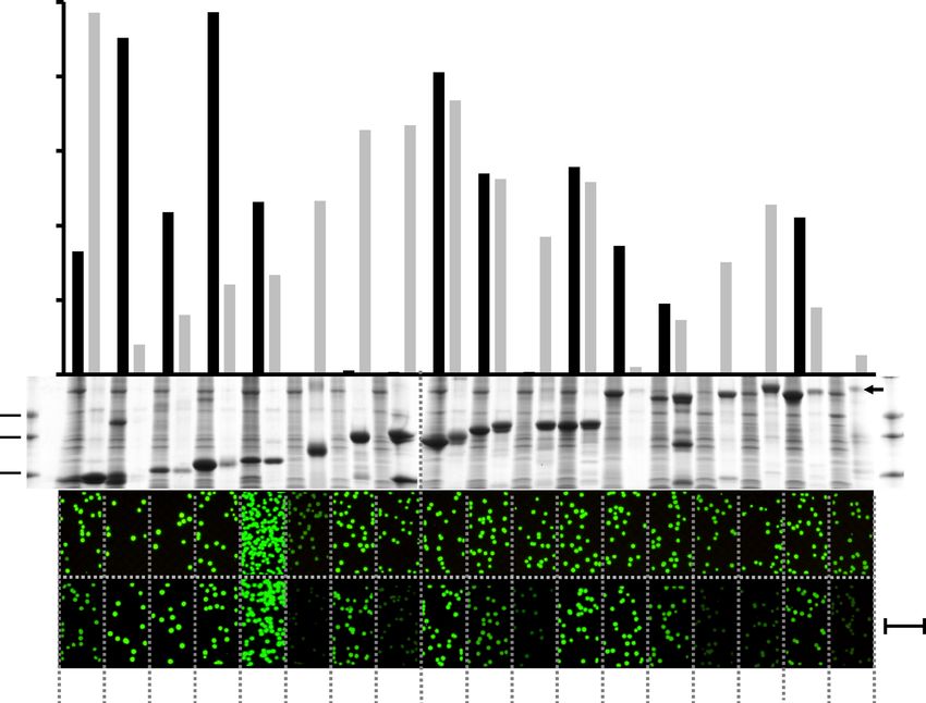
Figure 4. In vitro protein quantification and in vivo protein expression and solubility screens in E. coli.
Protein quantification of eighteen P. aerophilum test proteins (see Supplementary Table S1) expressed as
N-terminal fusions with ccGFP S11 v1 from the strong T7 promoter (bar graph, top). The ccGFP fragment
complementation assay fluorescence of soluble (black bars) and unfolded pellet fractions (gray bars) using
ccGFP 1–10 v2 (top). Arbitrary fluorescence units (A. U.). SDS-PAGE of the corresponding soluble (S), and
pellet fractions (P) (middle). Note that protein #8, tartrate dehydratase β-subunit, shows a second lower band
at ~ 13 kDa. #14, nirD protein, shows secondary bands at ~ 27 kDa and ~ 13 kDa. Original pictures of the 2 SDS-
PAGE gels showing the soluble and pellet fractions of 18 test proteins are included as Supplementary Figs. S9,
S10. In vivo solubility and expression screen using split ccGFP (lower). The same P. aerophilum test proteins
cloned with a C-terminal ccGFP S11 v1 tag on tet promoter plasmid, in E. coli BL21 (DE3) strain carrying a pET
plasmid for expression of ccGFP 1–10 v2. Fluorescence images of colonies on plates after total expression screen
by coinduction of the tagged constructs and ccGFP 1–10 v2 (upper row of colonies); or after soluble expression
screen by transient expression of the tagged constructs followed by expression of the ccGFP 1–10 v2 (lower row
of colonies). Fluorescence images of colonies were cropped from the same pictures. Note 1 cm scale bar (lower
right) illustrating the size of the colonies.
Use of the split ccGFP system for in vitro protein quantification. We measured fluorescence progress curves
for complementation of purified SR–ccGFP S11 v1 and ccGFP 1–10 v2 in 200 µl reactions in a microtiter plate
(Fig. 3). We avoided potential higher-order kinetic effects by initiating the complementation using a high con-
centration and large molar-excess of ccGFP 1–10 (800 pmol). Progress curves over a wide concentration range
could be superimposed by linear scaling (Fig. 3a). Over the range of S11 analyte tested (1.56–200 pmol) it was
not necessary to wait until the reactions approached their asymptotic limit (~ 6 h) to generate calibration curves.
For example, linear calibration curves were easily generated at 1 h (Fig. 3b), or even as soon as 6 min (Fig. 3c)
after the start of complementation. Progress curves were also measured for SR-ccGFP S11 v1 vs. either ccGFP
1–10 v1 (Supplementary Fig. S3a) or ccGFP 1–10 v3 (Supplementary Fig. S4a). After subtraction of the blank
progress curves due to formation of intrinsic fluorescence (no SR-ccGFP S11) (Supplementary Figures S3b, S4b),
calibration curves could be generated (Supplementary Figures S3c,d, S4c,d). The efficiency of complementation
was measured as a function of pH (see Supplementary Fig. S5). The complementation rate was highest above
pH 7.0, decreasing linearly with decreasing pH. Below pH 5.0 complementation was inefficient. Abosrption and
emission spectrum of the SR-ccGFP S11 v1 complemented with ccGFP 1–10 v3 showed a strong peak at 501 nm
Scientific Reports | (2021) 11:18440 | https://doi.org/10.1038/s41598-021-98149-8 6
Vol:.(1234567890)www.nature.com/scientificreports/
Figure 5. (a) Normalized progress curves for complementation of cognate and non-cognate ccGFP and GFP
fragments. Rapid complementation of cognate fragments (upper curves). ccGFP 1–10 v2 and SR-ccGFP S11 v1
(upper solid line); GFP 1–10 OPT and SR-GFP S11 M3 (upper dotted line). GFP fragments are from the original
split GFP1 derived from A. victoria. Weak complementation between non-cognate fragments (bottom curves).
Complementation between ccGFP 1–10 v2 and its non-cognate fragment SR-S11 M3 is not detectable and the
trace is at the baseline (bottom solid line). Complementation of GFP 1–10 OPT with the non-cognate fragment
ccGFP S11 v1 (lower dotted line) is ~ 4% of the final value for the cognate GFP S11 fragment (upper dotted line).
Complementation was initiated by mixing 800 pmol of each 1–10 fragment with 50 pmol of S11 fragment in
200 µl reaction wells. (b) Amino acid sequence alignment showing sequences of ccGFP S11 v1 and GFP S11 M3.
and 515 nm, respectively (Supplementary Fig. S6) indicating that the split ccGFP system has similar fluorescent
properties compared to the full length ccGFP E6.
Expression and solubility screens of 18 control proteins from P. aerophilum. To test the utility
of the split ccGFP screen for quantifying protein expression in vitro, 18 control proteins (see Supplementary
Table S1) with different expression and solubility levels from P. aerophilum, carrying the C-terminal ccGFP S11
v1 tag, were expressed in E. coli at 37 °C from pET vectors using the strong T7 promoter, and split into soluble
FP1, facilitating comparison with
and pellet fractions. The same proteins had previously been used to test split G
the performance of the new ccGFP (this work). Aliquots of the soluble fractions and solubilized denatured inclu-
sion bodies, processed to allow direct comparison (see “Methods”) were complemented with ccGFP 1–10 v2 and
the final fluorescence values were measured (Fig. 4, top). The final fluorescence was reflective of the amount of
the corresponding protein in the soluble and inclusion body fractions as revealed by SDS-PAGE (Fig. 4, mid-
dle). Since several of the urea-solubilized inclusion bodies visibly aggregated soon after dilution in the assay
buffer, the successful complementation implies that the ccGFP 1–10 fragment rapidly binds the S11 tag during
the dilution step before the formation of insoluble aggregates, committing the chromophore to form regardless
of the subsequent solubility of the complex. We previous observed rapid binding of complementary split GFP
fragments1.
Scientific Reports | (2021) 11:18440 | https://doi.org/10.1038/s41598-021-98149-8 7
Vol.:(0123456789)www.nature.com/scientificreports/
Resin on column
Thrombin
cleavage
CFP 1-10
ccGFP 1-10 discard Talon
Thrombin
wash bead
cleavage site CFP
ccGFP wash
6His SR 430 ex. 488 ex.
Resin on column 480 em. 520 em.
CFP S11
ccGFP S11
Talon beads
Flow-through
430 ex. 488 ex.
480 em. 520 em.
430 ex. 488 ex.
480 em. 520 em.
Figure 6. Double-labeling experiment with split C
FP22 and ccGFP (this work). Sulfite reductase with an
N-terminal 6HIS and ccGFP S11 v1 tag, and a C-terminal CFP S11 M3 tag was complemented with an excess of
CFP 1–10 OPT and ccGFP 1–10 v2 (left). Talon metal affinity beads were added to capture the complex, washed
and the beads imaged to show CFP and GFP bound (middle). After cleavage of the protein on the beads by
added thrombin, the beads were washed and the resin and flow through imaged to reveal where the CFP and
GFP localized (right). As expected, the ccGFP was retained on the beads (upper right), while the CFP was found
in the flow-through (lower right).
Estimating total protein expression in vivo in living bacterial cells. To estimate total protein expression in living
E. coli, the C-terminally ccGFP S11 v1-tagged protein (expressed from the moderately strong AnTET regu-
lated promoter) and the ccGFP 1–10 v2 detector protein (expressed from the very strong IPTG inducible T7
promoter) were co-expressed (see “Methods”). Referring to Fig. 4, upper row of fluorescent colonies, the fluo-
rescence can be easily detected regardless of the solubility of the protein (as estimated from the SDS-PAGE of
the soluble and pellet fractions of the same protein expressed alone from the strong T7 promoter (Fig. 4, SDS
gel (middle)). As previously noted for split GFP1, this is consistent with a model where the 1–10 fragment can
rapidly bind the S11 tag as soon as it appears in the cell, committing the complex to folding and chromophore
formation regardless of the subsequent fate (soluble or aggregated) of the S11-tagged protein of interest. As
expected, colonies expressing protein #6 (polysulfide reductase subunit) are fainter than colonies expressing the
other 17 control proteins, because its expression leads to the accumulation of large amounts of a red product,
absorbing the blue 488 nm excitation light.
Estimating soluble protein expression in vivo in living bacterial cells. To estimate soluble expression in an E.
coli colony assay, the S11-tagged proteins were expressed first from the moderate AnTET regulated promoter,
then the expression was shut off (see “Methods”). After resting for 1 h to allow the proteins to remain soluble
or become aggregated according to their intrinsic properties, and for any remaining AnTET inducer to dif-
fuse out, the ccGFP 1–10 v2 detector protein was then expressed from the strong T7 promoter to help insure
a molar excess of the 1–10 protein. Under these conditions, the 1–10 fragment should only bind soluble and
accessible S11 tagged protein molecules. Referring to Fig. 4, (lower row of fluorescent colonies), with the excep-
tion of protein #7, nucleoside diphosphate kinase (see below), the fluorescence is reflective of soluble expression
as estimated from the SDS-PAGE of the soluble and pellet fractions of the same protein expressed alone from
the stronger T7 promoter (Fig. 4, SDS gel (middle), and Supplementary Table 1). The in vivo solubility of the
protein #7 is higher from the moderate AnTET regulated promoter (based on colony fluorescence, Fig. 4 bot‑
tom row of colonies) compared to its expression from the strong T7 promoter (SDS gel lanes, Fig. 4, middle, and
Supplementary Table 1). This is consistent with our earlier o bservation1 using SDS-PAGE that showed protein
#7 tagged with GFP S11 M3 is partially soluble as expressed from the moderate pTET AnTET promoter, and
Scientific Reports | (2021) 11:18440 | https://doi.org/10.1038/s41598-021-98149-8 8
Vol:.(1234567890)www.nature.com/scientificreports/
insoluble expressed from pET T7 promoter1. Taken together, the behavior of the 18 control proteins tagged with
the ccGFP S11 v1 is consistent with earlier findings with the optimal GFP S11 M3 fragment1, suggesting that the
optimized ccGFP S11 v1 tag does not strongly perturb the solubility behavior of the fusion proteins.
Testing cross‑complementation between GFP and ccGFP split protein fragments. To test the
ability of ccGFP fragments to recognize GFP fragments and vs. versa, complementation reactions were set up
between the non-cognate pairs, i.e. ccGFP 1–10 v2 with GFP S11 M 31, and GFP 1–10 O PT1 with ccGFP S11 v1
(Fig. 5a). Complementation reactions were also set up between cognate pairs of fragments i.e. ccGFP 1–10 v2
with ccGFP S11 v1, and GFP 1–10 OPT with GFP S11 M3. Referring to Fig. 5a, under the conditions of the assay,
the GFP 1–10 OPT fragment weakly complements with ccGFP S11 v1, yielding a fluorescence value after ~ 12 h
and ~ 4% that of its cognate interaction with GFP S11 M3. Notably this reaction had not reached completion at
12 h. CFP 1–10 O PT22 also weakly complements with ccGFP S11 v1 but with lower efficiency compared to GFP
1–10 OPT (Supplementary Fig. S7). In contrast, under the same conditions, ccGFP 1–10 v2 does not detectably
complement with GFP S11 M3 (Fig. 5a). Amino acid sequences of ccGFP S11 v1 and GFP S11 M3 were aligned
with each other in Fig. 5b.
Double‑labeling experiments. To ascertain the utility of the new split ccGFP in cyan and green fluores-
cent protein double-labeling experiments, we tagged the soluble protein sulfite reductase from P. aerophilum
with an N-terminal ccGFP S11 v1, and a C-terminal GFP S11 M3 fragment22. The construct had an N-terminal
6His tag for capture by metal affinity resin, followed by a thrombin tag for selective cleavage and release from
the bead. Referring to Fig. 6, after mixing the tagged complex with CFP 1–10 O PT22 and ccGFP 1–10 v2, the
fluorescent complex was captured on Talon resin. Imaging the washed beads with the appropriate excitation and
emission filters revealed the expected cyan and green fluorescence. The complemented CFP was recovered in the
wash after cleavage by thrombin, while the ccGFP was retained on the resin via the 6His tag (Fig. 6).
Discussion
The optimized ccGFP 1–10 v2 contains 32 mutations relative to the wild type ccGFP protein, and 9 mutations
compared to the folding optimized full-length ccGFP E6 (Fig. 1). Most of the mutations are likely required for
the enhanced phenotypes, as unlike conventional error prone mutagenesis where mutants are not subjected to
recombination, the DNA shuffling (fragmentation and homologous recombination of the 30 best performing
clones per cycle of evolution) used here typically result in ‘backcrossing as you go’, i.e. removal of non-essential
or neutral stochastic mutations and amplification of beneficial mutations as seen previously31,32. Optimized
ccGFP S11 v1 contains three mutations relative to the starting ccGFP S11 E6 fragment (Fig. 1). These significantly
improve complementation with the 1–10 fragment (Fig. 2c). The autofluorescence phenotype in ccGFP 1–10
v1 and v3 is intriguing. Apparently, the chromophore residues in the 1–10 fragment are capable of forming a
population of fluorescent states even without the S11 strand. Here, we find autofluorescence of the ccGFP 1–10
v1 can be eliminated by three mutations (D78Y, Q85R, and A109V yielding ccGFP 1–10 v2) without strongly
effecting complementation. Since backcrossing was not done after selecting this single triple mutant of ccGFP
1–10 v1, further work will be needed to ascertain whether all of the three mutations are required.
Various strategies have been used to apply selective pressure during the evolution of split fluorescent proteins,
based on the intended application. For example, starting from a superfolder red fluorescent protein ‘sfCherry’20,
Bo Huang et al. evolved an improved split sfCherry for CRISPR-based protein labeling21. Initially the authors
applied selective pressure by inserting a long disrupting peptide loop between strands 10 and 11 in the full-length
sfCherry prior to mutation and selection of brighter clones. When co-expressed from different constructs, the
complementation of the resultant mutant sfCherry 1–10 and S11 modestly improved, likely because the peptide
loop insertion did not apply sufficient selective p ressure21. In a subsequent paper the authors co-expressed the
sfCherry fragments as separate polypeptides during the mutation and s election26. Not surprisingly, these frag-
ments resulted in much brighter complementation relative to the starting sfCherry fragments and appeared
well-suited for co-expression in vivo, such as after CRISPR labeling. The authors also used the new split cherry
in one experiment to label synapse p artners26. Others have previously noted that versions of GFP 1–10 that are
prone to aggregation when expressed alone, can be rescued when co-expressed with GFP S111. Thus, split proteins
evolved for co-expression may perform adequately when used for routine protein labeling in cells constitutively
expressing the fragments, but may fail in the panoply of more stringent applications described in the introduc-
tion (above) where expression of the S11-tagged protein and 1–10 are separated in time and space. It remains to
be seen if this is a limitation of the split sfCherry fragments of the Huang lab. Thus in the present work, and in
our earlier work with split GFP1, we maximized selective pressure during evolution, transiently expressing the
1–10 fragment alone, and then shutting off expression to allow ample time for it to aggregate prior to subsequent
expression of the S11 fragment.
We had considered starting with soluble monomeric Azami green, rather than the insoluble ccGFP used here,
but decided against this as having a cysteine-free split protein system was an important goal for eliminating the
possibility of disulfide formation, and Azami green contains four cysteines. We anticipated that replacing the
cysteines with amino acids serine or alanine could reduce folding yield, potentially offsetting any advantages of
using Azami. Indeed, after we had initially evolved a soluble ccGFP, replacing the cysteines with alanine or serine
greatly reduced folding yield, and three additional rounds of mutagenic shuffling and selection were required
to restore it as ccGFP E6 which is highly soluble and fluorescent expressed at 37 °C. Nonetheless, after extensive
engineering of the ccGFP fragments, the resulting ccGFP 1–10 optima are ~ 50% soluble when expressed at 37 °C
in E. coli. It is tempting to postulate that starting with a more soluble fluorescent protein, other than the initially
soluble ccGFP used here, might have resulted in an even more soluble ccGFP 1–10. This is not necessarily the
Scientific Reports | (2021) 11:18440 | https://doi.org/10.1038/s41598-021-98149-8 9
Vol.:(0123456789)www.nature.com/scientificreports/
Autofluorescence Complementation rate
ccGFP 1-10 version (Fig. 2a) (Fig. 2b) Refolding yield (mg/ml) In vivo assay In vitro assay
ccGFP 1·10 v1 Yes Fast 0.85 No Yes
ccGFP 1-10 v2 No Fast 0.5 Yes Yes
ccGFP 1·10 v3 Yes Slower 2.5 No Yes
Table 1. Summary of three versions of ccGFP 1–10 and their suggested uses for in vivo and in vitro assays.
case. For example, in the case of sfGFP, (which is exceptionally soluble and which was specifically engineered
to resist misfolding and still fluorescent even when fused to misfolding proteins), the GFP 1–10 fragment was
initially mostly insoluble1. It required extensive evolution to make it ~ 50% soluble and capable of complementing
GFP S11 in trans1. Partial solubility of ccGFP 1–10 should not preclude its use in a variety of applications. Indeed,
optimized GFP 1–10 too is only ~ 50% soluble when expressed at 37°C1, and it is widely used by other groups in
both eukaryote and prokaryote cell types (vide supra, Introduction, this manuscript). Finally, in keeping with
the behavior of overexpressed proteins, both optimized GFP 1–10 and ccGFP 1–10 are more soluble expressed
at lower temperatures (data not shown), and as illustrated in “Methods” section (vide infra, this manuscript) the
inclusion bodies obtained by forcing overexpression at 37 °C can be a ready source of substantially pure 1–10
for in vitro applications after washing, unfolding in guanidine, and diluting ~ 20-fold in buffer. In the future,
additional increases in solubility of the 1–10 fragments might be achieved by further increasing the stringency
of selection by attaching partially soluble proteins during evolution, as was done for the S11 fragments here.
The split ccGFP 1–10 v2 and S11 v1 complement nearly threefold faster than the corresponding split GFP
fragments, reaching 80% completion in 2 h rather than 6 h (Fig. 5a). When quantifying S11-tagged proteins in
lysates (Fig. 4), it is not necessary to wait until the complementation reaction asymptotically approaches comple-
tion. Reproducible linear calibration curves can be generated at 1 h (Fig. 3b) or as early as 6 min (Fig. 3c). The
complementation is sufficiently robust to allow quantification of solubilized inclusion bodies (Fig. 4). In living
bacterial cells, transient expression of S11 tagged proteins followed by expression of the 1–10 fragment gives
colony fluorescence reflective of soluble protein (Fig. 4). In contrast, co-expression of S11 tagged protein along
with 1–10 produces fluorescence proportional to total expression. The results of protein solubility test for 18
control proteins from P. aerophilum using the split ccGFP 1–10 v2 and S11 v1are in good agreement with in vitro
solubility test and with those using our previously published split G FP1 indicating that ccGFP 1–10 v2 is efficient
for both in vivo and in vitro assays (Supplementary Table 1). The − 8 charged variant of ccGFP 1–10 v1, ccGFP
1–10 v3, yields more soluble protein compared to ccGFP 1–10 v1 during refolding (2.5 mg/ml vs. 0.85 mg/ml),
and might be preferred for in vitro assays, since it is a simple matter to eliminate the interference of autofluo-
rescence by subtracting a blank progress curve measured for the 1–10 alone (Supplementary Fig. S3). While the
charged variant exhibits slightly lower autofluorescence compared to ccGFP 1–10 v1 (Fig. 2a), it complements
more slowly than either ccGFP 1–10 v1 or v2 (Fig. 2). A detailed comparision of three versions of ccGFP 1–10
and their suggested uses is included in Table 1.
The new split ccGFP provides an additional detection tool to our existing split GFP, YFP, and CFP systems and
has great potential for multiplexed labeling. However, while ccGFP 1–10 does not complement with GFP S11,
GFP 1–10 and CFP 1–10 complement ccGFP S11 at about 4% or less the efficiency of its cognate interaction with
GFP S11 (Fig. 5, Supplementary Fig. S7). Interestingly, amino acid sequence alignment of ccGFP S11 v1 and GFP
S11 M3 showed ~ 25% sequence identity but over 50% sequence similarity (Fig. 5b). Despite this limitation, the
strong preference for cognate interactions should facilitate a variety of double-labeling or multiplex experiments
as illustrated in Fig. 6 for split CFP and ccGFP. Related work in our labs suggests it should be straightforward
to mutate the interface of the ccGFP S11 so that it is no longer recognized by GFP 1–10 but still recognized by
ccGFP 1–10. Future work will show whether ccGFP fragments might cross-complement with split c herry26.
Complementation of GFP 1–10 and S11 fragments appears to be complicated by an equilibrium between a
complementation-competent monomeric 1–10 and a quiescent dimeric 1–10 that does not complement S 1123.
Early work suggested that the GFP 1–10 dimer had a lower specific activity than the monomeric species for
complementation with S111, but results were not conclusive since the studies involved gel-purified monomers
and dimers, which likely underwent slow interconversion during subsequent experiments1. More recently, by
detailed kinetic analyses, Pinaud and co-workers showed that only the monomeric form of GFP 1–10 appears
to complement with S 1123. Whether autofluorescence of ccGFP 1–10 v1 depends on dimerization/oligomeriza-
tion of the 1–10 fragments, and whether ccGFP 1–10 exhibits the same dimer/monomer equilibrium seen for
split GFP, will be the subject of future studies aimed and further understanding the ccGFP folding and assembly.
ccGFP has much greater sequence homology to the tetrameric Discosoma sp. red fluorescent protein than to
the monomeric A. victoria GFP24, which may affect its folding mechanism. Measurement of the stability of the
GFP 1–10 fragments might be possible using deuterium exchange NMR spectroscopy, and stability and kinetic
folding studies of the full-length ccGFPs (carrying the corresponding 1–10 v1 or v2 and S11 optima mutations)
would offer insight into understanding how ccGFP v2 eliminates the autofluorescence phenotype. For example,
full length ccGFP carrying the 1–10 v2 mutations might have lower thermodynamic stability compared ccGFP
carrying the 1–10 v1 mutations, possibly destabilizing a folding intermediate leading to chromophore formation
in the 1–10. The split ccGFP mutations might also confer exceptional stability to the full-length scaffold, as has
been seen previously for engineered split fibronectin d omains33. Experiments to measure the stability, folding
kinetics, and to obtain crystal structures of the full-length variants are underway.
Scientific Reports | (2021) 11:18440 | https://doi.org/10.1038/s41598-021-98149-8 10
Vol:.(1234567890)www.nature.com/scientificreports/
Methods
Engineering a monomeric version of ccGFP for improved solubility and folding. Corynactis
californica GFP (ccGFP) wildtype protein was predicted to be a tetramer, predicted monomerizing mutations
(V127E, N192R, I194E) were introduced following published protocols based on structural homology with mon-
omeric Azami Green27. The gene ordered from Blue Heron contained two unexpected wobble mutations H120Q,
N121K (CAT to CAA, and AAT to AAA, respectively). This wildtype ccGFP full length protein was evolved to
improve solubility and folding by directed evolution. The DNA coding ccGFP was PCR amplified using vector
flanking primers and was subjected to DNA fragmentation and shuffling using published p rotocols1. The library
of DNA plasmids was transformed into E. coli BL21 (DE3) gold (Novagen) competent cells for protein expres-
sion. The 1.0 O
D600nm cell stock frozen library was diluted by two sequential 400-fold dilutions and 600 µl plated
on each of 5 supported nitrocellulose membranes resting on 150 mm dia. Bauer plates containing Luria–Bertani
(LB) agar supplemented with 50 µg kanamycin/ml media as previously published1. Cells were grown overnight
at 32 °C and proteins were expressed by transferring the membrane colony side up to LB agar plates containing
50 µg kanamycin/ml media and 1 mM isopropyl-β-d-1-thiogalactopyranoside (IPTG) for 3 h at 37 °C. Clones
(~ 30 to 40) displaying the brightest fluorescence (488 nm excitation/520 nm emission) were selected, grown
overnight and frozen as 20% glycerol/LB freezer stocks at − 80 °C. Pooled plasmid preps of these clones served
as templates for PCR for the next round of evolution. After six rounds of directed evolution, sequences of the
brighter 30–40 constructs were confirmed by DNA sequencing and the brightest clone was chosen for subse-
quent modification. The six cysteine residues were mutated to alanine or serine by primer-directed PCR (C20S,
C71A, C73S, C104S, C153S, C175A). The protein was subjected to another three rounds of directed evolution
using the same protocol as indicated and the final, brightest, monomeric clone named ccGFP E6 was chosen for
engineering the split version of the protein.
Engineering an efficient split ccGFP 1–10. Using ccGFP E6 as starting scaffold, protein was first split
into two fragments: ccGFP S11 E6 (amino acids 205–221, GDTVQLQEHAVAKYFTV) and ccGFP 1–10 E6
(amino acids 1–202, MSMSKQVLK•••RHKIEHRLVRS). ccGFP 1–10 E6 aggregated and was primarily found
in inclusion bodies. Neither refolded inclusion bodies nor soluble cell lysates fractions could associate with
ccGFP S11 E6 to form a full length, fluorescent protein. ccGFP 1–10 E6 was engineered using directed evolution
as previously published and d escribed1 with the following modifications. Briefly, the DNA library of shuffled
ccGFP 1–10 was ligated into our in-house e ngineered1 pTET-SpecR with ColE1 origin, (which expresses the tetR
regulator), where expression is regulated by addition of anhydrotetracycline (AnTET). The plasmid library was
transformed into E. coli BL21 (DE3) gold (Novagen) competent cells containing the sulfite reductase–ccGFP S11
E6 tagged protein on a modified p15A vector with kanamycin resistance m arker1 where the protein of interest is
inducible with IPTG. The 1.0 OD600nm cell stock frozen library was diluted by two sequential 400-fold dilutions
and 600 µl plated on each of 5 supported nitrocellulose membranes resting on 150 mm dia. Bauer plates contain-
ing Luria–Bertani (LB) agar supplemented with 50 µg kanamycin/ml and 50 µg spectinomycin/ml as previously
published1. Cells were grown overnight at 32 °C to keep colony sizes below 0.5 mm diameter, and proteins were
expressed by transferring the membrane colony side up to an LB agar plate containing 50 µg kanamycin/ml,
50 µg spectinomycin/ml and 350 ng/ml AnTET for 3 h at 37 °C to express the ccGFP 1–10 library. The membrane
was then transferred to an agar plate containing only kanamycin and spectinomycin for 1 h to allow the AnTET
to diffuse out of the colonies to shut off expression of the ccGFP 1–10 library. This strategy allows each mutant
protein to either remain soluble, or aggregate according to their inherent propensity. The membrane was then
moved onto a new LB/agar plate containing the same antibiotics plus 1 mM IPTG to induce expression of the
SR-ccGFP S11 E6 for complementation of any soluble (mutant) ccGFP 1–10. Clones exhibiting the most rapid
development of fluorescence, indicating soluble and functional ccGFP 1–10 mutants, were selected and stored
as freezer stocks at − 80 °C in 20% glycerol/LB medium. Brighter clones were grown in 1 ml LB liquid culture
containing 50 µg kanamycin/ml, 50 µg spectinomycin/ml to 0.3 O D600nm and induced with 350 ng/ml AnTET for
3 h at 37 °C to express just the ccGFP 1–10, and after sonication and centrifugation, 20 µl of the 350 µl soluble cell
extract fractions were screened for better complementation efficiency in an in vitro assay with an excess amount
of purified SR-ccGFP S11 E6. The best 30 candidates were pooled, plasmid prepared, then PCR-amplified using
flanking primers in the vector backbone surrounding the ccGFP 1–10 insert, and subjected to three additional
rounds of directed evolution. Mutations of the top 24 optima were confirmed by DNA sequencing, revealing the
population had converged on a small subset of solutions. All the brightest optima showed some autofluorescence
(development of fluorescence in the absence of ccGFP S11 E6). The brightest and fastest version was termed
ccGFP 1–10 v1. To eliminate the autofluorescence, the top pool from the final round was replated, and imaged
after ccGFP 1–10 expression but prior to ccGFP S11 expression, and imaged again after ccGFP S11 expression.
After alignment of the images in NIH Image, the brightest clones also exhibiting the largest dynamic range
(faintest prior to, and brightest after ccGFP S11 expression) were picked and sequenced. One such clone, ccGFP
1–10 v2, was essentially non-fluorescent after expression alone. Using alignment with the structural homolog
monomeric Azami G reen27, GFP 1–10 v3, with a net − 8 charge relative to ccGFP 1–10 v1, was made by primer-
directed mutagenesis, mutating neutral surfaces residues predicted to be on the surface of the protein to charged
residues such as glutamate and aspartate: S4E, N23D, T28E, Q41E, S43E, N142E, S153E, T162E.
Engineering an efficient ccGFP S11. The ccGFP S11 E6 fragment was expressed as a C-terminal fusion
with the ‘bait protein’ hexulose phosphate synthase (HPS) (see Supplementary Table S1), (previously shown to
become less soluble with various split fluorescent protein fragments as C-terminal fusions1) as HPS-ccGFP S11
E6, and ligated into our in-house engineered1 pTET-SpecR with ColE1 origin, (which expresses the tetR regula-
tor), where expression is regulated by addition of AnTET. HPS-ccGFP S11 fusions were amplified by PCR and
Scientific Reports | (2021) 11:18440 | https://doi.org/10.1038/s41598-021-98149-8 11
Vol.:(0123456789)www.nature.com/scientificreports/
shuffled using published protocols1. For each round of directed evolution, the library of HPS-ccGFP S11 (con-
taining mutants of ccGFP S11) was transformed into E. coli BL21 (DE3) gold (Novagen) competent cells express-
ing the ccGFP 1–10 v2 protein on a modified p15A vector with kanamycin resistance m arker1 where the protein
of interest is inducible with IPTG. Optima were screened in vivo using a sequential induction protocol as previ-
ously published1. Briefly, the 1.0 OD600nm cell stock frozen library was diluted by two sequential 400-fold dilu-
tions and 600 µl plated on each of 5 supported nitrocellulose membranes resting on 150 mm dia. LB agar Bauer
plates supplemented with 50 µg kanamycin/ml and 50 µg spectinomycin/ml as previously p ublished1. Cells were
grown overnight at 32 °C to keep colony sizes below 0.5 mm diameter, and proteins were expressed by transfer-
ring the membrane colony side up to an LB agar plate containing 50 µg kanamycin/ml, 50 µg spectinomycin/ml
and 350 ng/ml AnTET for 3 h at 37 °C to express the HPS-ccGFP S11 library. Each membrane was then trans-
ferred colony side up to an agar plate containing 50 µg kanamycin/ml, 50 µg spectinomycin/ml for 1 h to allow
the AnTET to diffuse out of the colonies, shutting off expression of the HPS-ccGFP S11. This sequential strategy
allowed any mutants that interfered with the solubility of the HPS to become insoluble prior to expression of the
ccGFP 1–10 in the next step. Next membranes were moved colony side up to an LB agar plate containing 50 µg
kanamycin/ml, 50 µg spectinomycin/ml and 1 mM IPTG for 2 h to induce expression of the complementary
ccGFP 1–10 v2 from the pET plasmid. Brighter clones were selected, grown at 37 °C to 0.3 O D600nm in 1 ml LB
liquid cultures containing 50 µg kanamycin/ml and 50 µg spectinomycin/ml, and induced at 37 °C for 3 h with
350 ng/ml AnTET only to express just the HPS-ccGFP S11. Note that the medium must not contain lactose, and
the cultures should not be allowed to overgrow prior to AnTET induction, otherwise the LacUV/T7 expression
of the unwanted ccGFP 1–10 from the second plasmid may leak. After sonication and centrifugation, 20 µl of the
350 µl soluble cell extract fractions (estimated to contain less than 30 pmol of HPS-ccGFP S11) were screened
for better complementation efficiency in an in vitro plate assay using a 96-well plate fluorimeter1 with a molar
excess of refolded ccGFP 1–10 v2 (180 µl of 0.75 mg/ml refolded ccGFP 1–10, ~ 800 pmol). Clones with the fast-
est complementation rates were selected, pooled, plasmid prepped, and subjected to the next round of evolution
and screening. After two rounds of directed evolution ccGFP S11 v1 was selected. HPS-ccGFP S11 v1 solubility
was improved relative to HPS-ccGFP S11 E6, and HPS-ccGFP S11 v1 complemented with ccGFP 1–10 v2 three-
fold faster and brighter than HPS-ccGFP S11 E6 for comparable amounts of soluble fusion protein.
Expression and refolding of ccGFP 1–10 and GFP 1–10 fragments. Different versions of ccGFP
1–10 and GFP 1–10 proteins were expressed and prepared as previously d escribed1. One liter LB cultures of
BL21(DE3) cells expressing ccGFP 1–10 or GFP 1–10 constructs were grown until an O D600nm of 0.5–0.7 was
reached, protein expression was induced with 1 mM IPTG and cells were harvested after 5 h of induction at
37 °C. The cell pellets were resuspended in TNG buffer and lysed by sonication on ice. Inclusion bodies contain-
ing were obtained by centrifugation at 20,000g for 30 min, washed, and aliquoted to 75 mg per 1.8 ml eppendorf
tube as previously d escribed1. To prepare 25 ml of ccGFP 1–10 or GFP 1–10 assay solution, one vial containing
75 mg of prepared inclusion body was unfolded with 1 ml of 9 M urea in TNG buffer, centrifuged at 15,000g for
10 min, and ~ 1 ml soluble fraction was rapidly diluted by adding 25 ml of TNG buffer. Refolded protein samples
were centrifuged at 15,000g for 10 min to remove crude precipitate (minor fraction) and the soluble solutions
were filtered through a 0.2 mm syringe filter and quantified using the Bio-Rad Protein Assay Reagent Kit (Bio-
Rad). To insure ccGFP 1–10 proteins were at equal concentration for subsequent experiments, protein samples
were concentrated using a tangential flow Amicon Ultra-15 centrifugal filter device (10 kDa cutoff; Millipore)
and protein concentrations were measured and diluted to a final concentration of ~ 0.75 mg/ml. To examine
pH dependence of the complementation of ccGFP 1–10 v2 with ccGFP S11 v1, refolded ccGFP 1–10 v2 was
dialyzed against various buffers at different pH values. The protein samples at different pH were then collected,
concentrated and diluted to a final concentration of ~ 0.75 mg/ml. In vitro complementation assays were set up
as described below and final fluorescence values were recorded using a Tecan Microplate Fluorescence Reader
(Tecan).
Expression and purification of sulfite reductase‑ccGFP S11 fusion protein. Sulfite reductase (SR)
from P. aerophilum was cloned and expressed as an N-terminal fusion with ccGFP S11 E6 WT and ccGFP S11
v1 fragments in a pET vector with a N-terminal His6 tag. Briefly, 1 L cultures of BL21(DE3) cells expressing SR
with different versions of ccGFP S11 were grown to OD600nm ~ 0.5 to 0.7 and induced with 1 mM IPTG for 4 h
at 37 °C. Cells were harvested and resuspended in TNG buffer and lysed by sonication on ice for 10 min at 70%
duty cycle. Cell lysates were then centrifuged at 15,000g for 30 min at 10 °C to remove cell debris and the super-
natant was incubated with pre-equilibrated Talon® cobalt metal affinity resin (Clontech) at room temperature
(22 °C) with gentle rocking for 1 h to allow proteins to bind to resin. The proteins bound to resin was separated
from unbound protein by centrifuging 3000g for 5 min, and the pelleted resin was washed three times with ten-
fold volume excess TNG buffer before it was packed into a gravity-flow column. The column then was washed
with 50 ml of TNG buffer followed by 50 ml of TNG buffer with 5 mM imidazole, and 20 ml of TNG buffer with
20 mM imidazole to remove unbound and non-specifically bound proteins, respectively. The purified proteins
were completely eluted with 30 ml of 150 mM imidazole in TNG buffer and protein solutions were dialyzed
against TNG buffer to eliminate imidazole. Protein was quantified using the Bio-Rad Protein Assay reagent Kit
(Bio-Rad) and was concentrated to a final concentration of 5 mg/ml using a tangential flow Amicon Ultra-15
centrifugal filter device (10 kDa cutoff; Millipore).
In vitro complementation assays. In vitro complementation assays of various combinations of the
ccGFP S11 and ccGFP 1–10 were done using previously described p rotocols1. Briefly, a 96-well white microplate
(Nunc-Immuno plate, Nunc) was first blocked with 0.5% bovine serum albumin (BSA) in TNG for 10 min. Puri-
Scientific Reports | (2021) 11:18440 | https://doi.org/10.1038/s41598-021-98149-8 12
Vol:.(1234567890)You can also read