In Sickness and in Health: The Immunological Roles of the Lymphatic System - Semantic ...
←
→
Page content transcription
If your browser does not render page correctly, please read the page content below
International Journal of
               Molecular Sciences

Review
In Sickness and in Health: The Immunological Roles of the
Lymphatic System
Louise A. Johnson

                                          MRC Human Immunology Unit, MRC Weatherall Institute of Molecular Medicine, University of Oxford,
                                          John Radcliffe Hospital, Headington, Oxford OX3 9DS, UK; louise.johnson@imm.ox.ac.uk

                                          Abstract: The lymphatic system plays crucial roles in immunity far beyond those of simply providing
                                          conduits for leukocytes and antigens in lymph fluid. Endothelial cells within this vasculature are dis-
                                          tinct and highly specialized to perform roles based upon their location. Afferent lymphatic capillaries
                                          have unique intercellular junctions for efficient uptake of fluid and macromolecules, while expressing
                                          chemotactic and adhesion molecules that permit selective trafficking of specific immune cell subsets.
                                          Moreover, in response to events within peripheral tissue such as inflammation or infection, soluble
                                          factors from lymphatic endothelial cells exert “remote control” to modulate leukocyte migration
                                          across high endothelial venules from the blood to lymph nodes draining the tissue. These immune
                                          hubs are highly organized and perfectly arrayed to survey antigens from peripheral tissue while
                                          optimizing encounters between antigen-presenting cells and cognate lymphocytes. Furthermore,
                                          subsets of lymphatic endothelial cells exhibit differences in gene expression relating to specific func-
                                          tions and locality within the lymph node, facilitating both innate and acquired immune responses
                                          through antigen presentation, lymph node remodeling and regulation of leukocyte entry and exit.
                                          This review details the immune cell subsets in afferent and efferent lymph, and explores the mech-

                                   anisms by which endothelial cells of the lymphatic system regulate such trafficking, for immune
                                          surveillance and tolerance during steady-state conditions, and in response to infection, acute and
Citation: Johnson, L.A. In Sickness
                                          chronic inflammation, and subsequent resolution.
and in Health: The Immunological
Roles of the Lymphatic System. Int.
J. Mol. Sci. 2021, 22, 4458.              Keywords: lymphatic; dendritic cell; inflammation; migration; lymph node; T-cell; trafficking; high
https://doi.org/10.3390/ijms22094458      endothelial venule; adhesion molecule; chemokine

Academic Editors: Sinem Karaman,
Maike Frye and Katarzyna Koltowska
                                          1. Introduction
Received: 12 March 2021                        Immunology is at the cutting edge of medical research, and the exponential increase in
Accepted: 18 April 2021                   knowledge of this rapidly developing field has led to better understanding and improved
Published: 24 April 2021
                                          treatment of infectious diseases, autoimmune disorders and cancer. However, the vital
                                          roles that the lymphatic system plays in this exciting field are frequently overlooked, from
Publisher’s Note: MDPI stays neutral
                                          normal day-to-day immune surveillance to aiding immunological responses to pathogenic
with regard to jurisdictional claims in
                                          challenges as they arise, and resolving subsequent inflammation.
published maps and institutional affil-
                                               The lymphatic system comprises lymphatic vessels, organs that contain lymphoid tis-
iations.
                                          sue such as the spleen, lymph nodes, Peyer’s patches and thymus, and the lymph fluid that
                                          flows throughout. Unlike the blood system, the lymphatics are not a circulatory network:
                                          instead, cells and fluid move through in one direction (Figure 1A). Excess interstitial fluid,
                                          macromolecules and leukocytes exit the tissue via blind-ended afferent lymphatic vessels
Copyright: © 2021 by the author.          and enter draining lymph nodes via the subcapsular sinus. Lymph nodes are critical for
Licensee MDPI, Basel, Switzerland.        immune surveillance, providing a highly organized hub in which blood-derived naïve
This article is an open access article
                                          lymphocytes might encounter antigen and antigen-presenting cells borne in the afferent
distributed under the terms and
                                          lymph. After percolating through the lymph node, fluid and lymphocytes return to the
conditions of the Creative Commons
                                          blood circulation through efferent lymphatic vessels and the thoracic duct.
Attribution (CC BY) license (https://
creativecommons.org/licenses/by/
4.0/).

Int. J. Mol. Sci. 2021, 22, 4458. https://doi.org/10.3390/ijms22094458                                       https://www.mdpi.com/journal/ijms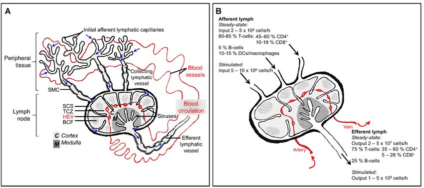
blood [4,7,8]. As with many areas of research in biological sciences, our understanding
improved massively upon the advent of monoclonal antibodies, which permitted more
thorough characterization of lymphocyte subsets, as well as other leukocytes such as den-
dritic cells (DCs), macrophages and neutrophils. Clearly, there is selectivity as to which
leukocytes are permitted to enter the lymphatic system and this review details the molec-
Int. J. Mol. Sci. 2021, 22, 4458 2 of 33
ular regulation of this, particularly that imposed by endothelial cells of high endothelial
venules, lymphatic vessels and lymph node sinuses.
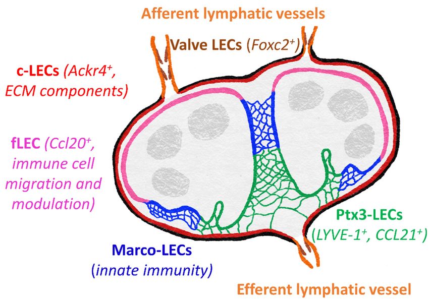
Figure 1.
Figure 1. The
The interplay
interplay between
between the the blood
blood circulation
circulation and
and lymphatic
lymphatic system,
system, and
and immune
immune cellcell composition
composition of of afferent and
afferent and
efferent lymph. (A) In peripheral tissue, fluid and macromolecules that have leaked from the blood,
efferent lymph. (A) In peripheral tissue, fluid and macromolecules that have leaked from the blood, as well as leukocytes, as well as leukocytes,
leave the interstitium and enter initial lymphatic capillaries. This lymph fluid (movement of which is indicated by blue
leave the interstitium and enter initial lymphatic capillaries. This lymph fluid (movement of which is indicated by blue
arrows) then passes into larger collecting vessels which contain intraluminal valves and are invested with contracting
arrows) then passes into larger collecting vessels which contain intraluminal valves and are invested with contracting
smooth muscle cells (SMC), finally entering the lymph node at the subcapsular sinus (SCS). Fluid and low molecular
smooth muscle cells
weight particles (
Int. J. Mol. Sci. 2021, 22, 4458                                                                                         3 of 33

                                   this, particularly that imposed by endothelial cells of high endothelial venules, lymphatic
                                   vessels and lymph node sinuses.

                                   2. In a Healthy Steady-State
                                        The immune system of a non-inflamed, non-infected individual may be described as
                                   “resting” but it is by no means static, with cell migration a constant feature throughout
                                   the blood and lymphatic networks. Studies on rodents, sheep and humans have revealed
                                   that afferent lymph draining normal healthy skin typically contains approximately 10-15%
                                   macrophages or DCs whereas efferent lymph contains only lymphocytes. Meanwhile,
                                   around 40% peripheral blood lymphocytes (mostly B-cells) are non-circulating cells and
                                   thus do not enter lymphoid tissue (Figure 1B), [4,9–12].

                                   2.1. Entering Lymph Nodes from the Blood
                                        Blood-borne lymphocytes enter the cortex of lymph nodes through specialized post-
                                   capillary venules termed high endothelial venules (HEVs), found in all secondary lymphoid
                                   organs with the exception of the spleen (reviewed in [13]. The high endothelial cells (HECs)
                                   of this vasculature are easily distinguishable from other endothelial cells by their raised,
                                   cuboidal morphology. In addition to expressing pan-endothelial cell markers such as CD31
                                   and vascular endothelial cadherin (VE-cadherin) [14], HECs have a unique transcriptional
                                   profile to permit selective lymphocyte recruitment [15] in a multi-step adhesion cascade,
                                   involving tethering, rolling, adhesion and transmigration [16–20]. The cuboidal structure
                                   of HECs results in a more irregular lining of these venules, increasing turbulence in blood
                                   flow. Consequently, HECs have greater adhesiveness for circulating lymphocytes and
                                   a collision with the vessel wall will result in a loose attachment which may last several
                                   seconds [18,21,22]. A similar collision occurring with normal blood vessel endothelial cells
                                   of the microcirculation would cause the lymphocyte to immediately rebound or adhere
                                   very briefly before being swept on by the force of the blood flow.
                                        Initial tethering of lymphocytes is supported by 6-sulpho sialyl LewisX motifs dec-
                                   orating O-glycans and N-glycans of sialomucins, a family of sulphated, fucosylated and
                                   sialylated glycoproteins termed peripheral node addressins (PNADs), which are displayed
                                   on HECs and bind the lymphocyte homing receptor L-selectin (CD62L). Rolling lympho-
                                   cytes are then activated, in part through the sheer force of blood flow but also through G
                                   protein-coupled chemokine receptors [23]. CC-chemokine ligand 21 (CCL21), immobilized
                                   on the luminal HEC surface by heparan sulphate, binds CC-chemokine receptor 7 (CCR7)
                                   on both naïve T-cells and B-cells [24–28], as well as on plasmacytoid DCs (pDCs) [29], The
                                   CXC-chemokine receptor CXCR4 also contributes to lymph node homing through binding
                                   CXCL12, which is broadly expressed on HEVs [30]. A further chemokine–receptor pair,
                                   CXCL13 and CXCR5, has been shown to regulate B-cell entry to both lymph nodes and to
                                   Peyer’s patches, lymphoid nodules in the small intestine [30,31].
                                        Curiously, chemokines are not always simply synthesized by HECs and secreted
                                   or presented apically. Although CCL21 is expressed in HEVs of murine lymph nodes,
                                   human HEVs lack CCL21 mRNA, and this chemokine, like CCL19, CXCL12 and CXCL13,
                                   is produced by stromal cells within the lymph node [15,32–35]. Additionally, chemokines
                                   may be transported in afferent lymph from peripheral tissues [36], particularly during
                                   inflammation (discussed later in this review). Subsequently, chemokines are bound at
                                   the basolateral surface of HECs, internalized and then presented on the apical surface
                                   following transcytosis by molecules such as the atypical chemokine receptor ACKR1 [37].
                                        The “inside-out” signaling induced by ligation of chemokine receptors with their
                                   cognate ligands in the tethered lymphocyte induces conformational changes of the cell
                                   adhesion molecule LFA-1 (αLβ1 integrin) [38,39]. Activated integrin LFA-1 on naïve T- and
                                   B-cells binds ICAM-1 and ICAM-2 on the endothelium, mediating firmer binding. There is
                                   also some contribution of VCAM-1, particularly in the smallest HEVs, through interacting
                                   with VLA-4 (α4β7 integrin) [40] following rapid (< 0.1s) chemokine-induced clustering
                                   of VLA-4 within lymphocyte-endothelial contact zones [41]. Additionally, in mesenteric
Int. J. Mol. Sci. 2021, 22, 4458 4 of 33
lymph nodes, VLA-4+ lymphocytes engage with the mucosal addressin adhesion molecule
MAdCAM-1 [19,20,38].
Integrin activation of HEV-homing T-cells is further amplified by the bioactive lipid
mediator sphingosine-1-phosphate (S1P), triggering signaling through its G-protein cou-
pled receptor S1P1 . This endogenous sphingolipid is produced by sphingosine kinases
Sphk1 and Sphk2, expressed in most eukaryotic cells [42], with lymph node lymphatic
endothelial cells (LECs) providing a major source of S1P [43]. A study using S1P1 −/− CD4+
T-cells showed that such cells rolled equally well as S1P1 + T-cells but exhibited reduced
firmer adherence, suggesting that S1P1 is required for optimal integrin activation [44].
After crawling on the HEC surface for several minutes, lymphocytes rapidly transmi-
grate across the endothelium and crawl along the highly organized stromal networks of the
lymph node, guided by stromal-derived chemokines [45–47]. For additional information
on intranodal positioning of leukocytes, the reader is referred to [48]. In brief, T-cells
localize to T-cell-rich areas of the paracortex, guided by the chemokines CCL21 and CCL19,
whereas B-cells enter B-cell follicles in the cortex, through CXCL13 as well as CCL21 and
CCL19. Here, these lymphocytes await possible encounters with their cognate antigens, to
be delivered to them from the periphery via afferent lymphatic vessels.
2.2. Egress from Peripheral Tissues via Afferent Lymphatics
In peripheral tissues, blind-ended capillaries of afferent lymphatic vessels form an
extensive network, removing excess fluid that has leaked out from the blood vasculature
into the interstitium and returning it to the blood circulation, but only after thoroughly
immune surveillance in downstream lymph nodes. In addition to soluble molecules, lymph
carries leukocytes: predominantly T-cells (80–90%) and dendritic cells (DCs), (5–15%), with
smaller numbers of monocytes, macrophages, B-cells and granulocytes [8,49], (Figure 1B).
The majority of afferent lymph-borne T-cells are CD4+ effector-memory T-cells, with naïve
T-cells representing only a minor subset [50,51].
In the steady state, continuous recirculation of memory T-cells through peripheral tis-
sues facilitates enhanced immunosurveillance and rapid responses to reinfection [50,52–54],
while egress of DCs from tissues is essential for maintaining peripheral tolerance [55]. DCs
capture and process soluble molecules, either self-antigens (for example, those derived
from dying cells) or non-self-antigens such as harmless environmental molecules, for
presentation via MHC class I and class II molecules. However, DC maturation is not
triggered as such antigens are at a low dose and there is an absence of “danger” signals [56].
Therefore, when such antigen is presented to the corresponding T-cells by these regula-
tory or tolerogenic DCs in draining lymph nodes, the autoreactive T-cells are deleted and
subsequently unresponsive to future similar antigenic challenges.
2.2.1. Functionally Specialized Architecture of Lymphatic Capillaries
Most leukocytes enter the proximal half of initial lymphatic capillaries, which are
also the likely sites at which fluid accesses the afferent lymphatic system [57–59]. To
aid fluid drainage, these blind-ended capillaries lack a continuous basement membrane
or smooth muscle cells, unlike larger collecting vessels (Figure 2). The endothelial cells
themselves have a highly distinctive oakleaf shape that permits them to interdigitate
and form loose discontinuous junctions [60]. Neighboring cells are “buttoned” together
with the adherens junction protein vascular endothelial cadherin (VE-cadherin) and tight
junction proteins occludin, endothelial cell selective adhesion molecule (ESAM), junctional
adhesion molecule-A (JAM-A), zonula occludens-1 (ZO-1) and claudin-5. CD31 and
lymphatic vessel endothelial receptor for hyaluronan (LYVE-1) are expressed on the flaps
in between, which form openings (0.5–1 µm) through which migrating leukocytes can
squeeze. These button-like junctions contrast with the continuous “zipper-like” junctions
in the collecting lymphatic vessels and blood vessels, which are believed to function more
as conduits rather than entry sites.Int.
Int.J.J.Mol.
Mol.Sci. 2021,22,
Sci.2021, 22,4458
x FOR PEER REVIEW 55 of
of 33
34
Figure2.2.Functional
Figure Functionalspecialization
specializationof of LEC
LEC junctions.
junctions. (A–C)
(A–C) Blind-ended
Blind-ended capillaries
capillaries of theofinitial
the initial
lym-
lymphatics
phatics contain
contain interdigitating
interdigitating oakleaf-shaped
oakleaf-shaped endothelial
endothelial cells
cells (A) held(A)together
held together with discon-
with discontinuous
tinuousjunctions,
button button junctions, where and
where adherens adherens and tight proteins
tight junctional junctional proteins
provide the provide
points ofthe points of
anchorage an-
(red)
chorage (red) in between flaps (B) through which fluid, macromolecules and migratory leukocytes
in between flaps (B) through which fluid, macromolecules and migratory leukocytes enter without
enter without compromising the integrity of the endothelium (C) [60]. Once inside the vessel, leu-
compromising
kocytes activelythe integrity
crawl alongofthe
theluminal
endothelium (C)
surface of[60]. Once inside
endothelial cells,the vessel,
passing leukocytes
into actively
pre-collector and
crawl along the luminal surface of endothelial cells, passing into pre-collector and
collector vessels. (D–E) Collector vessels LECs are of a cuboidal shape, connected by continuous collector vessels.
(D,E)
zipperCollector
junctionsvessels
(D) andLECs are of a cuboidal
surrounded shape,
by basement connectedand
membrane by continuous
smooth muscle zipper junctions
cells (D)
(grey), with
and surrounded
intraluminal by basement
valves membrane
to aid passive transitand smooth muscle
of migratory immunecellscells
(grey),
(E).with intraluminal valves to
aid passive transit of migratory immune cells (E).
2.2.2. Chemotactic Guidance
2.2.2. Chemotactic Guidance
As with leukocyte recruitment via HEVs, CCL21 and CCR7 are crucial for egress of
As with leukocyte recruitment via HEVs, CCL21 and CCR7 are crucial for egress
both T-cells and DCs from peripheral tissue [24,61–66], Table 1, whereas CCL19 is dispen-
of both T-cells and - DCs from peripheral tissue [24,61–66], Table 1, whereas CCL19 is
sable [67,68]. CCR7 effector T-cells remain resident within peripheral tissues as sentinels,
dispensable [67,68]. +CCR7− effector T-cells remain resident within peripheral tissues as
in contrast to CCR7 recirculating memory T-cells and immunosuppressive CD4+ regula-
sentinels, in contrast to CCR7+ recirculating+ memory T-cells and immunosuppressive CD4 +
tory subsets [69,70]. Curiously, both CD8 and+CD4+ T-cells + express CCR7 and yet CD4+
regulatory subsets [69,70]. Curiously, both CD8 and CD4 T-cells express CCR7 and yet
cells exit skin more efficiently than CD8+ cells [61,62,71,72]. It is possible that CCR7 func-
CD4+ cells exit skin more efficiently than CD8+ cells [61,62,71,72]. It is possible that CCR7
tion may be modulated by signals mediated by other receptors, as has been described in
function may be modulated by signals mediated by other receptors, as has been described
DCs [73]. Additionally, as T-cells are not completely absent from the lymph nodes of
in DCs−/−[73]. Additionally, as T-cells are not completely absent from the lymph nodes of
CCR7 mice [65], compensatory mechanisms must exit, either to mediate cell entry from
CCR7−/− mice [65], compensatory mechanisms must exit, either to mediate cell entry from
the blood or from peripheral tissue.
the blood or from peripheral tissue.
CCL21 is expressed constitutively by LECs [74,75], secreted, and immobilized to hep-
CCL21 is expressed constitutively by LECs [74,75], secreted, and immobilized to
aran
heparansulphates, thus
sulphates, forming
thus forminga asteeply
steeplydecaying
decayingperilymphatic
perilymphatic gradient which guides
gradient which guides
CCR7 + DCs towards lymphatic capillaries from distances of up to 90 µm [68]. Somewhat
CCR7+ DCs towards lymphatic capillaries from distances of up to 90 µm [68]. Somewhat
surprisingly, LEC-produced
surprisingly, LEC-produced heparan
heparan sulphates
sulphates areare dispensable
dispensable for
for the
theformation
formation ofofthe
the
interstitial CCL21 gradient [76]. Following specific genetic abrogation of heparan
interstitial CCL21 gradient [76]. Following specific genetic abrogation of heparan sulphate sulphate
production by
production by lymphatic
lymphatic endothelium,
endothelium, there
there isisaamodest
modestreduction
reductionininlevels
levelsofofCCL21
CCL21 atInt. J. Mol. Sci. 2021, 22, 4458 6 of 33
at the lymphatic capillary but the CCL21 gradient anchored to mesenchymal heparan
sulphates remains intact. Subsequent direct contact between DCs and LECs stimulates
endothelial calcium fluxes that in turn trigger local release of CCL21 from intracellular
depots within the trans-Golgi network and intracellular vesicles [77]. Such acute chemokine
release might attract a migrating leukocyte to enter the lymphatic vessel via a specific
entry portal [78], where physical pushing is sufficient to open the button-like junction
between LECs [60], with successive migratory cells using the same pre-formed portal in an
integrin-independent manner [79,80]. Entry of DCs is also guided by the LEC-expressed
GPI-anchored protein semaphorin 3A, through stimulating actomyosin contraction within
the DC uropod by interacting with the receptor components Plexin A1 and Neuropilin-1, to
facilitate squeezing through the button junctions into the lymphatic lumen [81]. Once inside
afferent lymphatic capillaries, DCs must actively crawl in a semi-directed manner, again
guided by CCL21 gradients [78,82] until reaching collector vessels, whereupon lymph flow
is faster and transport is passive.
A second chemotactic pathway regulating egress or retention of T-cells in peripheral
tissue is lymphatic endothelial-derived S1P [43] and its receptor, S1P1 , which is highly
expressed on recirculating CD4+ T-cells [83]. The immunosuppressive drug FTY720 (also
known as fingolimod) is phosphorylated in vivo and acts as an agonist for S1P receptors,
leading to downregulation of expression and subsequently suppressing function [84].
Such pharmacological agonism of S1P1 has been demonstrated to impair entry of CD4+
T-cells into afferent lymphatics [83] while S1P1 over-expression in CD8+ T-cells prevented
retention in intestine, kidney, salivary gland and skin [85].
Table 1. Summary of the molecules known to be involved in lymphatic trafficking of leukocytes.
Homeostasis Additional Molecules Involved under
Inflammatory Conditions
Migration into and through afferent CCL21:CCR7 [24,61–66] DCs and T-cells LYVE-1:HA:CD44 [91–95] DCs
lymphatics S1P:S1P1 [43,83–85] T-cells VCAM-1:VLA4 [96–99] DCs and
Semaphorin 3A:Plexin A1 + neutrophils
neuropilin-1 [81] DCs ICAM-1:LFA-1 [96–103] DCs, T-cells and
MR:L-selectin [86] T-cells neutrophils
MR:CD44 [87,88] T-cells ALCAM [104,105] DCs
CLEVER-1 [89,90] T-cells L1CAM [106] DCs
CD99 [99,107] DCs and neutrophils
CD31 [107] DCs
CXCL12:CXCR4 [107–113] DCs
CD69 [114] T-cells
E-selectin [99] Neutrophils
CXCL8 [99] Neutrophils
CX3CL1:CX3CR1 [115,116] DCs
ACKR2, scavenging CCL2 and CCL5
[117–120] Antigen-presenting cells and
T-cells
CCL21:CCR7 [121] Neutrophils (as well as
DCs and T-cells)
ACKR4, scavenging CCL19 [122]
Antigen-presenting cells
CCL2:CCR2 [123] DCs and Langerhans
cells
ROCK [124] DCs
Entry to lymph node via afferent CCL21 and CCL19, via CCR7 CD209 [130] Neutrophils
lymphatics and migration within sinuses [24,25,28,65,125] DCs and T-cells
MR:CD44 [88] T-cells
β1, β2, β7 and/or αv integrins [126]
T-cells
CCL1:CCR8 [127] DCs
PLVAP [128] T-cells
ACKR4 and CCR7 ligands [129] DCsInt. J. Mol. Sci. 2021, 22, 4458 7 of 33
2.2.3. Adhesion Molecules
In addition to chemotactic cues, certain adhesion molecules have been implicated
in regulating entry into the afferent lymphatic capillaries under steady-state conditions
(Table 1). Macrophage mannose receptor (MR, CD206), a C-type lectin expressed on tissue
macrophages, immature DCs, lymphatic endothelium and lymph node sinuses, has been
reported to bind leukocyte-expressed L-selectin [86] and CD44 [87,88], mediating CD4+ and
CD8+ T-cell egress from the skin. Similarly, CLEVER-1 (Common lymphatic endothelial and
vascular endothelial receptor-1) has been shown to mediate transmigration of peripheral
blood mononuclear cells across cultured lymphatic endothelial cell monolayers, although
the leukocyte-expressed binding partners are yet to be defined [89,90]. Integrins and
selectins do not appear to be involved in leukocyte entry into afferent lymphatics under
steady state conditions [80,83]. It is important to note, however, that the majority of studies
on T-cell trafficking have been carried out using adoptively transferred T-cells derived
from lymph nodes or spleens of donor mice and may not necessarily behave in a similar
manner to those of endogenous recirculating lymphocytes, or truly represent steady-state,
non-inflamed conditions.
2.3. Entering Lymph Nodes via Afferent Lymphatics
Afferent lymphatics bear lymph fluid, antigens and leukocytes to tissue-draining
lymph nodes, accessing these highly ordered structures via the subcapsular sinus (SCS),
situated below the surrounding collagen-rich capsule. Lymph nodes provide two levels of
filtration: firstly, in the SCS, providing innate immunological surveillance; and secondly in
the parenchyma, where adaptive responses may be generated. In the SCS, lymph-borne
cells and solutes are sampled by resident innate immune cells such as CD169+ macrophages
and CD11b+ DCs [131], which identify potentially dangerous particles through pattern
recognition receptors (PRRs), for retention and processing in the sinus. The second level of
filtration is more selective, permitting only low molecular weight particles (Int. J. Mol. Sci. 2021, 22, 4458 8 of 33
revealed niche-specific functional specialization and specific markers of LECs within the
lymph node, core subsets of which are common to both mouse and human [130,136]. These
five main subsets include Foxc2+ valve LECs, Ackr4+ SCS ceiling LECs, Ccl20+ SCS floor
LECs, Marco-LECs and Ptx3-LECs (Figure 3). There are also two transitional populations:
Int. J. Mol. Sci. 2021, 22, x FOR PEER REVIEW 9 of 34
bridge cells, linking ceiling LECs and floor LECS, and transitional zone LECs, between
floor LECs and Marco-LECs.
Figure 3.3. Subsets
Subsetsofoflymphatic
lymphatic vasculature
vasculature within
withinthethe
lymph node.
lymph Specialized
node. genegene
Specialized expression pro-
expression
files revealed
profiles revealedfivefive
major distinct
major LEC
distinct LECsubsets
subsetsininboth
bothmouse
mouseand
andhuman
humanlymph
lymph nodes
nodes [136]. These
include
include valve
valve cells
cells of
of the afferent and
the afferent and efferent
efferent lymphatic
lymphatic vessels,
vessels, SCS
SCS ceiling
ceiling LECs
LECs (c-LECs)
(c-LECs) and
and
floor
floor LECs (f-LECs), and
LECs (f-LECs), and medullary
medullary sinus
sinus Marco-LECs
Marco-LECs and and Ptx3-LECs.
Ptx3-LECs. Genes
Genes of the c-LECs
of the c-LECs and
and
Ptx3-LECs suggest roles in maintaining the structure of the lymph node, whereas f-LEC appear spe-
Ptx3-LECs suggest roles in maintaining the structure of the lymph node, whereas f-LEC appear
cialized for regulating lymph-borne immune cell entry and antigen presentation, with Marco-LECs
specialized for regulating lymph-borne immune cell entry and antigen presentation, with Marco-
important in innate immunity and response to pathogens. Ptx3-LECs also express numerous genes
LECs
to important
suggest in innate immunity
responsiveness and response to pathogens.
to inflammation-induced remodeling Ptx3-LECs also express
and expansion numerous
of medullary and
genes to suggest
paracortical sinuses. responsiveness to inflammation-induced remodeling and expansion of medullary
and paracortical sinuses.
ACKR4++ ceiling LECs are most similar to valve cells, expressing extracellular matrix
ACKR4 and
components ceiling LECstoare
proteins most similar
maintain to contacts
cell-cell valve cells, expressing
[136,130]. ACKR4extracellular
itself is anmatrix
atyp-
components and proteins to maintain cell-cell contacts [130,136]. ACKR4 itself +is an atypical
ical chemokine receptor, playing a vital role in regulating entry of+ CCR7 DCs to the
chemokine receptor, playing a vital role in regulating entry of CCR7 DCs to the lymph
lymph node by scavenging CCR7 ligands from the sinus lumen and creating functional
node by scavenging CCR7 ligands from the sinus lumen and creating functional CCL21
CCL21 gradients across the SCS floor [129]. Floor LECs appear more immunologically
gradients across the SCS floor [129]. Floor LECs appear more immunologically active,
active, expressing transcripts for adhesion molecules (ICAM-1, VCAM-1, Glycam1, MAd-
expressing transcripts for adhesion molecules (ICAM-1, VCAM-1, Glycam1, MAdCAM1)
CAM1) and chemokines (CCL20, CXCL4, CXCL1) suggestive of roles in leukocyte migra-
and chemokines (CCL20, CXCL4, CXCL1) suggestive of roles in leukocyte migration, as
tion, as well as immune cell function modulators (CXCL4, CSF1, BMP2/SMAD), MHC
well as immune cell function modulators (CXCL4, CSF1, BMP2/SMAD), MHC class II
class II presentation (CD74, H2-Ab1) and tolerance (PDL1) [130,136,137]. Lymph from the
presentation (CD74, H2-Ab1) and tolerance (PDL1) [130,136,137]. Lymph from the SCS
SCS then passes through medullary sinuses, coming into contact with Marco-LECs. These
then passes through medullary sinuses, coming into contact with Marco-LECs. These
cells share gene expression patterns with myeloid cells, which may indicate a role in in-
cells share gene expression patterns with myeloid cells, which may indicate a role in
nate immune functions. MARCO (macrophage receptor with collagenous structure) is a
innate immune functions. MARCO (macrophage receptor with collagenous structure) is
scavenger
a scavengerreceptor,
receptor,also
alsoexpressed
expressedby bymedullary
medullary macrophages
macrophages and aiding phagocytic
and aiding phagocytic
clearance
clearance ofof both Gram-positive and
both Gram-positive Gram-negative bacteria
and Gram-negative bacteria [138,139].
[138,139]. As
As yet,
yet, it is not
it is not
known whether it performs a similar function
known whether it performs a similar function in LECs. in LECs.
Gene
Gene expression
expressionin inPtx3-LECs
Ptx3-LECsisismostmostsimilar to to
similar that of LECs
that from
of LECs peripheral
from peripheraltissues
tis-
(although Ptx3 itself is not reported to be highly expressed in peripheral
sues (although Ptx3 itself is not reported to be highly expressed in peripheral LECs) LECs) and both
share certain
and both sharemorphological features, features,
certain morphological includingincluding
blind ends specialized
blind for fluidfor
ends specialized uptake
fluid
[60,136,140,141]. Ptx3-LEC express LYVE-1, which is a widely used lymphatic marker
[142] and has been shown to promote DC entry to lymphatic vessels in mouse dermis [91],
discussed later in this review. Macrophage mannose receptor MR is also expressed both
in peripheral LECs and Ptx-LECs, orchestrating lymphocyte egress from tissues through
interacting with the leukocyte receptor CD44 [88], as well as binding microbes and pro-Int. J. Mol. Sci. 2021, 22, 4458 9 of 33
uptake [60,136,140,141]. Ptx3-LEC express LYVE-1, which is a widely used lymphatic
marker [142] and has been shown to promote DC entry to lymphatic vessels in mouse
dermis [91], discussed later in this review. Macrophage mannose receptor MR is also
expressed both in peripheral LECs and Ptx-LECs, orchestrating lymphocyte egress from tis-
sues through interacting with the leukocyte receptor CD44 [88], as well as binding microbes
and promoting phagocytosis [143]. Additionally, expression of Ccl21 has been detected in
Ptx3-LECs [136]. Whether LYVE-1 and MR perform similar functions in Ptx3-LECs as they
do during exit from peripheral tissue remains to be demonstrated but it is possible that
these molecules and CCL21 facilitate egress of CCR7+ naïve B- and T-cells, in addition to
S1P [43,144].
The aforementioned Ptx3 is a member of the pentraxin family, which act as soluble
PRRs, binding pathogens and damaged self-proteins, promoting phagocytosis and me-
diating activation of complements [145]. However, Ptx3-LECs of the central medullary
and paracortical sinuses may also play roles in stabilizing the extracellular matrix (ECM),
as Ptx3 also binds collagen and fibrinogen-domain containing proteins (including ECM
components), [146]. Furthermore, Ptx3-LECs display the transcript for IαI (inter-alpha-
trypsin inhibitor) heavy chain 5. IαI heavy chain proteins can interact with both Ptx3
and hyaluronan (HA) [147–149], and thus it is tempting to speculate that links between
LYVE-1, HA, IαI heavy chain 5 and Ptx3 may provide further structural integrity. Another
Ptx3-LEC gene, Reln, encodes reelin, an ECM glycoprotein that serves as an important
regulator of lymphatic vasculature development in the periphery, contributing to cross-talk
between collecting lymphatic vessels and smooth muscle cells [150]. It seems likely that
reelin signaling is involved during lymph node development too, and potentially during
remodeling such as that induced during prolonged inflammatory stimuli.
2.3.2. Tolerance
Although the maintenance of peripheral tolerance is traditionally ascribed to lymphatic-
migratory CD8α+ DCs that cross-present self-antigen [151], lymph node-resident LECs
also play an important role. Such LECs express numerous peripheral tissue antigens and
can mediate deletion of specific cognate CD8+ T-cells [152]. SCS floor LECs are the most
likely of lymph node LECs to be the key players in this, firstly as their location gives
them access to lymph-borne tissue-derived antigens and secondly, because single cell
analysis has shown that they express high levels of genes suggestive of effective antigen
presentation [136]. Both human and mouse LECs express MHC class I and MHC class II
molecules [153]. However, in addition to MHC-antigen complexes, effective activation of
T-cells requires antigen-independent signals, provided by co-stimulatory molecules such
as CD40, CD80 and CD86. These molecules are virtually absent from LECs, even following
inflammatory stimuli. Instead, lymph node-resident LECs express inhibitory receptors
such as PD-L1 (CD274) and PD-L2 (CD273), which engage counter-receptors on activated
CD8+ T-cells to dampen the immune response. The reader is directed to reviews by [154]
and [135] for further details on the immunomodulatory roles of LECs.
2.3.3. The Fibroblastic Reticular Network
Once leukocytes have migrated through the floor of the SCS, further infiltration into
the paracortex is supported by fibroblastic reticular cells (FRCs) [125]. These specialized
fibroblasts synthesize and organize fibers of extracellular matrix components, including
collagen, forming a conduit network [141,155,156]. This permits lymph fluid flow while
providing a scaffold for leukocyte migration. In addition to secreting CCL19 and CCL21,
FRCs also express podoplanin [157], a heavily O-glycosylated type I protein widely used as
a LEC marker in peripheral tissue [74,158,159]. Interactions between podoplanin and the
C-type lectin-like receptor 2 (CLEC-2) [160], found on the surface of DCs and up-regulated
upon maturation, are critical for efficient transit in the FRC network [161,162]. Additionally,
podoplanin has been shown to form a complex with CCL21 that is subsequently shed [163],
potentially contributing further to the CCL21 gradient. For more detail on recent advancesInt. J. Mol. Sci. 2021, 22, 4458 10 of 33
into our understanding on DC migration through the FRC network, the reader is referred
to [164].
2.3.4. Regulation of Lymph Node Function by Afferent Lymph
Afferent lymph also plays a crucial role in ensuring the maintenance of normal lymph
node function. Studies in mice have shown that when lymph nodes are deprived of
lymph following occlusion of the afferent lymphatics, HEVs become flat walled, lumi-
nal expression of PNAd is lost and HEV-specific genes including Glycam1 are down-
regulated [165,166]. Moreover, these modified HEVs support minimal lymphocyte extrava-
sation, while macrophages disappear from the SCS. Subsequent investigations demon-
strated a crucial role for lymph-borne CCR7+ CD11c+ DCs in HEV maintenance, supporting
HEV formation and function by secreting VEGF and lymphotoxin, as well as stimulating
CCL21 production in FRCs [167,168].
2.4. Exiting Lymph Nodes via Efferent Lymphatics
Naïve lymphocytes that do not encounter their cognate antigen migrate through
medullary sinuses and the subcapsular region, before exiting the lymph node via efferent
lymphatic vessels. Evidence that this egress is non-random but rather controlled originally
came from studies by Binns and colleagues, who injected labelled sheep lymphocytes into
neonatal pigs and labelled pig lymphocytes into fœtal sheep, and then assessed the route
of exit [169,170]. Pig lymphocytes exited via the efferent lymph in the recipient sheep while
sheep lymphocytes left via the blood from the porcine lymph nodes. Thus, the stromal
cells native to the recipient animal dictated the migratory route taken by lymphocytes.
Regulation of this step is still poorly understood, although now it is known that the
“choice” between retention or egression of both B- and T-cells is made by G-protein coupled
receptors and their ligands. Lymphocyte retention is promoted by Gαi-coupled receptors
(primarily CCR7) whereas S1P1 signaling overrides this, mediating entry into cortical sinus
central branches of the lymph node [17,43,84,171–173]. Additionally, adhesion molecules
CLEVER-1 and MR are expressed on efferent lymphatic vessels and blockade of these has
been shown to reduce T-cell binding to lymph node sinuses in adhesion assays using frozen
sections of lymph node [86,90], although these findings have yet to be confirmed in vivo.
In animals (such as humans and rodents) in which lymph nodes occur in chains,
naïve T-cells can migrate from one lymph node to another, as an efferent vessel of one
lymph node serves as an afferent lymphatic for the next [174], until returning to the blood
circulation via the thoracic duct and subclavian veins, into the vena cava. Thus, under
normal conditions of homeostasis, some leukocytes recirculate through the body whereas
others are resident in both peripheral and lymphoid tissue, so that all are poised to mount
a swift immune response when necessary.
3. Acute Inflammation and Infection
One of the striking effects of inflammation or infection is the increase in numbers
of DCs egressing from the affected tissue and entering afferent lymphatic vessels [175],
orchestrated by chemokine receptors and adhesion molecules. The lymphatic system pro-
vides another layer of molecular control in the form of chemokines and counter receptors,
summarized in (Table 1).
3.1. Cell Migration in Peripheral Tissue and Entry into Afferent Lymphatics
3.1.1. DCs and T-Cells
Inflammation is an essential and complex response to biological, chemical or physical
stimuli, which must be carefully regulated and targeted, to remove potential threats to the
body but without causing excessive damage to healthy tissue [176]. In the acute phase,
the infectious agent or foreign material must be cleared, along with any dead or dying
cells that have been damaged by the injury. Inflammation-associated cytokines including
TNFα, IL-1β and IL-6 produced in the affected tissue stimulate recruitment of leukocytesInt. J. Mol. Sci. 2021, 22, 4458 11 of 33
such as monocytes, lymphocytes and neutrophils from the blood. As mentioned above,
most DCs do not circulate through the blood and instead are inherent in peripheral tissues,
especially in the skin and mucosal surfaces, where threats from pathogens and other
environmental agents are greatest. Upon maturation, DCs change from phagocytes to
professional antigen-presenting cells, with an altered chemokine receptor repertoire that
promotes increased cell motility and tropism for lymph chemokines [177]. All DC subsets
and other antigen-presenting cells such as macrophages, express an array of PRRs, which
detect molecular patterns of invading microorganisms or endogenous “danger” signals
such as those from damaged cells. Intracellular and cell surface PRRs sense a wide range
of molecules, including proteins, carbohydrates, lipids and nucleic acids. Of the many
pro-inflammatory stimuli that can induce maturation, most appear to act through TNFα
and IL-1β [178–180]. Maturation results in several phenotypic changes to enhance uptake
of antigen and subsequent presentation in draining lymph nodes, including upregulation
of MHC class II and co-stimulatory molecules CD80, CD83 and CD86. Additionally,
mature DCs downregulate expression of chemokine receptors CCR1, CCR2, CCR5 and
CXCR1, required for pro-inflammatory chemotaxis in the tissues, while exhibiting enhanced
expression of CCR7 [177].
Migratory leukocytes, however, are not the only cells undergoing phenotypic changes
upon inflammatory stimuli. The lymphatic endothelium also adopts a radically different
transcriptional profile, to selectively enhance lymphatic trafficking of specific immune
cells [96,97]. ICAM-1 and VCAM-1 expression increases dramatically following TNFα
stimulation, and antibody blockade or genetic deletion of these cell adhesion molecules
or their integrin ligands LFA-1 and VLA4 significantly reduces exit of DCs from the skin
and lymphatic migration to draining lymph nodes [96,100,101], thus impairing their ca-
pacity to induce CD8+ T-cell priming [98]. ICAM-1 accumulates on microvilli projections
surrounding adherent DCs in a manner dependent upon the conformational change of β2
integrin on the DC, promoting DC crawling over the lymphatic endothelial basolateral
surface and transendothelial migration [102]. Formation of such endothelial transmigratory
cups is induced through ligation of the lymphatic endothelial receptor LYVE-1 engaging
with its ligand hyaluronan (HA) [91], organized in a dense 400–500 nm thick glycocalyx
on the DC surface by the leukocyte receptor CD44 [92]. Disruption of LYVE-1:HA inter-
actions by gene deletion, antibody blockade or depletion of the HA glycocalyx impair
DC adhesion, transmigration and lymphatic trafficking, resulting in diminished antigen-
specific T-cell immune responses in draining lymph nodes [91]. Additionally, binding
of HA to LYVE-1 induces disruption of the VE-cadherin-lined endothelial cell junctions,
thus further aiding diapedesis [93]. Given that CD44-anchored HA extends beyond the
sphere of interaction of smaller surface molecules such as β2 integrin (with an extracellular
domain of 20 mm, [181,182]) or even selectins (50–100 nm, [183,184]), it is tempting to
speculate that LYVE-1:HA interactions form the first adhesive contacts between migra-
tory DCs and lymphatic endothelium. Additionally, as has been shown with tumour
cell glycocalyces [185], the extended dimensions of CD44:HA:LYVE-1 complexes might
constrain the lateral diffusion of smaller molecules, corralling them to promote clustering
and subsequent integrin-mediated adhesion.
At present, it is unknown whether lymph-migrating T-cells also employ integrins
when exiting inflamed peripheral tissue or indeed possess a HA-rich glycocalyx. In
addition to the molecules facilitating homeostatic trafficking, such as MR [86–88] and
CLEVER-1 [89,90], it is likely that specific leukocyte subsets are recruited by a number
of additional receptors, including ALCAM (CD166) [104,105], L1CAM (CD171) [106],
CD99 and CD31 [107]. E-selectin (CD62E) and P-selectin (CD62P) are also upregulated in
inflamed lymphatic endothelium, albeit transiently in the case of the former [96,97], and
the functional significance of this has yet to be demonstrated.Int. J. Mol. Sci. 2021, 22, 4458 12 of 33
3.1.2. Chemokines
In addition to adhesion molecules, increased immune cell recruitment to lymph
nodes via afferent lymphatic vessels is further supported by chemokines. CCL21 is not
only a homeostatic chemokine but is also upregulated during acute inflammation in both
mouse and human lymphatic endothelia [75,186–188]. Enhanced secretion of CCL21 from
LECs triggers integrin activation on DCs, stimulating DC translymphatic migration by
chemotaxis [186].
As well as promoting inflammation-induced lymphatic trafficking of DCs, CCL21
and CCR7 also support efficient egress of CCR7+ T-cells from acutely inflamed peripheral
tissue. CCR7 deficiency in splenocytes adoptively transferred into inflamed skin (following
application of Complete Freund’s Adjuvant, CFA) resulted in reduced trafficking of CD4+ T-
cells and B-cells by around 80% and 70% respectively [69]. Additionally, in a Th1-mediated
antigen-specific delayed type hypersensitivity (DTH) model in mice, CCR7 deficiency was
found to cause an accumulation of regulatory T-cells (T-regs) in the inflamed skin, which
was accompanied by enhanced suppression of inflammation [70]. As the ratio between
effector T-cells and T-regs at inflamed sites is a critical determinant for the outcome of
the inflammatory response [189] egress of T-cell subsets must be carefully regulated, and,
given that some T-cells are permitted to migrate in a CCR7-independent manner, other
chemokines are clearly involved in such extra levels of selectivity.
As under steady-state conditions, S1P plays a crucial role in mediating retention of
T-cells in inflamed sites, and tissue concentrations of this lipid mediator increase after
initiation of an inflammatory response induced by alloantigen or viral antigen, coupled
with a decrease in concentration of its precursor, ceramide [83]. This is coincidental with an
increase in the early leukocyte activation marker and C-type lectin, CD69, on virus-specific
CD8+ T-cells [114]. CD69 directly interacts with S1P1 , resulting in mutual inhibition of
surface expression and S1P1 degradation, coupled with transcriptional downregulation.
Thus, such T-cells are retained in the skin and long-lived adaptive immune memory may
be generated in peripheral tissue, to guard against future invasion by the same pathogen.
Lymphatic endothelium responds to inflammatory stimuli by increasing synthesis and
secretion of a variety of chemotactic factors in addition to CCL21 and S1P. These include
CCL1, CCL2, CCL5, CCL20, CXCL12 and CX3CL1 (chemotactic for DCs, monocytes and
T-cells bearing the receptors CCR8, CCR2, CCR5, CXCR4 and CX3CR1 respectively) and
CXCL1, CXCL2, CXCL5 and CXCL8 (neutrophil chemokines, acting through CXCR1 and
CXCR2) [96,97,99]. Expression of CXCL12 in resting lymphatic endothelium is sparse
and the CXCL12-CXCR4 chemokine-receptor pair have been shown to be redundant in
steady-state T-cell trafficking [83]. In contrast, expression of CXCL12 mRNA has been
shown to be induced in response to TNFα in cultured human dermal LECs [107], as
well as at the protein level on lymphatic vessels within mouse dermis following hapten
application [108]. Furthermore, CXCR4 is highly expressed in cutaneous MHC class
II+ DCs, and a pharmacological antagonist (4-F-benzoyl-TN14003) to CXCR4 impairs
lymph node migration of both dermal DCs and Langerhans cells by up to 50% during
the sensitization phase of contact hypersensitivity. Like CCL21, CXCL12 binds heparan
sulphate through a basic region at the N-terminus and hence may establish haptotactic
gradients in tissues [109,110]. Lymphatic-derived CXCL12 may also serve to enhance DC
survival, as CXCR4 activation has been shown to increase DC viability [111].
Like CXCL12, CX3CL1 is largely absent from resting lymphatic endothelium but is
dramatically induced following stimulation with pro-inflammatory cytokines, particularly
TNFα, through de novo RNA and protein synthesis in both primary LECs and in intact
vessels within mouse and human dermis [96,115]. CX3CL1 is an unusual chemokine in that
it is synthesized as a large (373 amino acids) type I integral membrane protein, comprising
an extracellular domain that contains a novel C-X-X-X-C chemokine motif and an extended
mucin-like stalk [190]. Expressed on several cell types, including activated vascular en-
dothelium, CX3CL1 can exist in either membrane-anchored or soluble forms [190,191]. The
membrane-bound form of CX3CL1 supports extravasation from the blood through induc-Int. J. Mol. Sci. 2021, 22, 4458 13 of 33
ing shear-resistant adhesion of leukocytes to blood endothelium, in a manner independent
of activation of either integrins or Gαi proteins [190,192–195]. Additionally, soluble forms
of CX3CL1 (generated by proteolytic cleavage with the disintegrin and metalloproteinases
ADAM10 and ADAM17) promote conventional integrin-mediated chemotaxis [196,197].
The sole receptor, CX3CR1, is widely expressed by leukocytes, including CD14+ cells
of the monocyte/macrophage/DC lineage and subsets of tissue-resident DCs and epi-
dermal Langerhans cells [194,198,199]. In inflamed lymphatic endothelium, CX3CL1 is
rapidly shed from the cell surface in a predominantly basolateral direction, promoting
transmigration of monocyte-derived DCs across monolayers of primary LECs in vitro and
supporting trafficking to draining lymph nodes in vivo [115]. However, dual blockade
of CX3CL1 and CCL21 does not yield an additive effect in transmigration assays, and
targeting CX3CL1:CX3CR1 interactions by neutralizing antibodies does not result in a
“log-jam” of DCs around lymphatic capillaries, as has been reported following disruption
of CCL21:CCR7 [63,64] or ICAM:LFA1 interactions [96]. This suggests that CCL21 and
CX3CL1 act sequentially, with CX3CL1 providing guidance for DC migration through
the interstitium by fluid phase gradients, while CCL21 forms haptotactic gradients and
facilitates docking to lymphatic endothelium.
LECs release basolateral CD9+ CD63+ exosome-rich endothelial vesicles, both consti-
tutively and also in greater numbers in response to pro-inflammatory cytokines, leading to
the formation of peri-lymphatic halos around initial vessels [116]. Proteomic analysis of
these exosomes has revealed that the cargo proteins are predominantly associated with a
motility-promoting function, such as chemokines, actin cytoskeleton regulatory proteins,
motor proteins and adhesion molecules, and include membrane-anchored CX3CL1. These
exosomes enhance formation of cellular protrusions in human monocyte-derived DCs and
promote their transit across lymphatic endothelium, in a CX3CL1-dependent manner. Such
exosome-driven transmigration is in co-operation with CCL21, and, as CCL2 and CCL5 are
also enriched within these lymphatic-derived exosomes, it is likely that they selectively aid
lymphatic homing of multiple leukocyte subsets [116].
As blood and lymphatic capillaries are so closely apposed within peripheral tissues,
it is possible that lymphatic endothelial-derived chemokines act in a paracrine fashion to
recruit more monocytes and T-cells from the blood after transfer to the luminal face of
blood vascular endothelium [200]. However, only CCL21 and CX3CL1 have been found to
be secreted basolaterally, while CCL2, CCL5 and CCL20 are secreted predominantly from
the luminal surface, into the afferent lymph [74,115,186]. Furthermore, local accumulation
of such inflammatory chemokines has been shown to be detrimental to efficient leukocyte
entry to lymphatics, with the atypical chemokine receptor ACKR2 playing a crucial role in
preventing such accrual (reviewed by [201]). ACKR2 exhibits some sequence homology
with other chemokine receptors and binds to at least 12 different CC chemokines. However,
it internalizes bound ligand and targets it for intracellular degradation, rather than con-
ventional signaling. ACKR2 is expressed in dermal LECs, as well as in other tissues which
perform a barrier function [117], ensuring that chemokines including CCL2 and CCL5 are
absent from the basolateral surface of lymphatic vessels and thus preventing congestion
around sites of entry [118]. Furthermore, ACKR2 is upregulated under inflammatory
conditions, permitting CCR2− CCR5− CCR7+ mature DCs (in preference to CCR2+ CCR5+
CCR7− immature DCs) to adhere to lymphatic endothelium [119,177]. Moreover, studies
using ACKR2-deficient mice have demonstrated the importance of this molecule in allow-
ing effective resolution of cutaneous inflammatory responses [120]. Efficient migration of
DCs and Langerhans cells from skin is also aided by an additional atypical chemokine
receptor, ACKR4, expressed by keratinocytes and a subset of LECs [122]. This receptor
scavenges stromal-derived CCL19, preventing de-sensitization of CCR7 and preserving
the responsiveness of DCs and Langerhans cells to CCL21.Int. J. Mol. Sci. 2021, 22, 4458 14 of 33
3.1.3. Neutrophils
Neutrophils are the first leukocyte population to be recruited from the blood into
inflamed and/or injured peripheral tissue, in response to cytokines, such as TNFα and
IL-1β, and chemokines such as CXCL8 (reviewed in [202]). They exhibit highly effective
anti-microbial activity, through a combination of phagocytosis and release of cytotoxic
granules and extracellular traps, before undergoing apoptosis within the tissue. Dead
neutrophils are subsequently cleared by macrophages, via the lymphatics. However, it
is now accepted that neutrophils are not always the short-lived infantrymen they were
originally believed to be, and lymph-migrating neutrophils (albeit the minority) have an
extended lifespan beyond the usual T1/2 ~ 6–12 h. In mice, following dermal immunization
with either Mycobacterium bovis bacillus Calmette-Guerin (BCG) or peptide antigen
(ovalbumin), neutrophils are the rapid responders responsible for capturing bacilli or
antigen and transporting them to draining lymph nodes via afferent lymphatics [203,204].
Ovalbumin-pulsed neutrophils display a DC-like phenotype, presenting antigens in an
MHC class II-dependent manner to induce proliferation of antigen-specific T-cells and
regulate adaptive immune responses [205].
Clearly, however, neutrophil entry into the lymphatics must be carefully regulated
and balanced, tailored to individual types of infection and inflammation. In a study of skin
inflammation elicited by topical application of CFA, lymph migratory neutrophils were
found to be exclusively CCR7+ , with CCR7−/− mice exhibiting around 60% fewer neu-
trophils in draining lymph nodes [121]. Moreover, entry of neutrophils to TNFα-stimulated
cremaster lymphatic vessels was almost completely abolished in CCR7−/− mice [206]. In
contrast, following intradermal administration of killed Staphylococcus aureus, lymph-borne
neutrophils did not require CCR7 for skin egress but rather CXCR4 and CD11b, (αM
integrin subunit, associating with β2), [207]. Curiously, pertussis toxin did not inhibit neu-
trophil migration to lymph nodes [207], suggesting that CXCR4 acts in a Gαi-independent
manner [112,113]. Additionally, ICAM-1 blockade did not reduce neutrophil egress from
skin in response to S. aureus, and thus CD11b may regulate lymphatic migration through
other interactions such as JAM-C, as has been shown in neutrophil extravasation [208,209].
β2 integrin was found to be similarly important in mediating lymphatic migration
of neutrophils following infection with M. bovis BCG, through both ICAM-1-dependent
and -independent mechanisms [99]. In vitro studies using primary human dermal LECs
and peripheral blood-derived neutrophils indicate involvement of additional adhesion and
junctional molecules in neutrophil adhesion and diapedesis, specifically E-selectin, CD99
and VCAM-1 [99].
Adhesion triggers local release of neutrophil elastase, matrix metalloproteinases
MMP8 and MMP9, and the arachidonate-derived chemorepellent lipid 12- hydroxye-
icosatetraenoate (12(S)HETE), which induce extremely targeted endothelial junctional
retraction [99,210]. These act as transient portals, permitting ~ 10-fold more rapid migra-
tion of neutrophils than DCs before resolving spontaneously, with no permanent damage
to the integrity of the lymphatic endothelium. It is interesting to note that although TNFα-
stimulated human LECs upregulate expression of various neutrophil chemokines, namely
CXCL2, CXCL5 and CXCL8 [96,97], these are secreted almost exclusively from the luminal
surface, with only CXCL8 directing transmigration [99].
3.1.4. Immunomodulation by Peripheral LECs
Adhesion interactions between DCs and LECs do not only serve to facilitate traf-
ficking but may also modulate DC function and subsequent immune responses. In vitro
studies have revealed that αMβ2 integrin:ICAM-1-mediated interactions between human
immature monocyte-derived DCs and TNFα-stimulated LECs resulted in a reduction in
surface levels of the co-stimulatory molecule CD86 on these DCs [211]. Subsequently,
such “LEC-educated” immature DCs exhibited an impaired ability to activate T-cells in
a mixed lymphocyte reaction. Crucially, this blunting of DC-mediated responses only
occurred in the absence of lipopolysaccharide (LPS), an example of a pathogen-derivedInt. J. Mol. Sci. 2021, 22, 4458 15 of 33
signal, suggestive of a role for LECs in preventing undesired immune reactions when there
is no threat of infection.
The findings from a later investigation in transgenic mice further support the con-
cept of immune regulation by LECs, where DC maturation was suppressed by the anti-
inflammatory effects of prostaglandin, secreted by LECs in inflamed skin [212]. The
transgenic mice used in this study exhibited a VEGF-C-induced expansion of lymphatic
vessels within the skin, which, in addition to the preponderance of immature, tolero-
genic DCs, established an immune-inhibitory microenvironment characterized by CD8+
T-cells with decreased effector function, increased numbers of T-regs, reduced levels of
inflammatory cytokines including TNFα, IL-6 and IFN-γ, and increased secretion of the
anti-inflammatory cytokine TGF-β1. Such a response could be expected to be most physi-
ologically relevant during resolution, following inflammation-induced expansion of the
lymphatic network and acting to limit chronic inflammation [213].
3.2. Intraluminal Crawling in Initial Lymphatic Capillaries
Upon entering afferent lymphatic vessels, leukocytes actively crawl in a semi-directed
manner along the luminal surface of initial capillaries, where the lymph flow alone
(0–30 µm/s) is insufficient to propel them towards the larger collecting vessels and drain-
ing lymph node [214–217]. Surprisingly, DCs crawl at an even slower rate than average
lymph flow in these small vessels, and downstream-directed DC migration is lymph flow
independent [82]. Instead, DC crawling is directed by CCL21 and CCR7, with intralym-
phatic CCL21 gradients induced by the low lymph flow. It is possible that CCL2:CCR2
may play a role at this stage too, as “cords” of DCs were visible inside lymphatic vessels
of CCR2-deficient mice, suggestive of a defect in intraluminal crawling rather than due
to impaired vessel entry [123]. Such lymphatic cords are also apparent when increased
levels of endogenous HA are incorporated into the CD44-held glycocalyx on DCs, by
antibody-induced potentiation of CD44:HA interactions [92]. Adhesion of these DCs to
lymphatic endothelium was found to be greatly enhanced; however, transit to draining
lymph nodes was impaired and such cells remained stuck in initial capillaries. As the
lymphatic HA receptor LYVE-1 is expressed on both the luminal and basolateral surfaces
of these lymphatic vessels [218], this may be indicative of a role for the LYVE-1:HA:CD44
axis in mediating intraluminal crawling, especially as CD44 is distributed predominantly
within the pro-adhesive uropod of crawling DCs [91,92].
In addition to chemotactic cues, DC crawling within lymphatic vessels is further sup-
ported by the Rho-associated protein kinase (ROCK), which promotes reorganization and
contraction of actomyosin filaments, thus permitting cellular contraction and de-adhesion
at the uropod [219]. ROCK plays a minor role in intralymphatic DC migration from rest-
ing tissues but is indispensable during tissue inflammation, mediating de-adhesion from
ICAM-1 [124], which is expressed on both basolateral and luminal surfaces of inflamed
lymphatic endothelium [96].
Like DCs, T-cell crawling within lymphatic vessels of inflamed mouse skin is depen-
dent upon interactions with LFA-1 integrin interactions with ICAM-1 [103]. Intriguingly,
intravital microscopy studies of inflamed mouse skin during hypersensitivity responses
have also revealed that antigen-experienced effector T-cells and DCs frequently interact
with one another within afferent lymphatic capillaries [220]. Such interactions between
polyclonal T-cells and adoptively transferred DCs that were not hapten-experienced were
found to be short-lived, with DCs interacting with multiple T-cells simultaneously, in an
MHC class II-independent manner. However, contacts between antigen-specific T-cells and
cognate antigen-bearing DCs required MHC class II and lasted much longer (>30 min in
over 60% of DC:T-cell contacts), typically forming between a DC and a single T-cell. Thus,
afferent lymphatic vessels may play a role in modulating and supporting adaptive immune
responses during leukocyte transit, even before they reach the lymph node.
Once leukocytes are in the larger pre-collector and collector lymphatic vessels, lymph
flow is much faster due to the presence of contracting smooth muscle cells surroundingYou can also read